Beta Tubulin Polyclonal antibody
Beta Tubulin Polyclonal Antibody for FC, IF, IHC, IP, WB, ELISA
Host / Isotype
Rabbit / IgG
Reactivity
human, mouse, rat and More (6)
Applications
WB, IP, IHC, IF, FC, ELISA
Conjugate
Unconjugated
322
Cat no : 10068-1-AP
Synonyms
"Beta Tubulin Antibodies" Comparison
View side-by-side comparison of Beta Tubulin antibodies from other vendors to find the one that best suits your research needs.
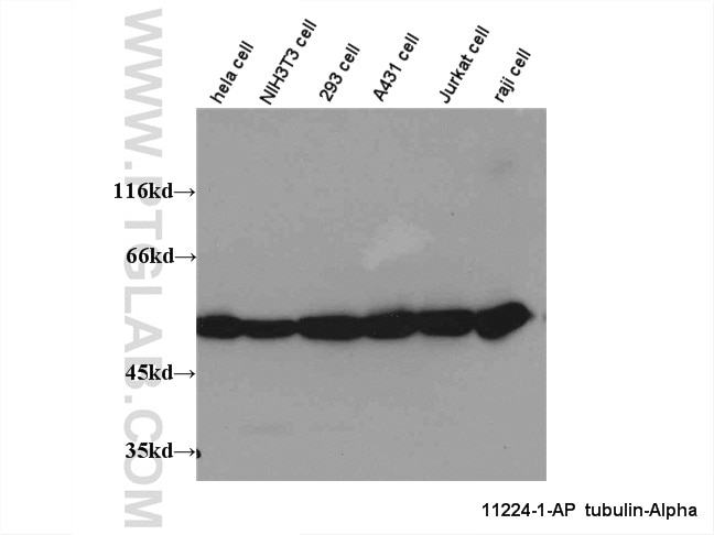
 at dilution of 1:8000 incubated at room temperature for 1.5 hours.")
 at dilution of 1:1000 incubated at room temperature for 1.5 hours.")
 with <a class='green' href='/productredirect?CatalogNo=HEK-293' target='_blank'>HEK-293</a> cells lysate 2320 ug.")
.")
.")
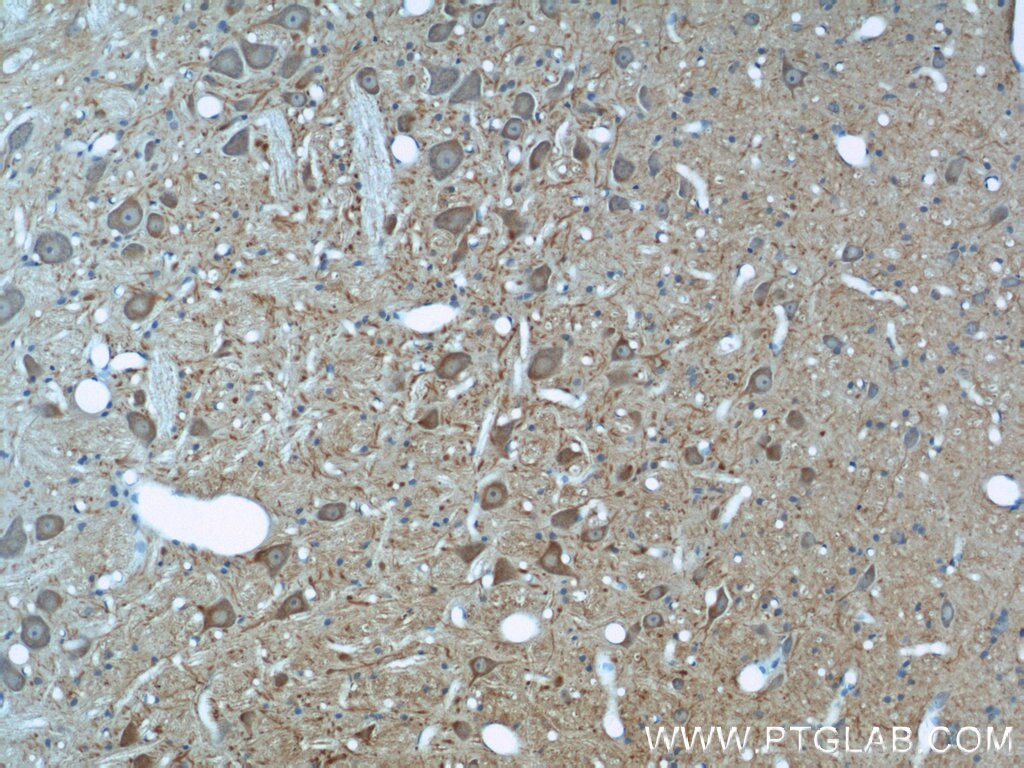
 at dilution of 1:200 (under 10x lens).")
 at dilution of 1:200 (under 40x lens).")
.")
.")
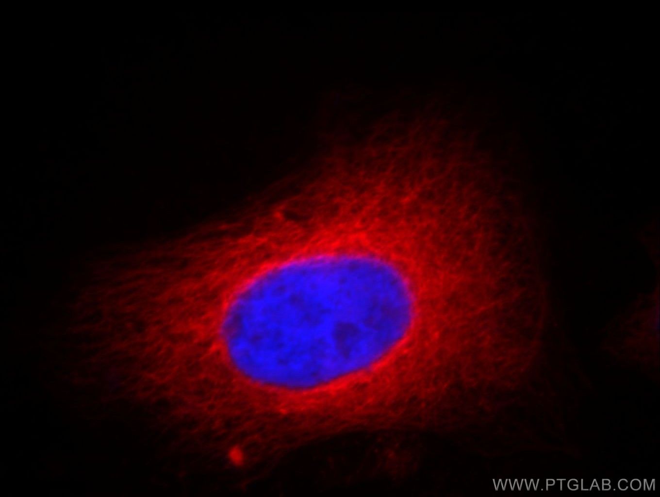
 fixed <a class='green' href='/productredirect?CatalogNo=C2' target='_blank'>C2</a><a class='green' href='/productredirect?CatalogNo=C12' target='_blank'>C12</a> cells using Beta Tubulin antibody (<a class='green' href='/productredirect?CatalogNo=10068-1-AP' target='_blank'>10068-1-AP</a>) at dilution of 1:400 and CoraLite®488-Conjugated AffiniPure Goat Anti-Rabbit IgG(H+L), <a class='green' href='/productredirect?CatalogNo=CL59' target='_blank'>CL59</a>4-Phalloidin (red).")
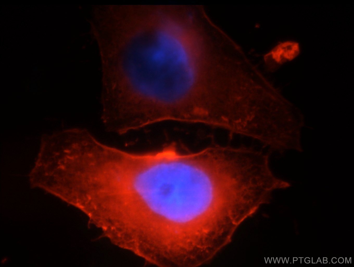
 fixed <a class='green' href='/productredirect?CatalogNo=HepG2' target='_blank'>HepG2</a> cells using <a class='green' href='/productredirect?CatalogNo=10068-1-AP' target='_blank'>10068-1-AP</a> (beta Tubulin antibody) at dilution of 1:50 and <a class='green' href='/productredirect?CatalogNo=CoraLite48' target='_blank'>CoraLite48</a>8-Conjugated AffiniPure Goat Anti-Rabbit IgG(H+L).")
 fixed <a class='green' href='/productredirect?CatalogNo=HepG2' target='_blank'>HepG2</a> cells using <a class='green' href='/productredirect?CatalogNo=10068-1-AP' target='_blank'>10068-1-AP</a> (beta Tubulin antibody) at dilution of 1:50 and <a class='green' href='/productredirect?CatalogNo=CoraLite48' target='_blank'>CoraLite48</a>8-Conjugated AffiniPure Goat Anti-Rabbit IgG(H+L).")
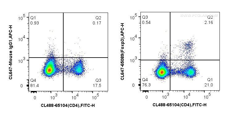
 and CoraLite®488-Conjugated AffiniPure Goat Anti-Rabbit IgG(H+L) at dilution 1:1000 (red), or 0.4 ug Control Antibody. Cells were fixed with 4% PFA and permeabilized with Flow Cytometry Perm Buffer (<a class='green' href='/productredirect?CatalogNo=PF0001' target='_blank'>PF0001</a>1-C).")
Tested Applications
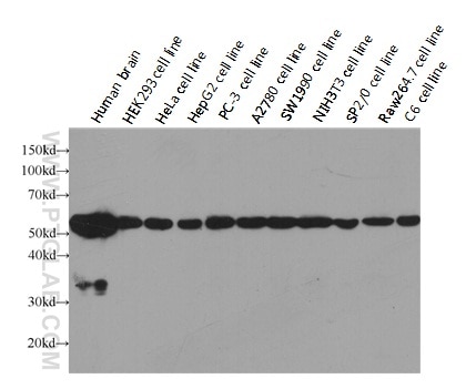
| Positive WB detected in | mouse cerebellum tissue, HeLa cells, mouse brain tissue, rat brain tissue, HEK-293 cells, Jurkat cells, HepG2 cells, A431 cells, NIH/3T3 cells |
| Positive IP detected in | HEK-293 cells |
| Positive IHC detected in | mouse brain tissue, human brain tissue, rat brain tissue Note: suggested antigen retrieval with TE buffer pH 9.0; (*) Alternatively, antigen retrieval may be performed with citrate buffer pH 6.0 |
| Positive IF detected in | C2C12 cells, HepG2 cells |
| Positive FC detected in | HeLa cells |
Recommended dilution
| Application | Dilution |
|---|---|
| Western Blot (WB) | WB : 1:500-1:2000 |
| Immunoprecipitation (IP) | IP : 0.5-4.0 ug for 1.0-3.0 mg of total protein lysate |
| Immunohistochemistry (IHC) | IHC : 1:20-1:200 |
| Immunofluorescence (IF) | IF : 1:200-1:800 |
| Flow Cytometry (FC) | FC : 0.40 ug per 10^6 cells in a 100 µl suspension |
| Sample-dependent, check data in validation data gallery | |
Published Applications
| WB | See 296 publications below |
| IHC | See 2 publications below |
| IF | See 12 publications below |
| IP | See 1 publications below |
Product Information
10068-1-AP targets Beta Tubulin in WB, IP, IHC, IF, FC, ELISA applications and shows reactivity with human, mouse, rat samples.
| Tested Reactivity | human, mouse, rat |
| Cited Reactivity | human, rat, mouse, zebrafish, pig, Caenorhabditis elegans, canine, silkworm, bovine |
| Host / Isotype | Rabbit / IgG |
| Class | Polyclonal |
| Type | Antibody |
| Immunogen | Beta Tubulin fusion protein Ag0117 相同性解析による交差性が予測される生物種 |
| Full Name | tubulin, beta 3 |
| Calculated molecular weight | 450 aa, 50 kDa |
| Observed molecular weight | 50-55 kDa |
| GenBank accession number | BC000748 |
| Gene symbol | TUBB3 |
| Gene ID (NCBI) | 10381 |
| RRID | AB_2303998 |
| Conjugate | Unconjugated |
| Form | Liquid |
| Purification Method | Antigen affinity purification |
| Storage Buffer | PBS with 0.02% sodium azide and 50% glycerol pH 7.3. |
| Storage Conditions | Store at -20°C. Stable for one year after shipment. Aliquoting is unnecessary for -20oC storage. |
Background Information
There are five tubulins in human cells: alpha, beta, gamma, delta, and epsilon. Tubulins are conserved across species. They form heterodimers, which multimerize to form a microtubule filament. An alpha and beta tubulin heterodimer is the basic structural unit of microtubules. The heterodimer does not come apart, once formed. The alpha and beta tubulins, which are each about 55 kDa MW, are homologous but not identical. Alpha, beta, and gamma tubulins have all been used as loading controls. Tubulin expression may vary according to resistance to antimicrobial and antimitotic drugs.
Protocols
| Product Specific Protocols | |
|---|---|
| WB protocol for Beta Tubulin antibody 10068-1-AP | Download protocol |
| IHC protocol for Beta Tubulin antibody 10068-1-AP | Download protocol |
| IF protocol for Beta Tubulin antibody 10068-1-AP | Download protocol |
| IP protocol for Beta Tubulin antibody 10068-1-AP | Download protocol |
| Standard Protocols | |
|---|---|
| Click here to view our Standard Protocols |
Publications
| Species | Application | Title |
|---|---|---|
Cell Complement Signals Determine Opposite Effects of B Cells in Chemotherapy-Induced Immunity. | ||
Nat Struct Mol Biol CRISPR-Cas9-based functional interrogation of unconventional translatome reveals human cancer dependency on cryptic non-canonical open reading frames | ||
Adv Sci (Weinh) EGFR-Induced and c-Src-Mediated CD47 Phosphorylation Inhibits TRIM21-Dependent Polyubiquitylation and Degradation of CD47 to Promote Tumor Immune Evasion | ||
Acta Pharm Sin B Nuciferine protects against high-fat diet-induced hepatic steatosis and insulin resistance via activating TFEB-mediated autophagy-lysosomal pathway. | ||
Acta Pharm Sin B Protocatechuic aldehyde protects cardiomycoytes against ischemic injury via regulation of nuclear pyruvate kinase M2. | ||
Autophagy Tumor-derived lactate promotes resistance to bevacizumab treatment by facilitating autophagy enhancer protein RUBCNL expression through histone H3 lysine 18 lactylation (H3K18la) in colorectal cancer |