CD206 Polyclonal antibody
CD206 Polyclonal Antibody for WB, IHC, IF-P, ELISA
Host / Isotype
Rabbit / IgG
Reactivity
human, mouse, rat and More (3)
Applications
WB, IHC, IF-P, ELISA
Conjugate
Unconjugated
526
Cat no : 18704-1-AP
Synonyms
Validation Data Gallery
Tested Applications
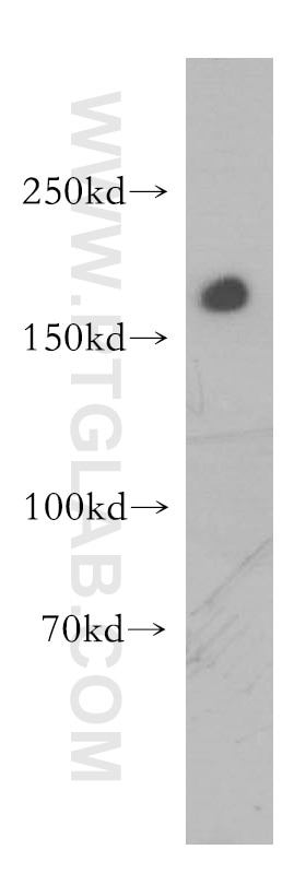
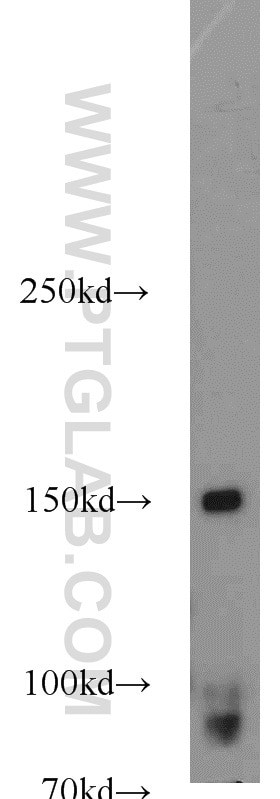
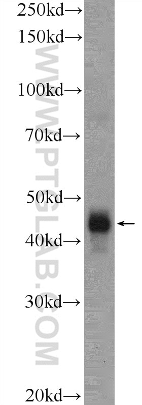
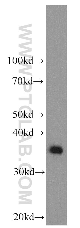
| Positive WB detected in | human placenta tissue, human kidney tissue, mouse liver tissue, rat liver tissue |
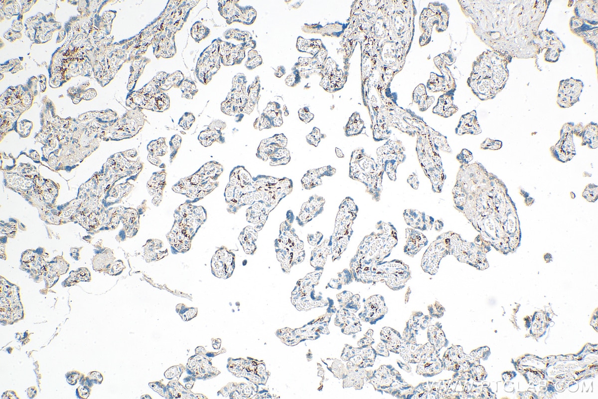
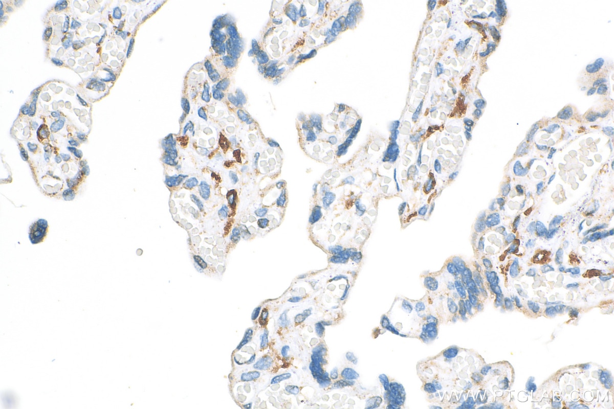
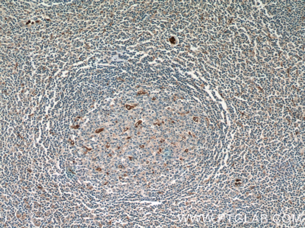
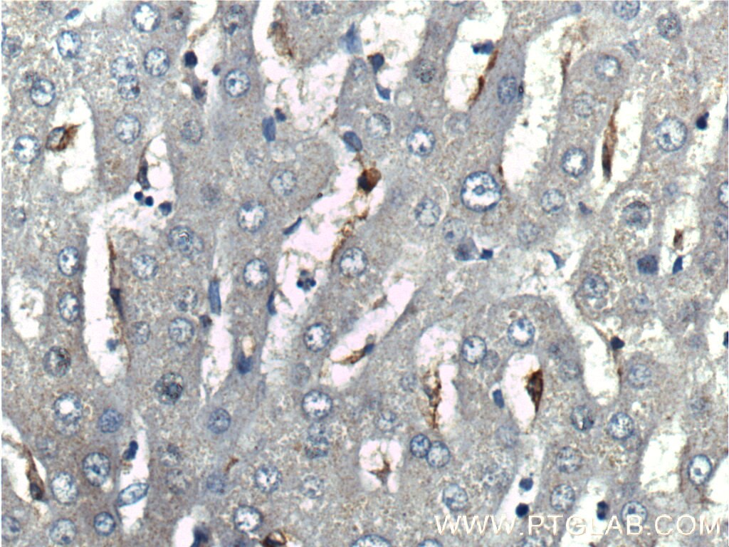
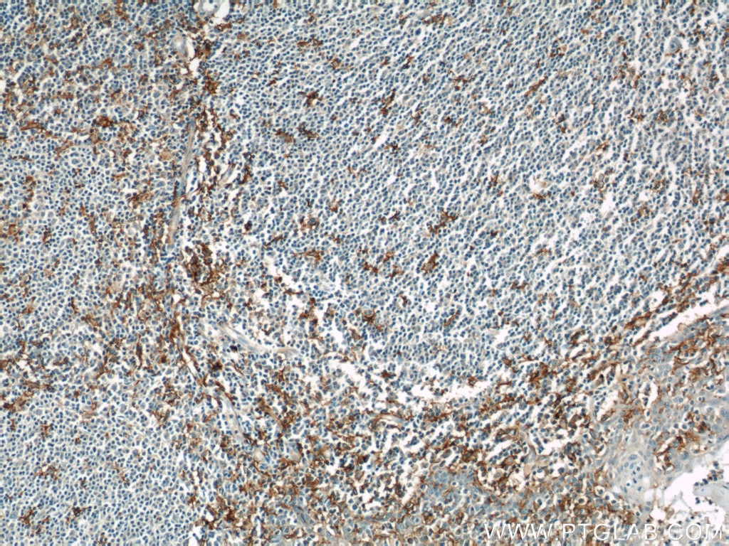
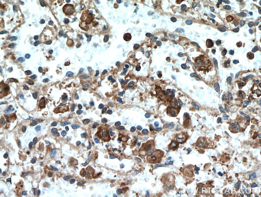
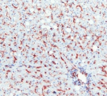
| Positive IHC detected in | human placenta tissue Note: suggested antigen retrieval with TE buffer pH 9.0; (*) Alternatively, antigen retrieval may be performed with citrate buffer pH 6.0 |
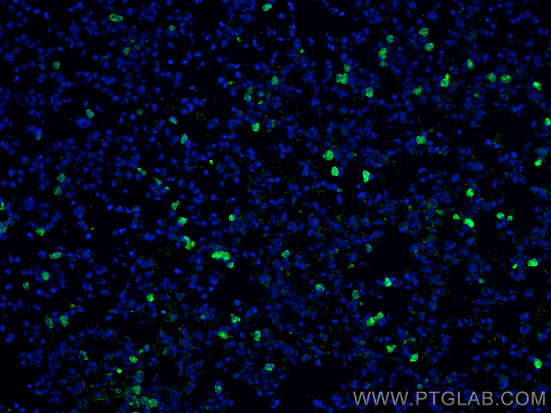
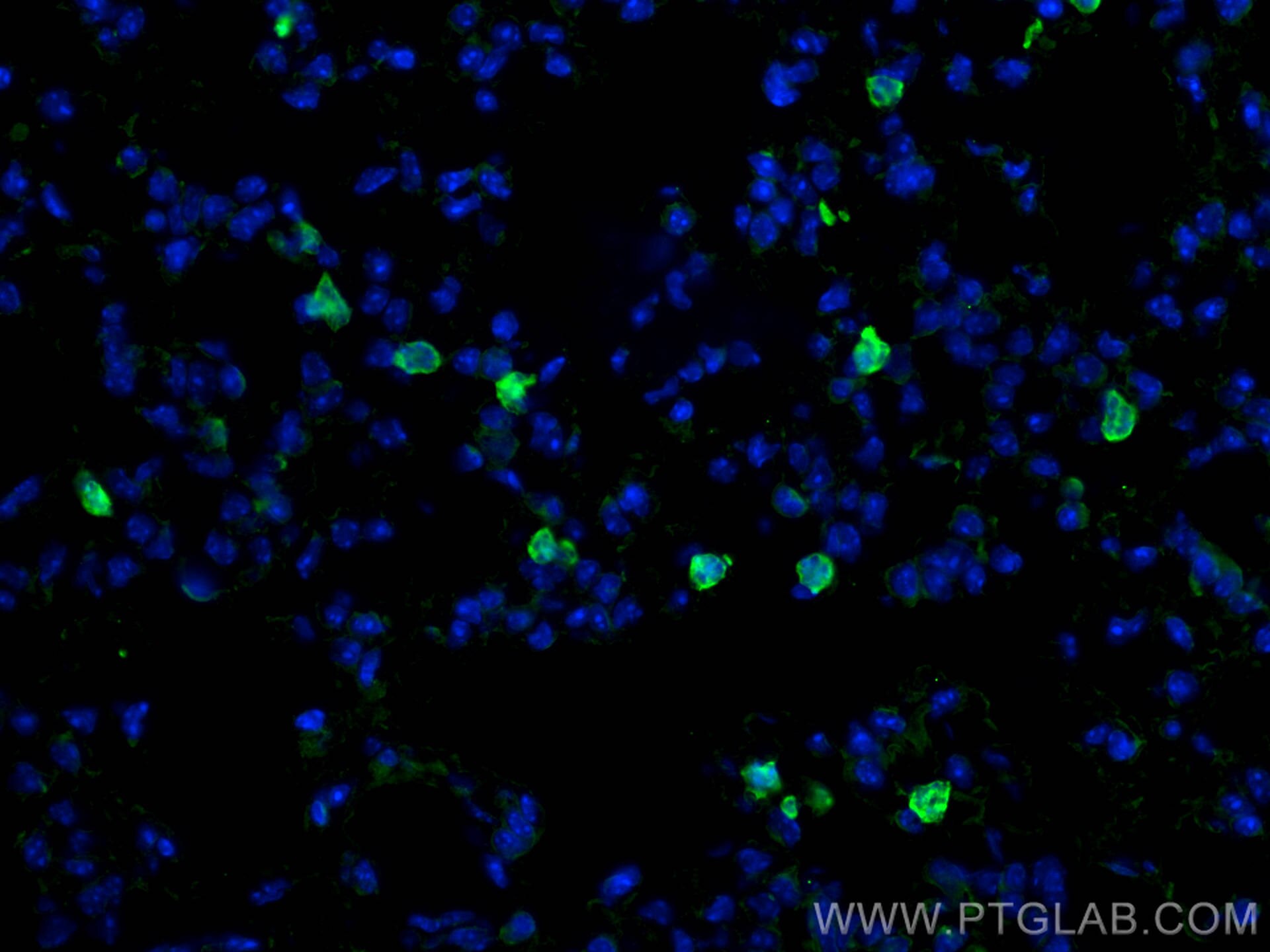
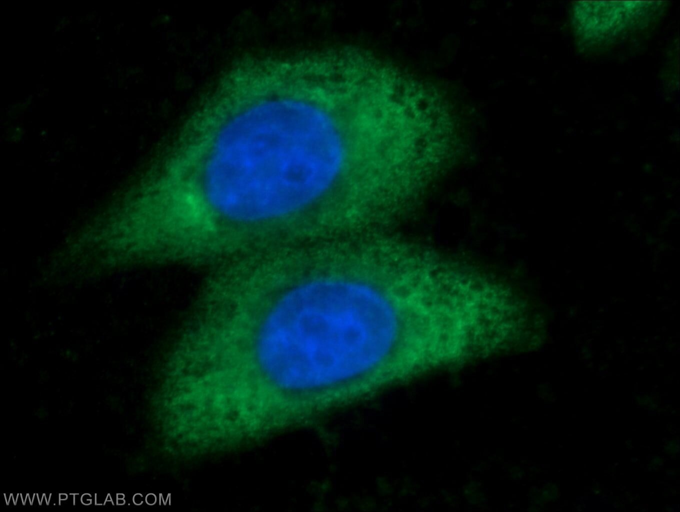
| Positive IF-P detected in | mouse lung tissue |
Recommended dilution
| Application | Dilution |
|---|---|
| Western Blot (WB) | WB : 1:500-1:1000 |
| Immunohistochemistry (IHC) | IHC : 1:2000-1:8000 |
| Immunofluorescence (IF)-P | IF-P : 1:200-1:800 |
| It is recommended that this reagent should be titrated in each testing system to obtain optimal results. | |
| Sample-dependent, Check data in validation data gallery. | |
Published Applications
| WB | See 190 publications below |
| IHC | See 110 publications below |
| IF | See 315 publications below |
| FC | See 25 publications below |
Product Information
18704-1-AP targets CD206 in WB, IHC, IF-P, ELISA applications and shows reactivity with human, mouse, rat samples.
| Tested Reactivity | human, mouse, rat |
| Cited Reactivity | human, mouse, rat, pig, rabbit, mussel |
| Host / Isotype | Rabbit / IgG |
| Class | Polyclonal |
| Type | Antibody |
| Immunogen | Peptide 相同性解析による交差性が予測される生物種 |
| Full Name | mannose receptor, C type 1 |
| Calculated molecular weight | 166 kDa |
| Observed molecular weight | 165-180 kDa |
| GenBank accession number | NM_002438 |
| Gene symbol | CD206 |
| Gene ID (NCBI) | 4360 |
| RRID | AB_10597232 |
| Conjugate | Unconjugated |
| Form | Liquid |
| Purification Method | Antigen affinity purification |
| Storage Buffer | PBS with 0.02% sodium azide and 50% glycerol pH 7.3. |
| Storage Conditions | Store at -20°C. Stable for one year after shipment. Aliquoting is unnecessary for -20oC storage. |
Background Information
Background
CD206 (macrophage mannose receptor 1) is a lectin-type endocytic receptor expressed on selected macrophages, dendritic cells, and non-vascular endothelium and plays a role in antigen processing and presentation, phagocytosis, and intracellular signaling.
1. What is the molecular weight of CD206?
The molecular size of full-length CD206 is 170-180 kDa, depending on the exact tissue-specific glycosylation pattern (PMID: 19427834). Additionally, CD206 can be cleaved off and a soluble form (sMR) lacking the tail, with a slightly lower molecular weight, can be released to the cell medium (PMID: 9722572).
2. What is the subcellular localization of CD206?
CD206 is a type I membrane protein composed of a large extracellular multidomain, a transmembrane domain, and a short cytoplasmic tail. It is present at the plasma membrane and in endosomes, as CD206 undergoes constant recycling between the plasma membrane and endosomal compartment.
3. Is CD206 post-translationally modified?
CD206 undergoes quite extensive post-translational modifications, predominantly N-linked glycosylation that affects ligand binding recognition and affinity (PMID: 22966131).
4. Can CD206 marker be used as a marker of M2 macrophages?
The activation of macrophages with various stimuli leads to their polarization into classical (M1) or alternatively activated (M2) subtypes spectrums and both subtypes differ in their regulatory and effector functions (PMID: 24669294). Pathogens and IFN-γ promote M1 polarization, while IL-4 released during parasite infections and allergen response promotes M2 polarization. Classically, the markers of M2 macrophages include CD206, as well as arginase-1 (ARG1; https://www.ptglab.com/products/ARG1-Antibody-16001-1-AP.htm), CD163 (https://www.ptglab.com/products/CD163-Antibody-16646-1-AP.htm), and thrombospondin 1 (TSP1/ THBS1; https://www.ptglab.com/products/TSP1-Antibody-18304-1-AP.htm).
5. How can you polarize macrophages into M2 direction?
One of the most commonly used methods is stimulation by the addition of IL-4 cytokine. We recommend using our animal-free human IL-4 (https://www.ptglab.com/products/recombinant-human-il-4.htm).
Protocols
| Product Specific Protocols | |
|---|---|
| WB protocol for CD206 antibody 18704-1-AP | Download protocol |
| IHC protocol for CD206 antibody 18704-1-AP | Download protocol |
| IF protocol for CD206 antibody 18704-1-AP | Download protocol |
| Standard Protocols | |
|---|---|
| Click here to view our Standard Protocols |
Publications
| Species | Application | Title |
|---|---|---|
Adv Mater Osteoimmunity-Regulating Biomimetically Hierarchical Scaffold for Augmented Bone Regeneration. | ||
Bioact Mater Reprogramming macrophages via immune cell mobilized hydrogel microspheres for osteoarthritis treatments | ||
Bioact Mater Highly active probiotic hydrogels matrixed on bacterial EPS accelerate wound healing via maintaining stable skin microbiota and reducing inflammation | ||
ACS Nano Glutamine Antagonist Synergizes with Electrodynamic Therapy to Induce Tumor Regression and Systemic Antitumor Immunity. | ||
Adv Sci (Weinh) Colorectal Cancer-Derived Small Extracellular Vesicles Promote Tumor Immune Evasion by Upregulating PD-L1 Expression in Tumor-Associated Macrophages. |
Reviews
The reviews below have been submitted by verified Proteintech customers who received an incentive forproviding their feedback.
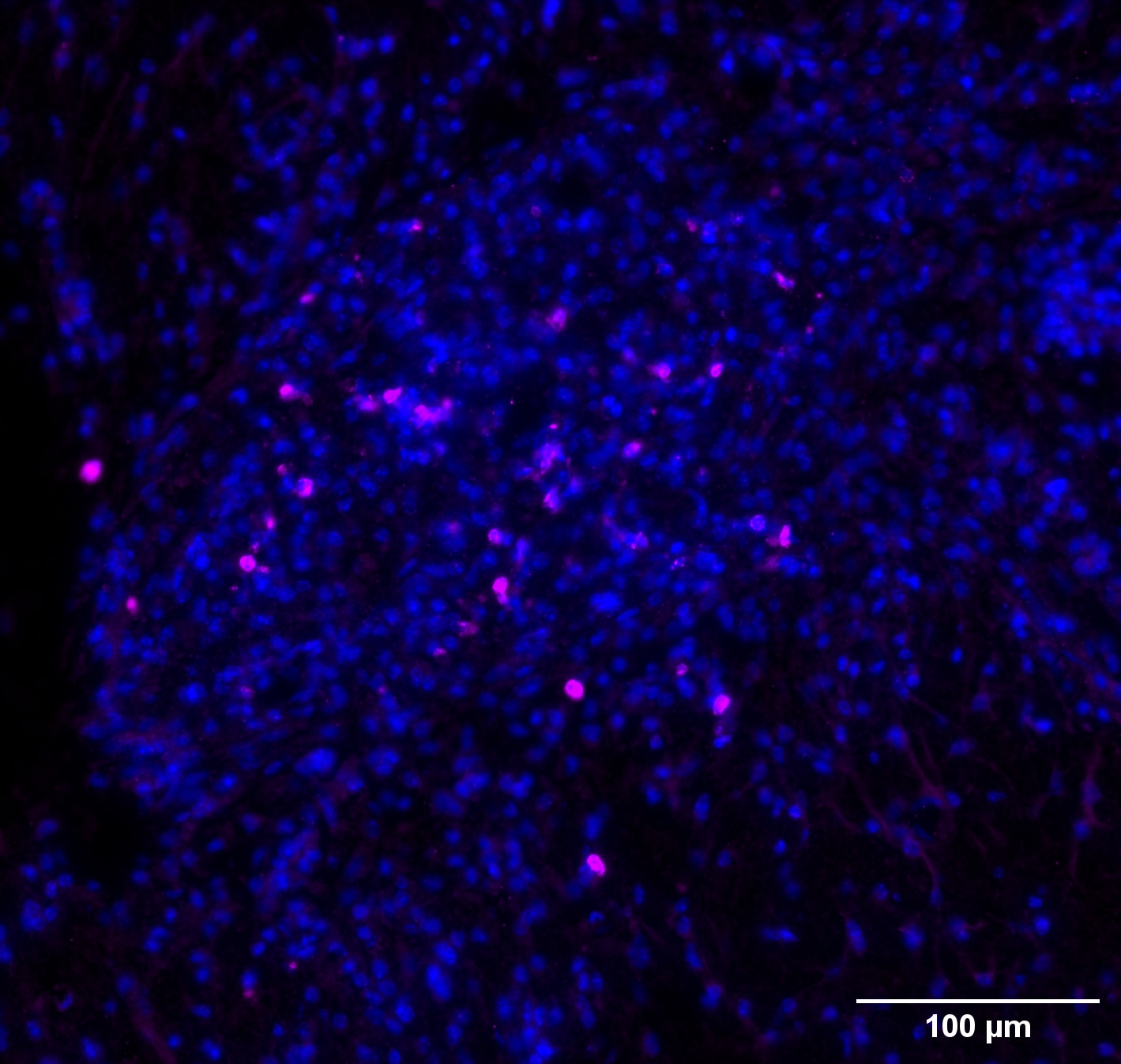
FH Marion (Verified Customer) (12-09-2024) | DAPI (blue) & CD206 (magenta) This antibody was tested to reveal CD206+/Iba-1+ cells in a murine AD model. Iba-1 staining is not presented here. The antibody works fine for IF on mouse tissue.
 |
FH Murali (Verified Customer) (07-28-2022) | This amazing antibody works well for human IHC staining.
 |
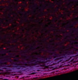
FH Uthra (Verified Customer) (10-14-2021) | The antibody works fine for IF.
 |
FH Nethaji (Verified Customer) (05-08-2019) | The antibody works fine for western and ICC.
|

