PER1 Polyclonal antibody
PER1 Polyclonal Antibody for WB, IHC, IF/ICC, ELISA
Host / Isotype
Rabbit / IgG
Reactivity
human, mouse, rat
Applications
WB, IHC, IF/ICC, ChIP, ELISA
Conjugate
Unconjugated
Cat no : 13463-1-AP
Synonyms
Validation Data Gallery
Tested Applications
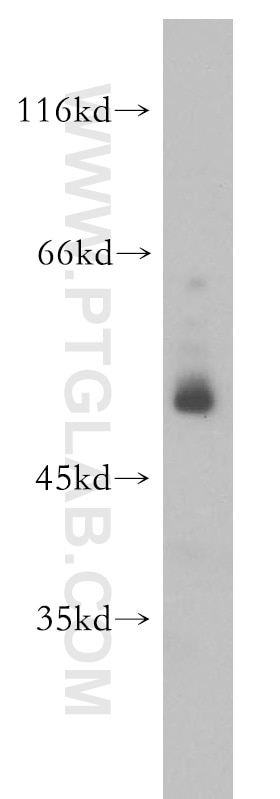
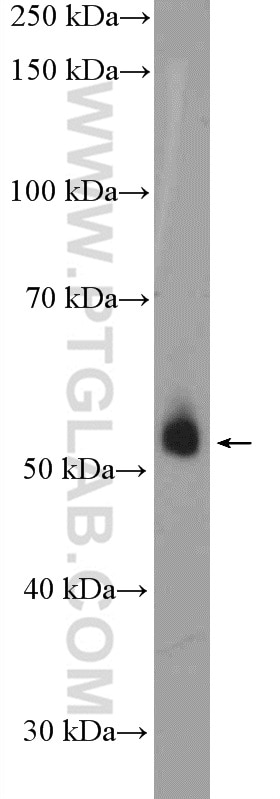
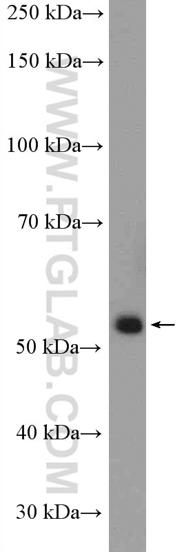
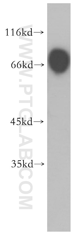
| Positive WB detected in | A549 cells, human brain tissue, mouse kidney tissue, mouse liver tissue |
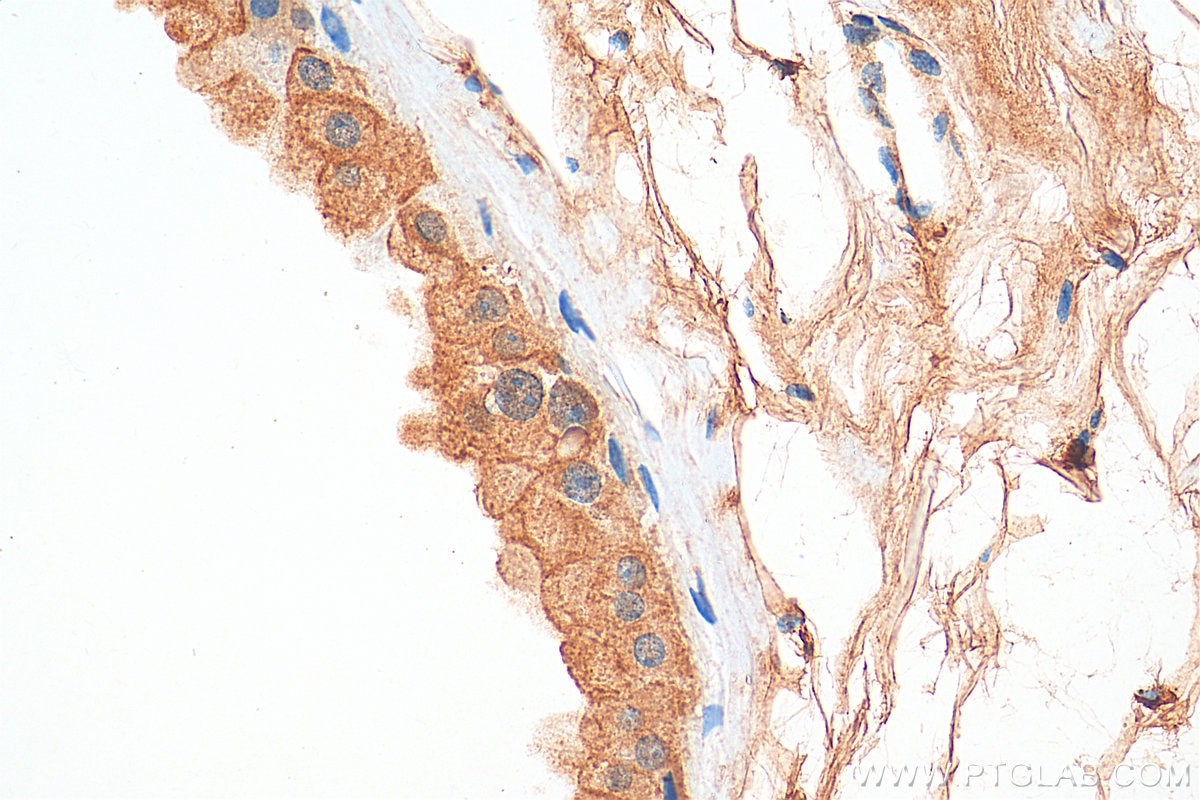
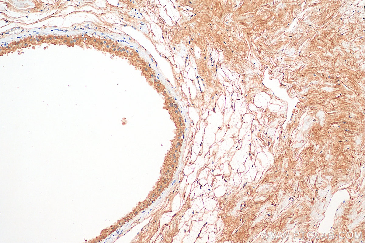
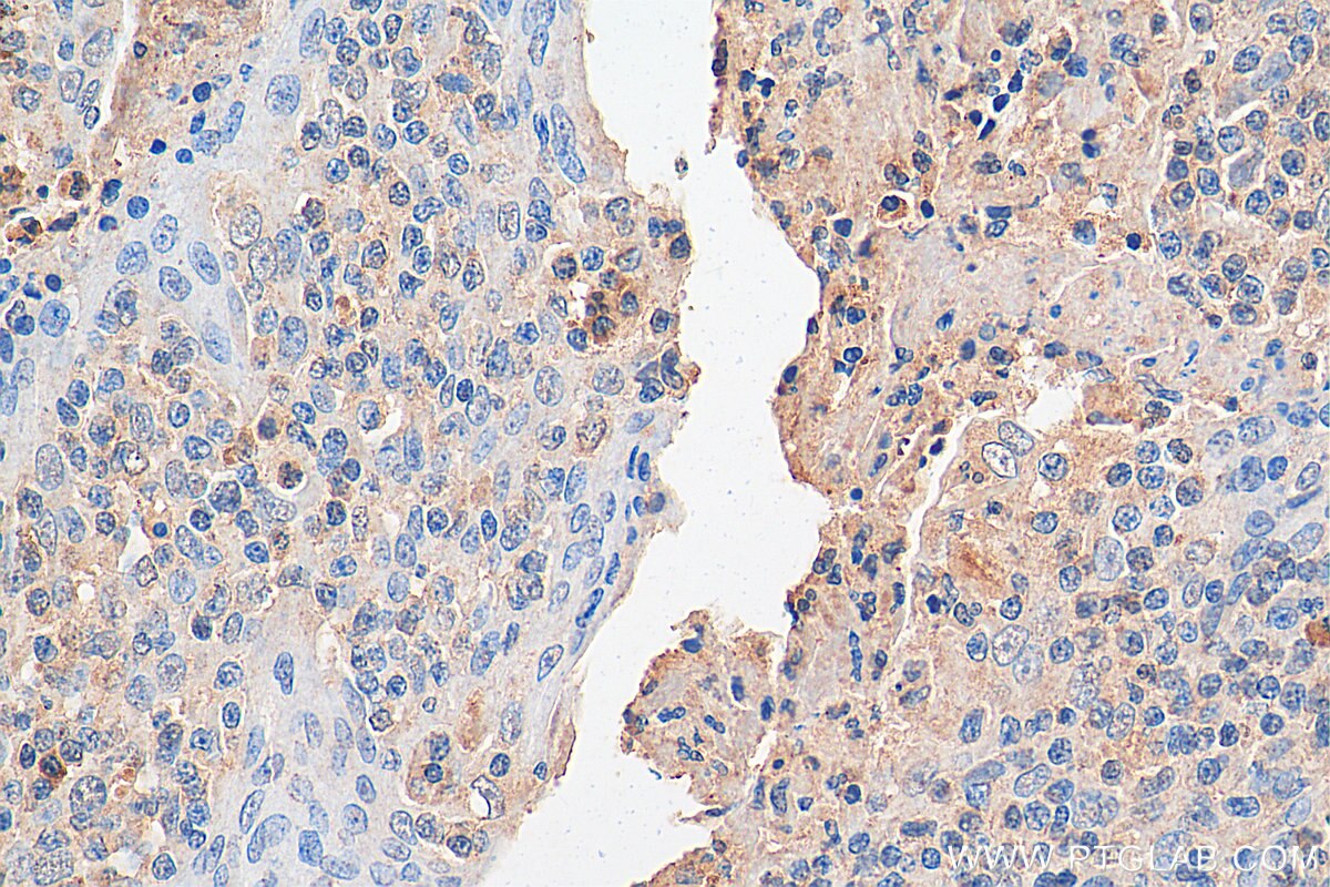
| Positive IHC detected in | human breast cancer tissue, human tonsillitis tissue Note: suggested antigen retrieval with TE buffer pH 9.0; (*) Alternatively, antigen retrieval may be performed with citrate buffer pH 6.0 |
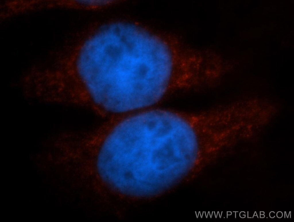
| Positive IF/ICC detected in | Hela cells |
Recommended dilution
| Application | Dilution |
|---|---|
| Western Blot (WB) | WB : 1:500-1:3000 |
| Immunohistochemistry (IHC) | IHC : 1:50-1:500 |
| Immunofluorescence (IF)/ICC | IF/ICC : 1:20-1:200 |
| It is recommended that this reagent should be titrated in each testing system to obtain optimal results. | |
| Sample-dependent, Check data in validation data gallery. | |
Published Applications
| WB | See 11 publications below |
| IHC | See 3 publications below |
| IF | See 1 publications below |
| ChIP | See 1 publications below |
Product Information
13463-1-AP targets PER1 in WB, IHC, IF/ICC, ChIP, ELISA applications and shows reactivity with human, mouse, rat samples.
| Tested Reactivity | human, mouse, rat |
| Cited Reactivity | human, mouse |
| Host / Isotype | Rabbit / IgG |
| Class | Polyclonal |
| Type | Antibody |
| Immunogen | PER1 fusion protein Ag4249 相同性解析による交差性が予測される生物種 |
| Full Name | period homolog 1 (Drosophila) |
| Calculated molecular weight | 1290 aa, 136 kDa |
| Observed molecular weight | 50-55 kDa |
| GenBank accession number | BC028207 |
| Gene symbol | PER1 |
| Gene ID (NCBI) | 5187 |
| RRID | AB_2161536 |
| Conjugate | Unconjugated |
| Form | Liquid |
| Purification Method | Antigen affinity purification |
| Storage Buffer | PBS with 0.02% sodium azide and 50% glycerol pH 7.3. |
| Storage Conditions | Store at -20°C. Stable for one year after shipment. Aliquoting is unnecessary for -20oC storage. |
Background Information
PER1 is a master regulator of circadian rhythm and functions in the nucleus to repress expression of the central circadian clock genes. The periodicity of PER1 abundance, nuclear translocation, and transcriptional repression is regulated by PER1 phosphorylation, ubiquitination, and proteasomal degradation [PMID: 21514639]. It is one component of the circadian clock mechanism which is essential for generating circadian rhythms. It can influences clock function by interacting with other circadian regulatory proteins and transporting them to the nucleus [PMID: 9856465, 20930143]. In addition to bands of the predicted molecular mass of 140 kDa, additional PER1 species of 55 and 45 kDa is detected that is predominantly nuclear [PMID: 11726537] .
Protocols
| Product Specific Protocols | |
|---|---|
| WB protocol for PER1 antibody 13463-1-AP | Download protocol |
| IHC protocol for PER1 antibody 13463-1-AP | Download protocol |
| IF protocol for PER1 antibody 13463-1-AP | Download protocol |
| Standard Protocols | |
|---|---|
| Click here to view our Standard Protocols |
Publications
| Species | Application | Title |
|---|---|---|
Cell Metab Dysfunctional circadian clock accelerates cancer metastasis by intestinal microbiota triggering accumulation of myeloid-derived suppressor cells | ||
Theranostics Development of human cartilage circadian rhythm in a stem cell-chondrogenesis model. | ||
J Transl Med Prognostic risk factors of serous ovarian carcinoma based on mesenchymal stem cell phenotype and guidance for therapeutic efficacy | ||
Int J Mol Sci Uncovering the Novel Role of NR1D1 in Regulating BNIP3-Mediated Mitophagy in Ulcerative Colitis | ||
J Agric Food Chem Capsaicin Attenuates Oleic Acid-Induced Lipid Accumulation via the Regulation of Circadian Clock Genes in HepG2 Cells. |
Reviews
The reviews below have been submitted by verified Proteintech customers who received an incentive forproviding their feedback.
FH Priya (Verified Customer) (01-05-2023) | The antibody works well. I have been using this antibody for human cardiomyocytes, human keratinocytes cell lines, and mouse tissues such as the liver and skin.
|
