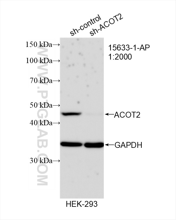
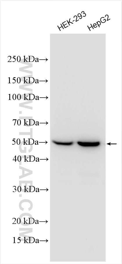
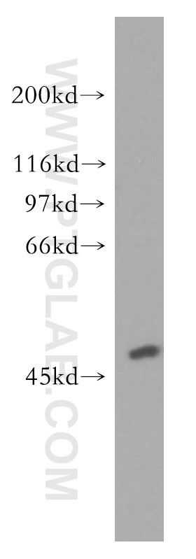
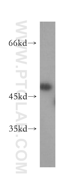
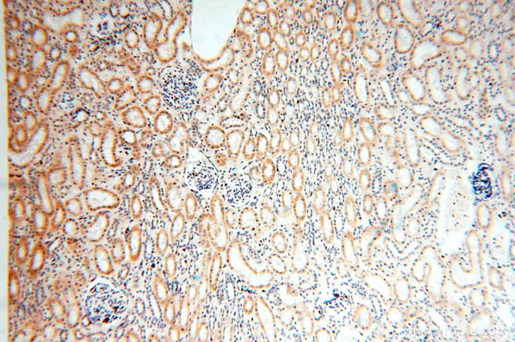
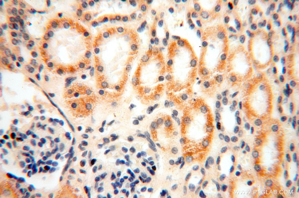
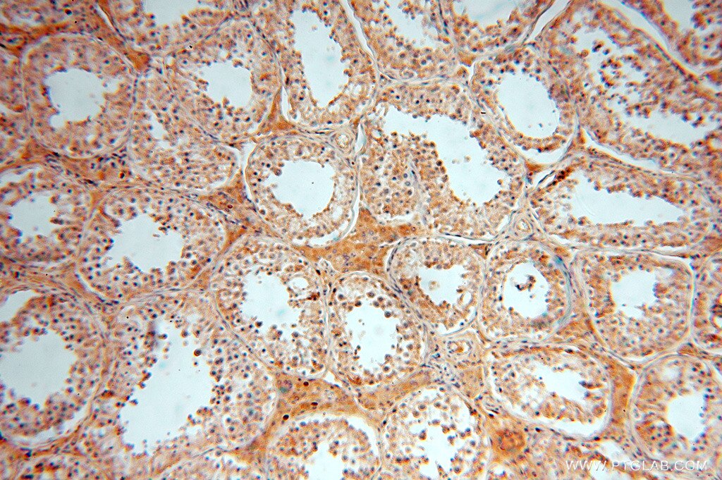
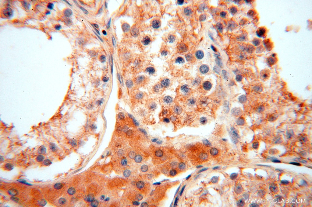
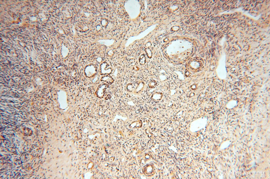
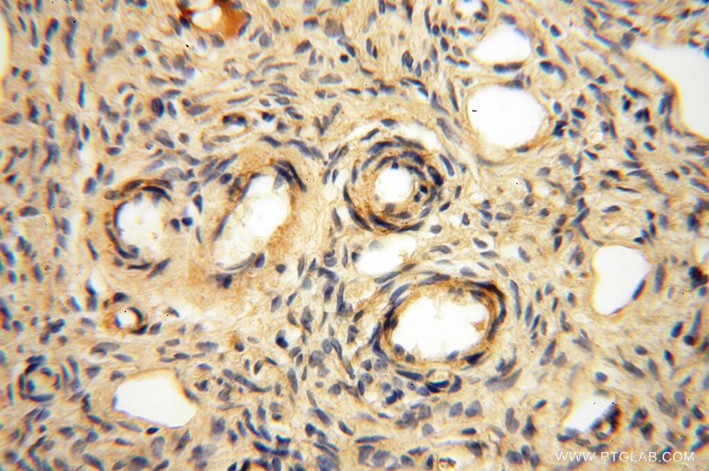
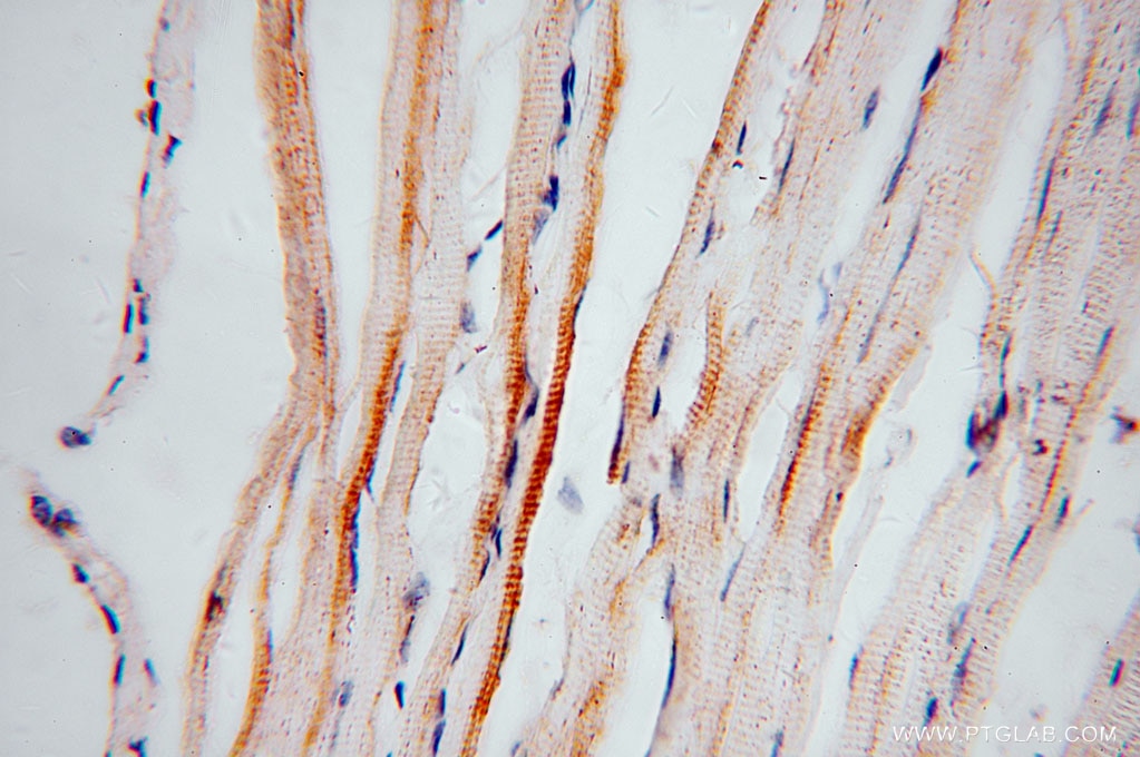
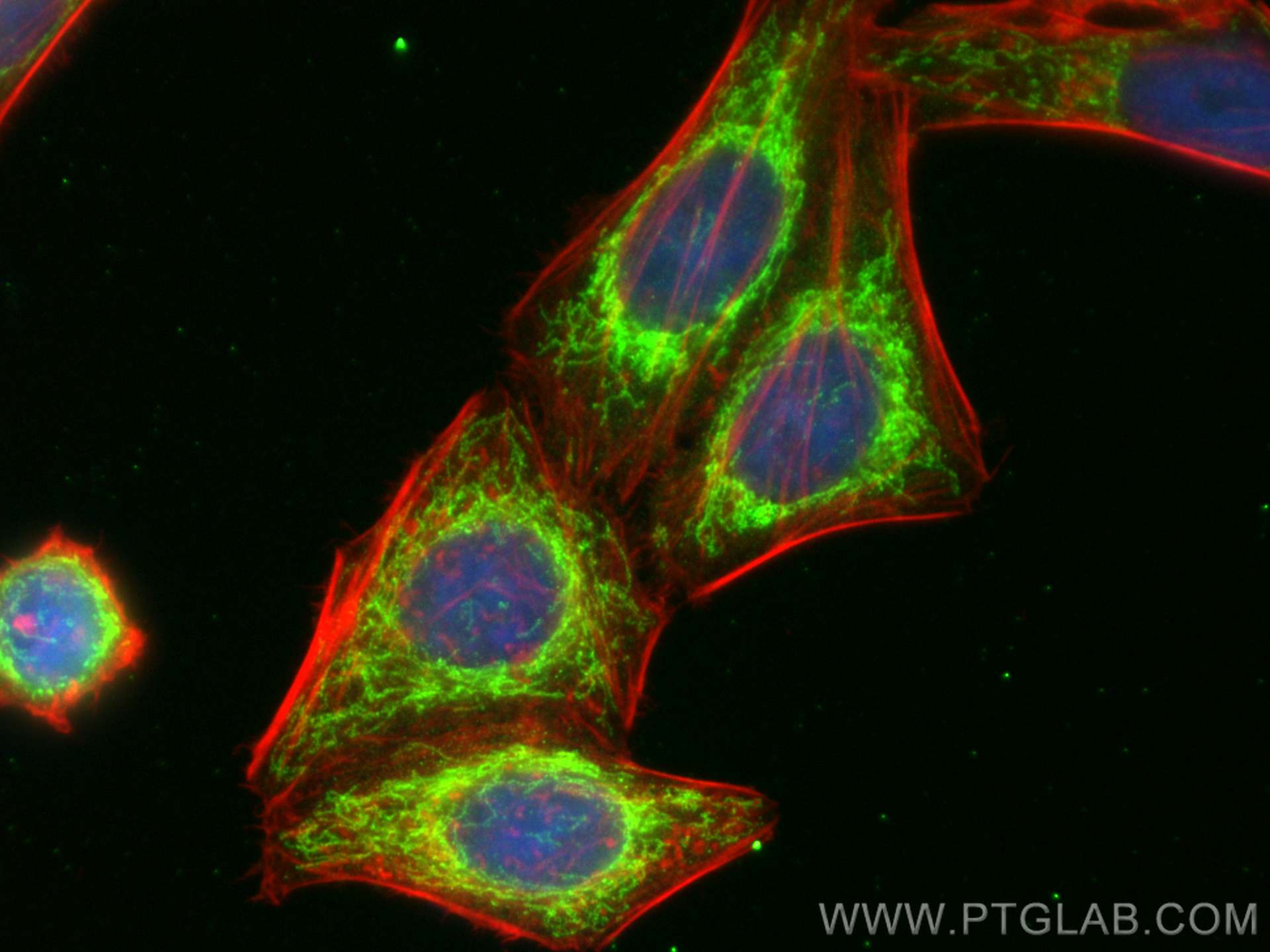
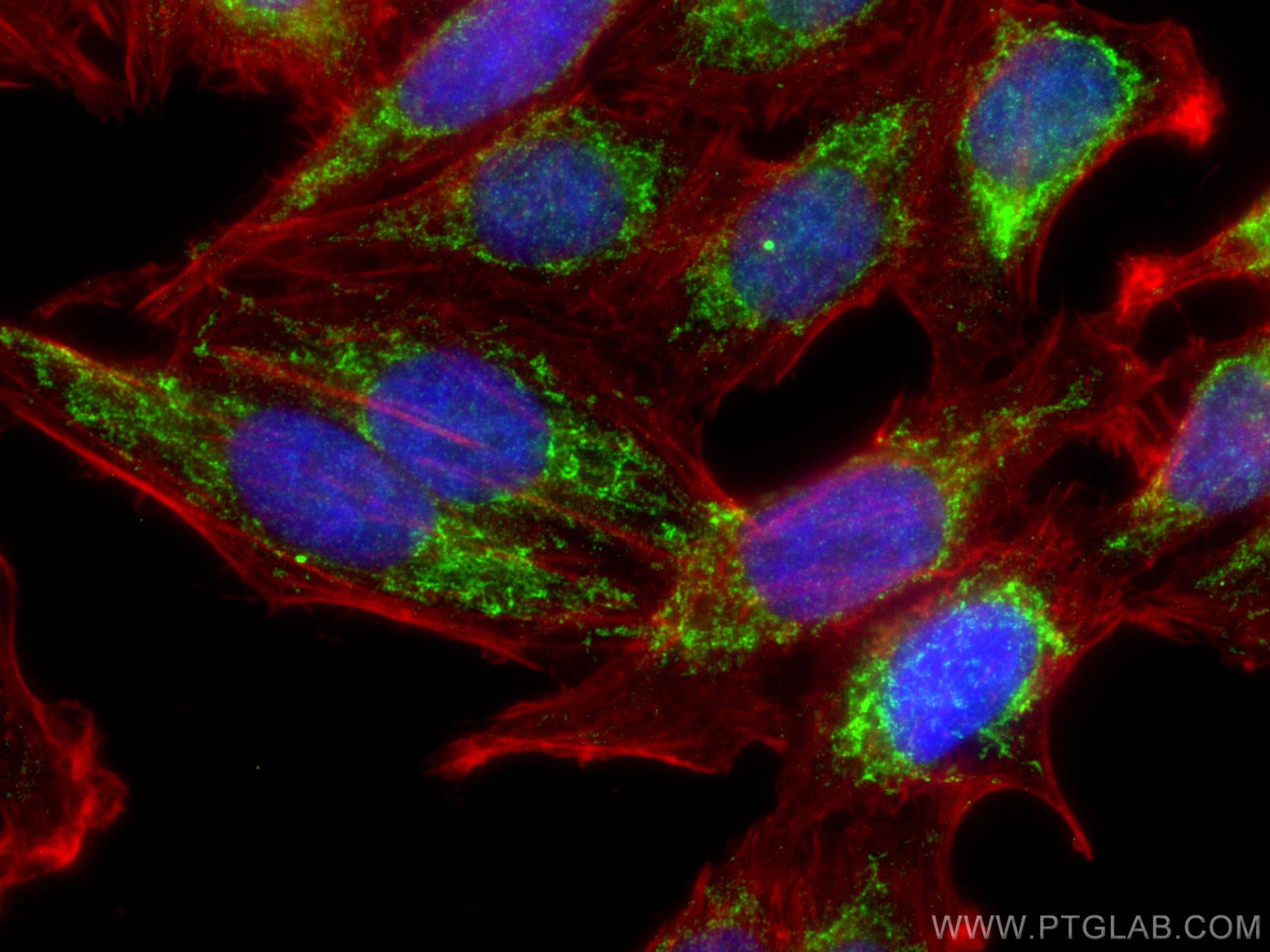
Validation Data Gallery
Tested Applications
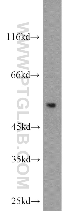
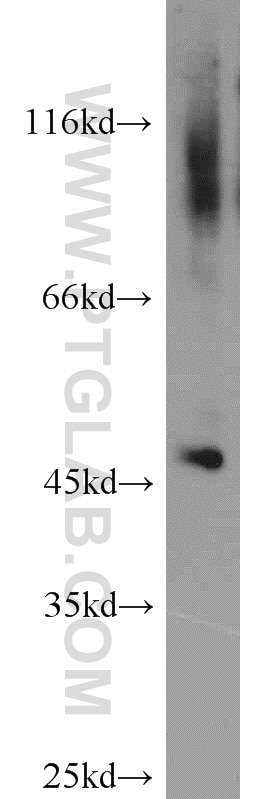
| Positive WB detected in | HEK-293 cells, human brain tissue, human kidney tissue, human testis tissue, mouse kidney tissue, HepG2 cells |
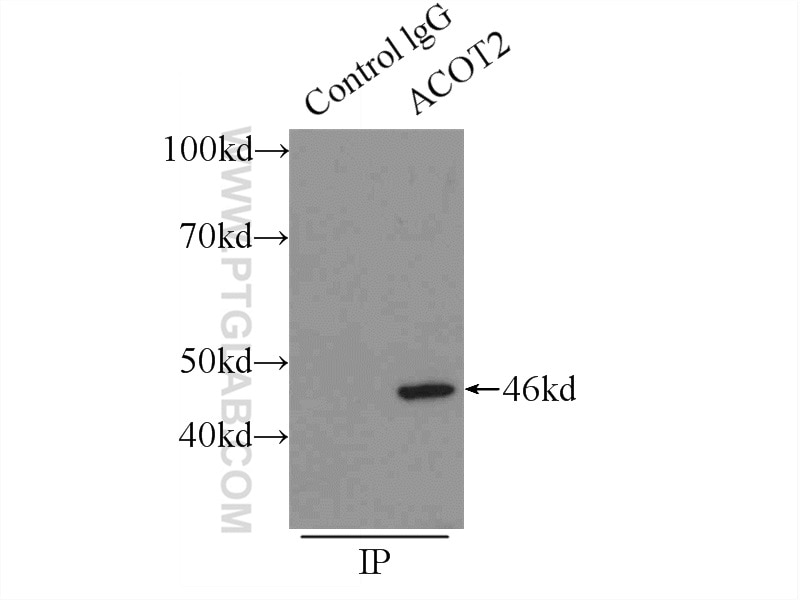
| Positive IP detected in | HepG2 cells |
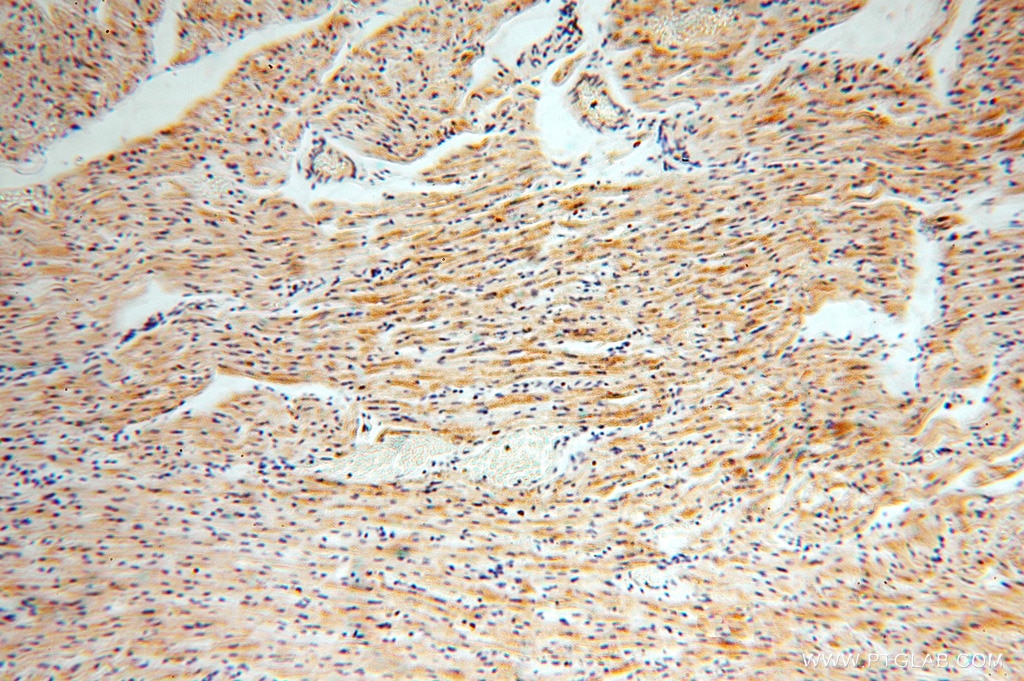
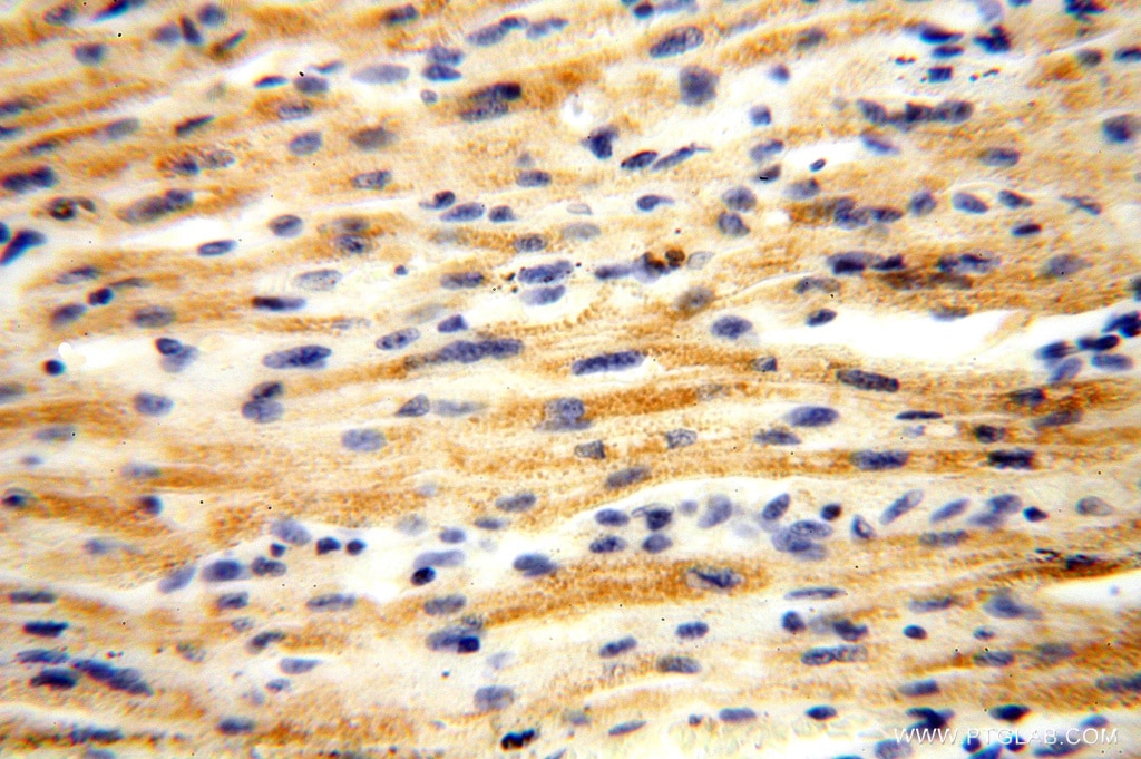
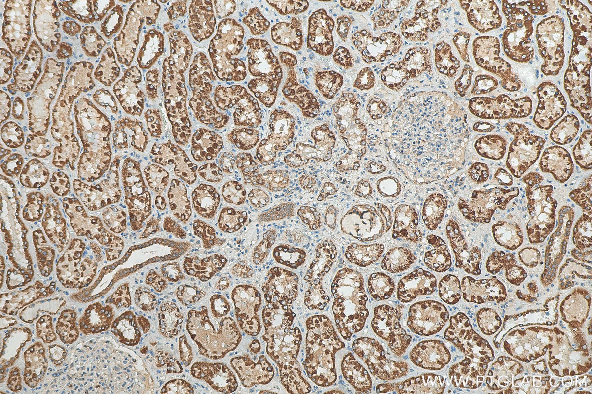
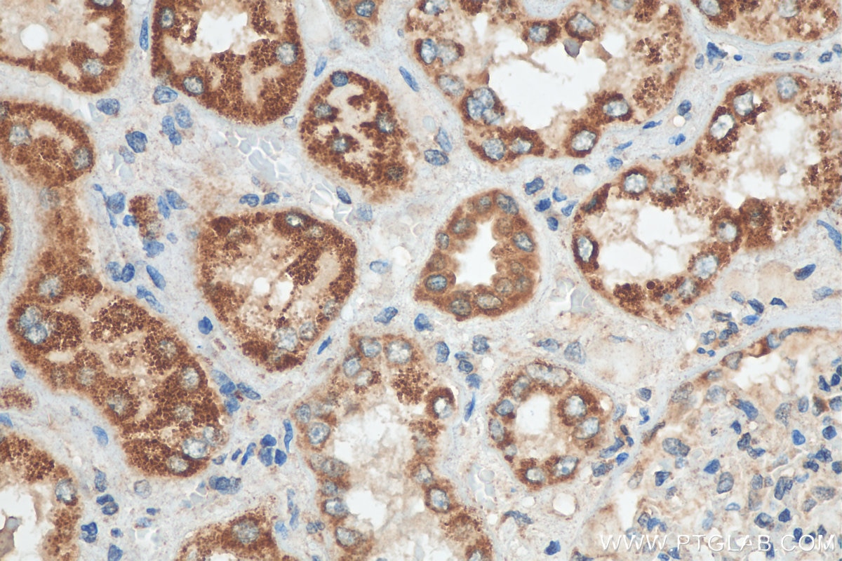
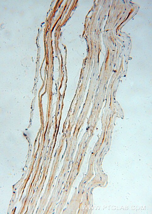
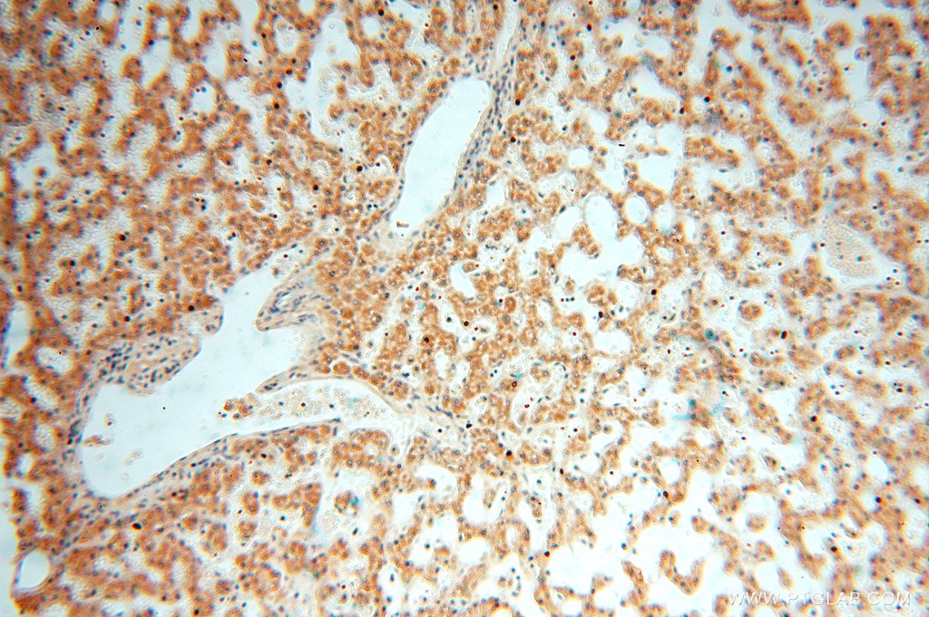
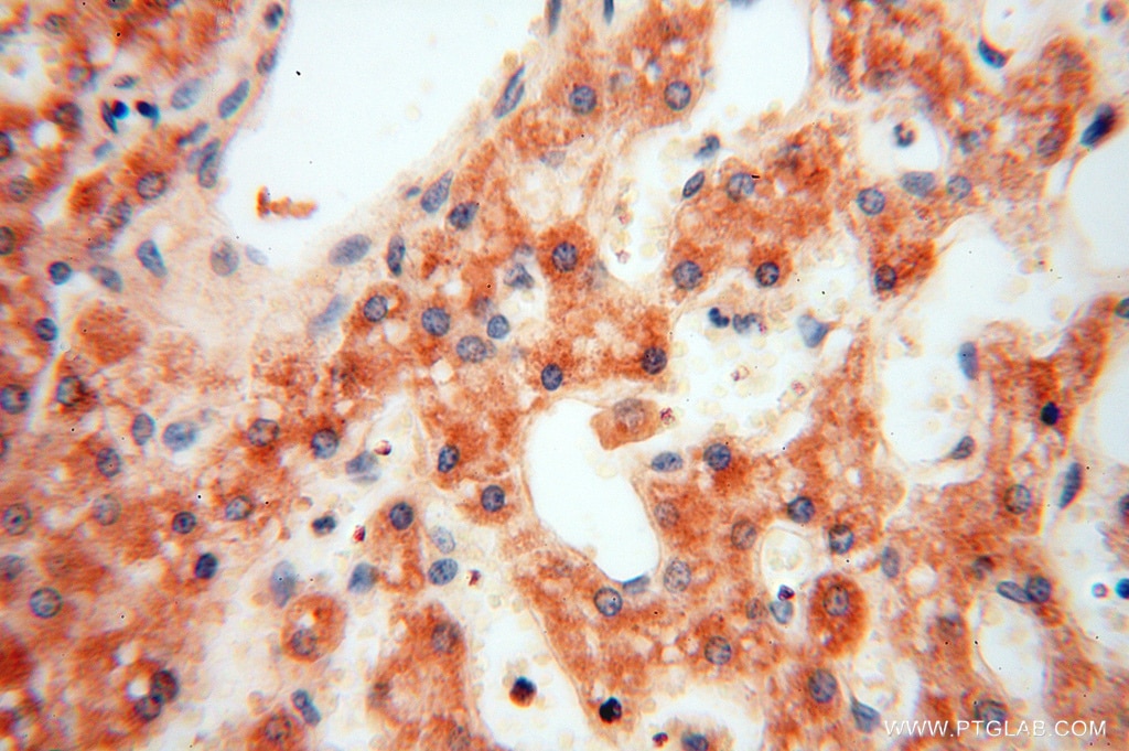
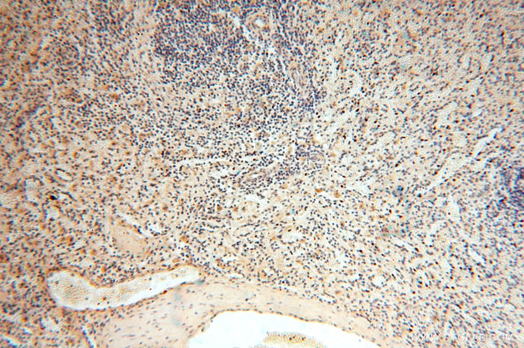
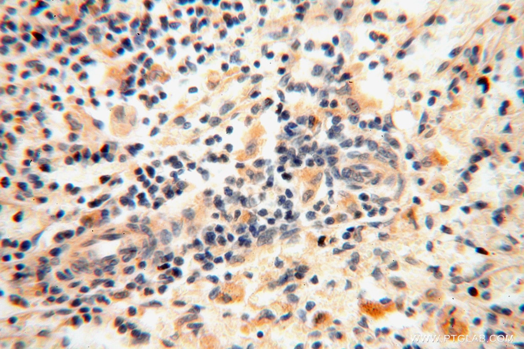
| Positive IHC detected in | human kidney tissue, human skeletal muscle tissue, human heart tissue, human testis tissue, human liver tissue, human spleen tissue, human ovary tissue Note: suggested antigen retrieval with TE buffer pH 9.0; (*) Alternatively, antigen retrieval may be performed with citrate buffer pH 6.0 |
| Positive IF/ICC detected in | HepG2 cells |
Recommended dilution
| Application | Dilution |
|---|---|
| Western Blot (WB) | WB : 1:500-1:1000 |
| Immunoprecipitation (IP) | IP : 0.5-4.0 ug for 1.0-3.0 mg of total protein lysate |
| Immunohistochemistry (IHC) | IHC : 1:50-1:500 |
| Immunofluorescence (IF)/ICC | IF/ICC : 1:50-1:500 |
| It is recommended that this reagent should be titrated in each testing system to obtain optimal results. | |
| Sample-dependent, Check data in validation data gallery. | |
Published Applications
| WB | See 8 publications below |
| IF | See 1 publications below |
Product Information
15633-1-AP targets ACOT2 in WB, IHC, IF/ICC, IP, ELISA applications and shows reactivity with human, mouse, rat samples.
| Tested Reactivity | human, mouse, rat |
| Cited Reactivity | human, mouse, rat |
| Host / Isotype | Rabbit / IgG |
| Class | Polyclonal |
| Type | Antibody |
| Immunogen | ACOT2 fusion protein Ag8093 相同性解析による交差性が予測される生物種 |
| Full Name | acyl-CoA thioesterase 2 |
| Calculated molecular weight | 483 aa, 53 kDa |
| Observed molecular weight | 46-53 kDa |
| GenBank accession number | BC006335 |
| Gene Symbol | ACOT2 |
| Gene ID (NCBI) | 10965 |
| RRID | AB_2221535 |
| Conjugate | Unconjugated |
| Form | Liquid |
| Purification Method | Antigen affinity purification |
| UNIPROT ID | P49753 |
| Storage Buffer | PBS with 0.02% sodium azide and 50% glycerol{{ptg:BufferTemp}}7.3 |
| Storage Conditions | Store at -20°C. Stable for one year after shipment. Aliquoting is unnecessary for -20oC storage. |
Background Information
Acyl-CoA thioesterase (Acot)2 localizes to the mitochondrial matrix and hydrolyses long-chain fatty acyl-CoA into free FA and CoASH. Acot2 is expressed in highly oxidative tissues and is poised to modulate mitochondrial FA oxidation (FAO) (PMID: 25114170). The structure of ACOT2 consists of two domains, N and C domains, and the active site of ACOT2 is located at the interface between the N and C domains (PMID: 19497300).
Protocols
| Product Specific Protocols | |
|---|---|
| WB protocol for ACOT2 antibody 15633-1-AP | Download protocol |
| IHC protocol for ACOT2 antibody 15633-1-AP | Download protocol |
| IF protocol for ACOT2 antibody 15633-1-AP | Download protocol |
| IP protocol for ACOT2 antibody 15633-1-AP | Download protocol |
| Standard Protocols | |
|---|---|
| Click here to view our Standard Protocols |
Publications
| Species | Application | Title |
|---|---|---|
Eur J Immunol YY1 control of mitochondrial-related genes does not account for regulation of immunoglobulin class switch recombination in mice. | ||
Life Sci Alliance High levels of TFAM repress mammalian mitochondrial DNA transcription in vivo | ||
Biomed Res Int Up-Regulated MicroRNA-27b Promotes Adipocyte Differentiation via Induction of Acyl-CoA Thioesterase 2 Expression. | ||
J Biol Chem Requirement of hepatic pyruvate carboxylase during fasting, high fat, and ketogenic diet | ||
EMBO Mol Med A coordinated multiorgan metabolic response contributes to human mitochondrial myopathy | ||
Hepatol Commun Dysregulation of lipid metabolism in the pseudolobule promotes region-specific autophagy in hepatitis B liver cirrhosis |
