• Featured Product
  • KD/KO Validated

ATP1A1-Specific Polyclonal antibody

ATP1A1-Specific Polyclonal Antibody for WB, IHC, IF/ICC, FC (Intra), ELISA

Host / Isotype

Rabbit / IgG

Reactivity

human, mouse and More (1)

Applications

WB, IHC, IF/ICC, FC (Intra), ELISA

Conjugate

Unconjugated

Cat no : 55187-1-AP

Synonyms

ATP1A1, ATP1A 1, Na(+)/K(+) ATPase alpha-1 subunit, Na(+)/K(+) ATPase 1, EC:7.2.2.13


"ATP1A1-Specific Antibodies" Comparison

View side-by-side comparison of ATP1A1-Specific antibodies from other vendors to find the one that best suits your research needs.
Filter:
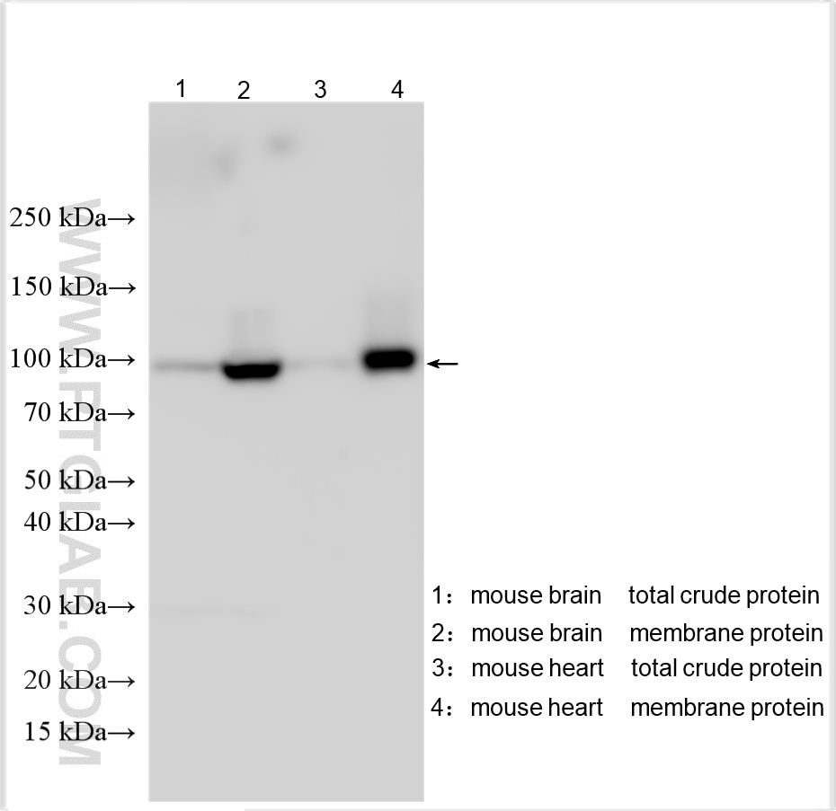
  • Western Blot (WB) analysis of various lysates using ATP1A1-Specific Polyclonal antibody (55187-1-AP)

    WB analysis using 55187-1-AP

    Various lysates were subjected to SDS PAGE followed by western blot with 55187-1-AP (ATP1A1-Specific antibody) at dilution of 1:4000 incubated at room temperature for 1.5 hours.

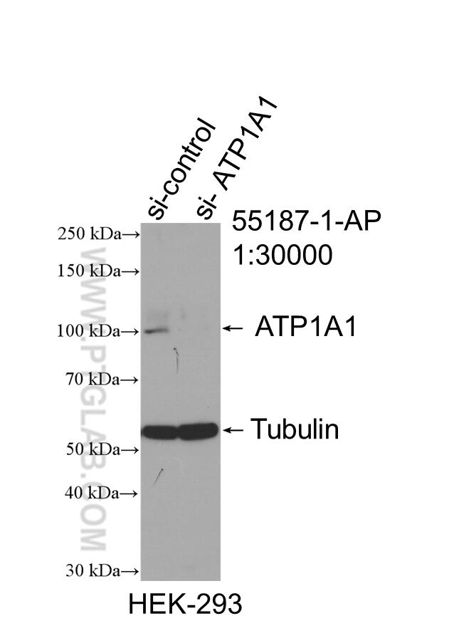
  • Western Blot (WB) analysis of HEK-293 cells using ATP1A1-Specific Polyclonal antibody (55187-1-AP)

    WB analysis of HEK-293 using 55187-1-AP

    WB result of ATP1A1-Specific antibody (55187-1-AP; 1:30000; incubated at room temperature for 1.5 hours) with sh-Control and sh-ATP1A1-Specific transfected HEK-293 cells.

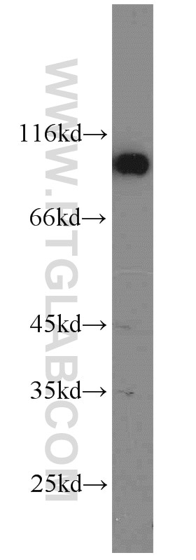
  • Western Blot (WB) analysis of mouse heart tissue using ATP1A1-Specific Polyclonal antibody (55187-1-AP)

    WB analysis of mouse heart using 55187-1-AP

    mouse heart tissue were subjected to SDS PAGE followed by western blot with 55187-1-AP (ATP1A1-Specific antibody) at dilution of 1:2000 incubated at room temperature for 1.5 hours.

  • Western Blot (WB) analysis of Neuro-2a cells using ATP1A1-Specific Polyclonal antibody (55187-1-AP)

    WB analysis of Neuro-2a using 55187-1-AP

    Neuro-2a cells were subjected to SDS PAGE followed by western blot with 55187-1-AP (ATP1A1-Specific antibody) at dilution of 1:2000 incubated at room temperature for 1.5 hours.

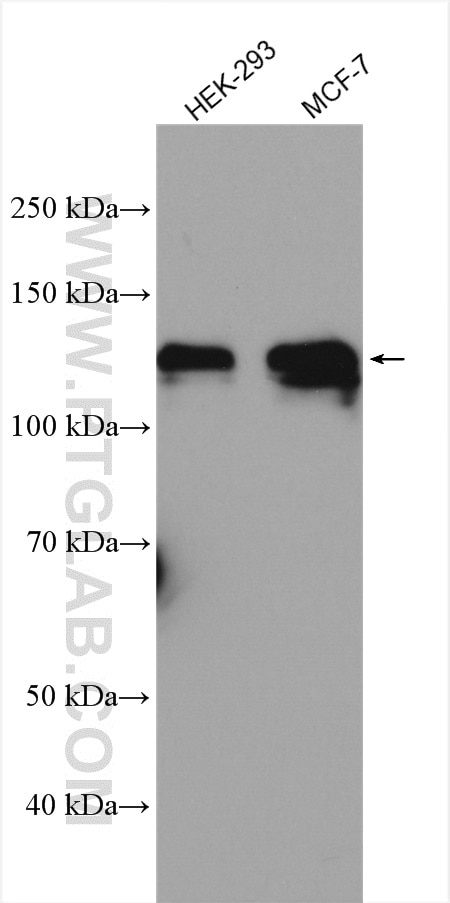
  • Western Blot (WB) analysis of MCF-7 cells using ATP1A1-Specific Polyclonal antibody (55187-1-AP)

    WB analysis of MCF-7 using 55187-1-AP

    MCF7 cells were subjected to SDS PAGE followed by western blot with 55187-1-AP (ATP1A1-Specific antibody) at dilution of 1:500 incubated at room temperature for 1.5 hours.

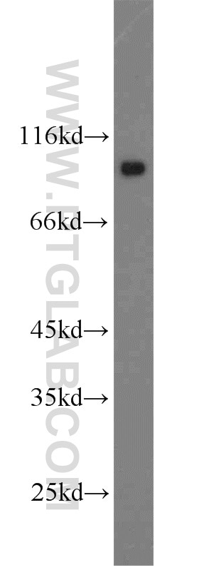
  • Western Blot (WB) analysis of HepG2 cells using ATP1A1-Specific Polyclonal antibody (55187-1-AP)

    WB analysis of HepG2 using 55187-1-AP

    HepG2 cells were subjected to SDS PAGE followed by western blot with 55187-1-AP (ATP1A1-Specific antibody) at dilution of 1:800 incubated at room temperature for 1.5 hours.

  • Western Blot (WB) analysis of HEK-293 cells using ATP1A1-Specific Polyclonal antibody (55187-1-AP)

    WB analysis of HEK-293 using 55187-1-AP

    HEK-293 cells were subjected to SDS PAGE followed by western blot with 55187-1-AP (ATP1A1-Specific antibody) at dilution of 1:1500 incubated at room temperature for 1.5 hours.

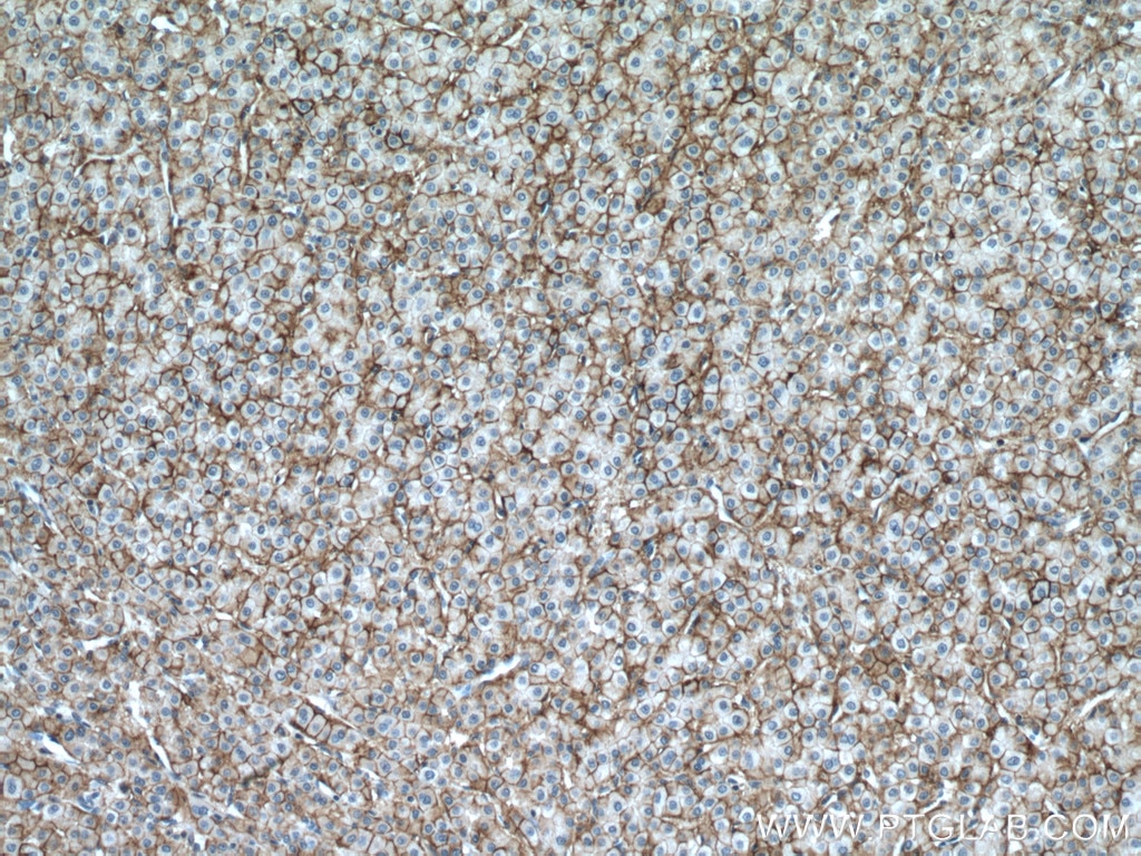
  • Immunohistochemistry (IHC) staining of human liver cancer tissue using ATP1A1-Specific Polyclonal antibody (55187-1-AP)

    IHC staining of human liver cancer using 55187-1-AP

    Immunohistochemical analysis of paraffin-embedded human liver cancer tissue slide using 55187-1-AP (ATP1A1-Specific antibody) at dilution of 1:200 (under 10x lens. Heat mediated antigen retrieval with Tris-EDTA buffer (pH 9.0).

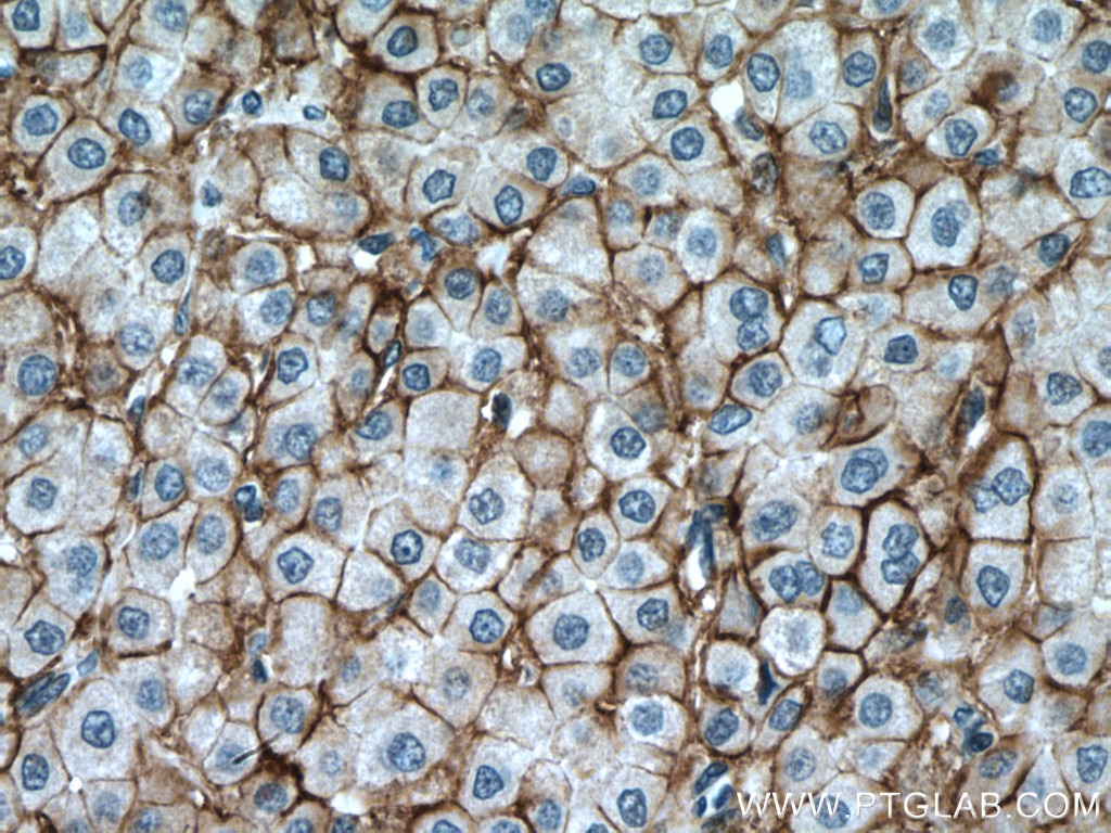
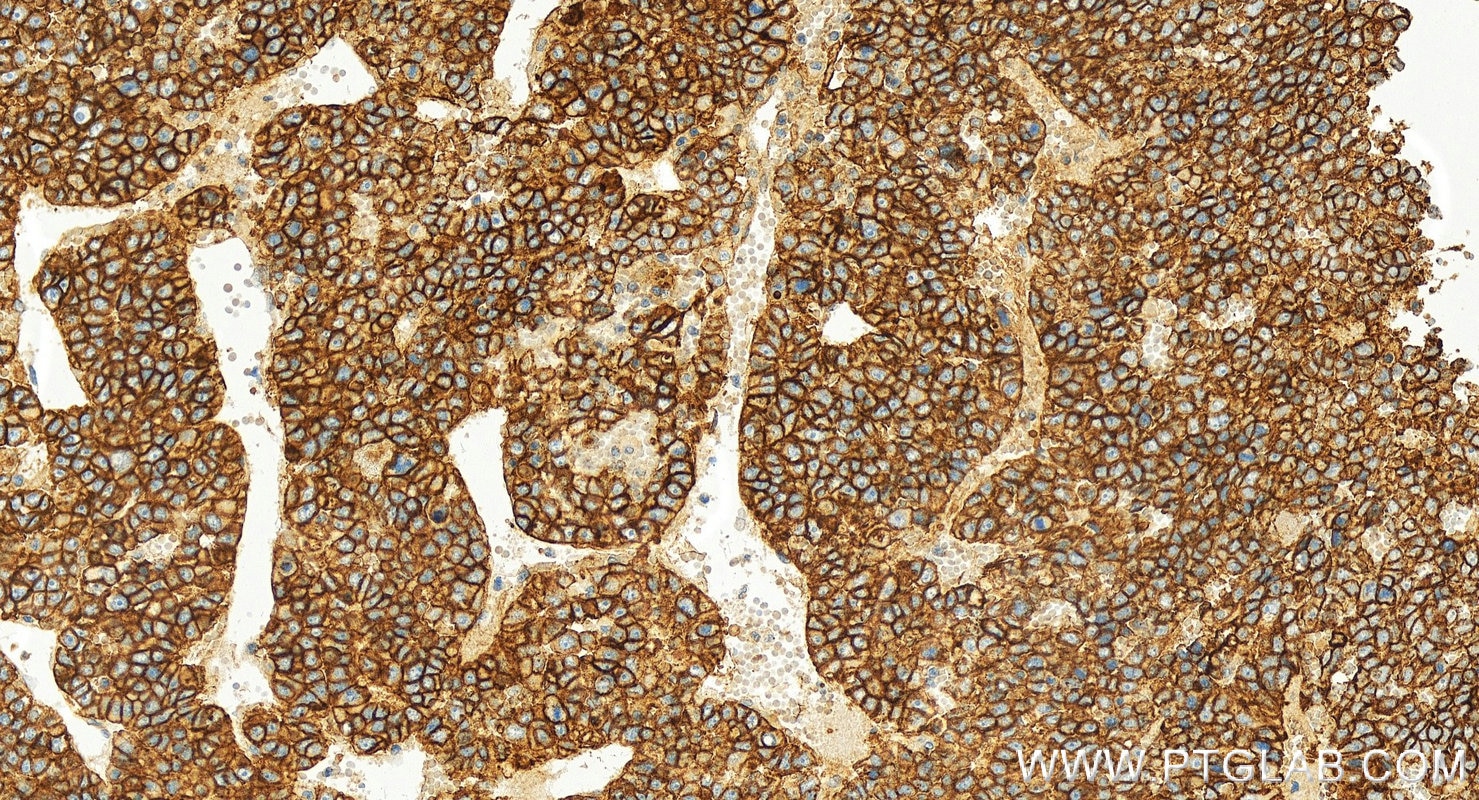
  • Immunohistochemistry (IHC) staining of human liver cancer tissue using ATP1A1-Specific Polyclonal antibody (55187-1-AP)

    IHC staining of human liver cancer using 55187-1-AP

    Immunohistochemical analysis of paraffin-embedded human liver cancer tissue slide using 55187-1-AP (ATP1A1-Specific antibody) at dilution of 1:200 (under 40x lens. Heat mediated antigen retrieval with Tris-EDTA buffer (pH 9.0).

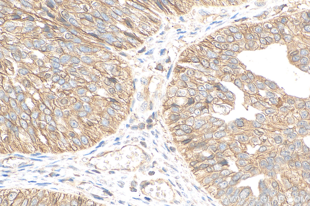
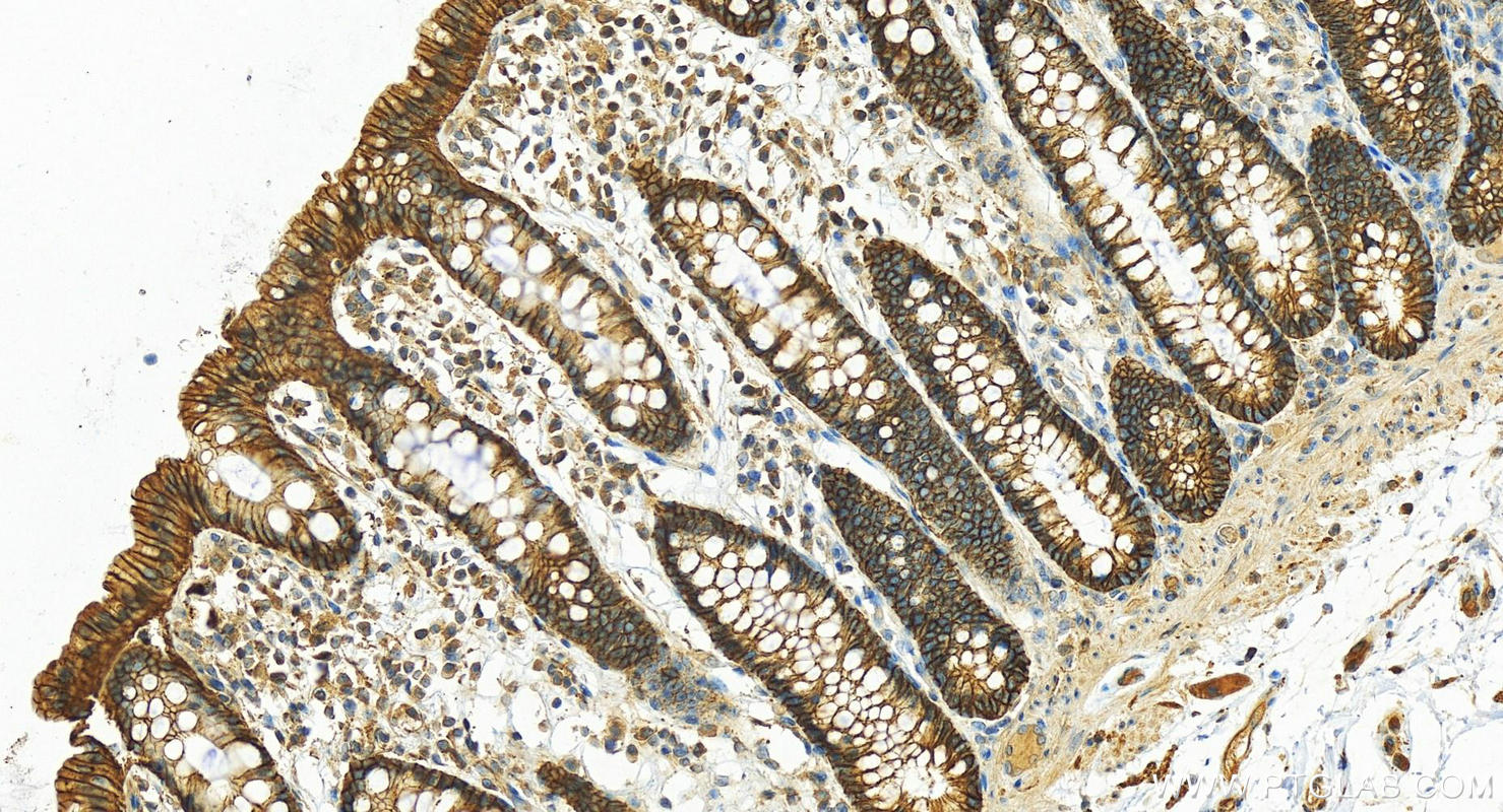
  • Immunohistochemistry (IHC) staining of human colon tissue using ATP1A1-Specific Polyclonal antibody (55187-1-AP)

    IHC staining of human colon using 55187-1-AP

    Immunohistochemical analysis of paraffin-embedded human normal colon slide using 55187-1-AP (ATP1A1-Specific antibody) at dilution of 1:800 (under 20x lens). Heat mediated antigen retrieval with Tris-EDTA buffer (pH 9.0).

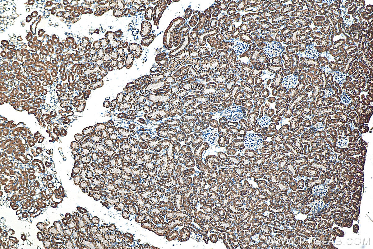
  • Immunohistochemistry (IHC) staining of mouse kidney tissue using ATP1A1-Specific Polyclonal antibody (55187-1-AP)

    IHC staining of mouse kidney using 55187-1-AP

    Immunohistochemical analysis of paraffin-embedded mouse kidney tissue slide using 55187-1-AP (ATP1A1-Specific antibody) at dilution of 1:400 (under 10x lens). Heat mediated antigen retrieval with Tris-EDTA buffer (pH 9.0).

  • Immunohistochemistry (IHC) staining of human kidney tissue using ATP1A1-Specific Polyclonal antibody (55187-1-AP)

    IHC staining of human kidney using 55187-1-AP

    Immunohistochemical analysis of paraffin-embedded human kidney tissue slide using 55187-1-AP (ATP1A1-Specific antibody) at dilution of 1:200 (under 10x lens). Heat mediated antigen retrieval with Tris-EDTA buffer (pH 9.0).

  • Immunohistochemistry (IHC) staining of mouse kidney tissue using ATP1A1-Specific Polyclonal antibody (55187-1-AP)

    IHC staining of mouse kidney using 55187-1-AP

    Immunohistochemical analysis of paraffin-embedded mouse kidney tissue slide using 55187-1-AP (ATP1A1-Specific antibody) at dilution of 1:200 (under 10x lens. Heat mediated antigen retrieval with Tris-EDTA buffer (pH 9.0).

  • Immunohistochemistry (IHC) staining of mouse kidney tissue using ATP1A1-Specific Polyclonal antibody (55187-1-AP)

    IHC staining of mouse kidney using 55187-1-AP

    Immunohistochemical analysis of paraffin-embedded mouse kidney tissue slide using 55187-1-AP (ATP1A1-Specific antibody) at dilution of 1:200 (under 40x lens. Heat mediated antigen retrieval with Tris-EDTA buffer (pH 9.0).

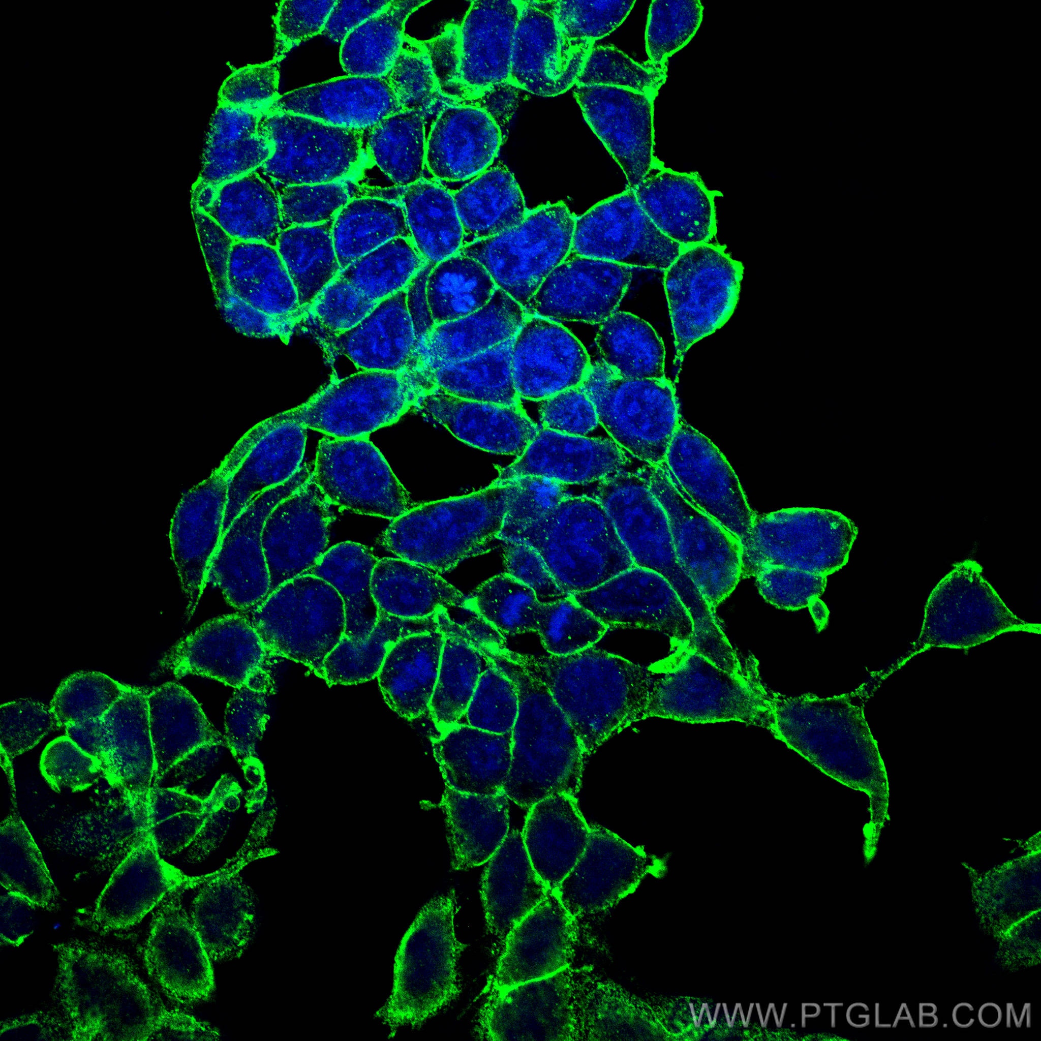
  • Immunofluorescence (IF) / fluorescent staining of HEK-293 cells using ATP1A1-Specific Polyclonal antibody (55187-1-AP)

    IF Staining of HEK-293 using 55187-1-AP

    Immunofluorescent analysis of (-20°C Methanol) fixed HEK-293 cells using ATP1A1-Specific antibody (55187-1-AP) at dilution of 1:1000 and CoraLite®488-Conjugated AffiniPure Goat Anti-Rabbit IgG(H+L).

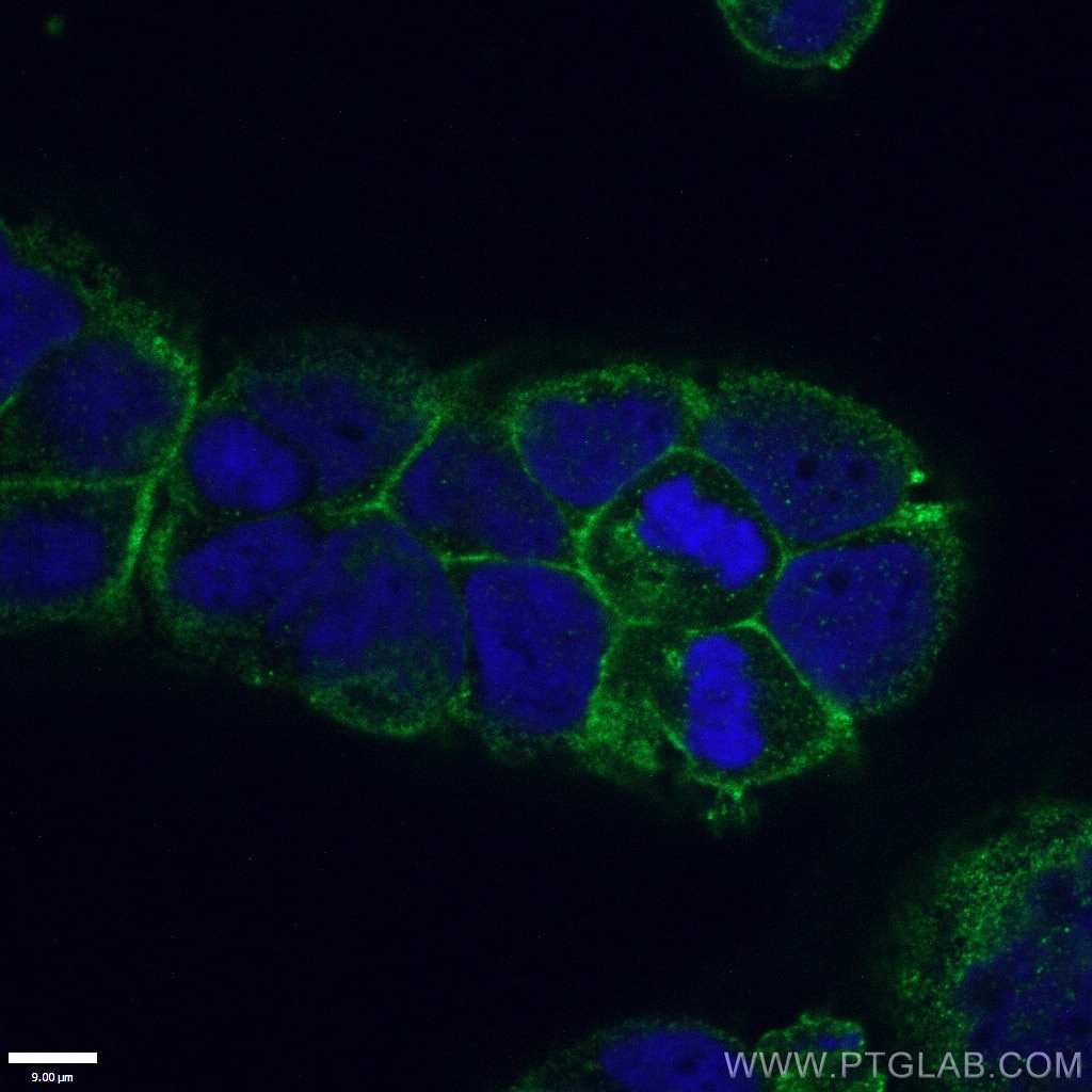
  • Immunofluorescence (IF) / fluorescent staining of Caco-2 cells using ATP1A1-Specific Polyclonal antibody (55187-1-AP)

    IF Staining of Caco-2 using 55187-1-AP

    Immunofluorescent analysis of (4% PFA) fixed Caco-2 cells using 55187-1-AP (ATP1A1-Specific antibody) at dilution of 1:100 and CoraLite488-Conjugated AffiniPure Goat Anti-Rabbit IgG(H+L).

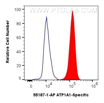
  • Flow cytometry (FC) experiment of HEK-293 cells using ATP1A1-Specific Polyclonal antibody (55187-1-AP)

    FC experiment of HEK-293 using 55187-1-AP

    1x10^6 HEK-293 cells were intracellularly stained with 0.4 ug ATP1A1-Specific Polyclonal antibody (55187-1-AP) and CoraLite®488-Conjugated AffiniPure Goat Anti-Rabbit IgG(H+L) (SA00013-2)(red), or 0.4 ug Rabbit IgG control Rabbit PolyAb (30000-0-AP) (blue). Cells were fixed with 4% PFA and permeabilized with Flow Cytometry Perm Buffer (PF00011-C).

☆大好評トライアルサイズ ¥20,000~! >>> 一次抗体特別価格キャンペーン開催中!


Tested Applications

Positive WB detected inHEK-293 cells, HepG2 cells, mouse heart tissue, MCF-7 cells, Neuro-2a cells
Positive IHC detected inhuman liver cancer tissue, mouse kidney tissue, human kidney tissue, human normal colon
Note: suggested antigen retrieval with TE buffer pH 9.0; (*) Alternatively, antigen retrieval may be performed with citrate buffer pH 6.0
Positive IF/ICC detected inHEK-293 cells, Caco-2 cells
Positive FC (Intra) detected inHEK-293 cells

Recommended dilution

ApplicationDilution
Western Blot (WB)WB : 1:1000-1:8000
Immunohistochemistry (IHC)IHC : 1:200-1:1000
Immunofluorescence (IF)/ICCIF/ICC : 1:500-1:2000
Flow Cytometry (FC) (INTRA)FC (INTRA) : 0.40 ug per 10^6 cells in a 100 µl suspension
It is recommended that this reagent should be titrated in each testing system to obtain optimal results.
Sample-dependent, Check data in validation data gallery.

Product Information

55187-1-AP targets ATP1A1-Specific in WB, IHC, IF/ICC, FC (Intra), ELISA applications and shows reactivity with human, mouse samples.

Tested Reactivity human, mouse
Cited Reactivityhuman, mouse, rat
Host / Isotype Rabbit / IgG
Class Polyclonal
Type Antibody
Immunogen Peptide 相同性解析による交差性が予測される生物種
Full Name ATPase, Na+/K+ transporting, alpha 1 polypeptide
Calculated molecular weight 113 kDa
Observed molecular weight 100-110 kDa
GenBank accession numberNM_000701
Gene symbol ATP1A1
Gene ID (NCBI) 476
RRIDAB_10859261
Conjugate Unconjugated
Form Liquid
Purification MethodAntigen affinity purification
Storage Buffer PBS with 0.02% sodium azide and 50% glycerol pH 7.3.
Storage ConditionsStore at -20°C. Stable for one year after shipment. Aliquoting is unnecessary for -20oC storage.

Background Information

ATP1A1 is the catalytic component of Na+/K+-ATPase which is a membrane bound enzyme primarily involved in generation of Na+ and K+ gradients across plasma membranes and in determination of cytoplasmic Na+ levels. ATP1A1 is a ubiquitously expressed membrane protein and often used as the marker or internal control for plasma membrane protein. This antibody is specific to ATP1A1.

Protocols

Product Specific Protocols
WB protocol for ATP1A1-Specific antibody 55187-1-APDownload protocol
IHC protocol for ATP1A1-Specific antibody 55187-1-APDownload protocol
IF protocol for ATP1A1-Specific antibody 55187-1-APDownload protocol
Standard Protocols
Click here to view our Standard Protocols

Publications

SpeciesApplicationTitle
humanIF

Autophagy

Hepatocyte CD36 modulates UBQLN1-mediated proteasomal degradation of autophagic SNARE proteins contributing to septic liver injury

Authors - Yanping Li
mouseWB

Hypertension

Renal Natriuretic Peptide Receptor-C Deficiency Attenuates NaCl Cotransporter Activity in Angiotensin II-Induced Hypertension.

Authors - Shuai Shao
mouseIF

Mol Biol Cell

Retinoschisin is linked to retinal Na/K-ATPase signaling and localization.

Authors - Karolina Plössl
humanIF

Sci Rep

Fluorescence- and magnetic-activated cell sorting strategies to separate spermatozoa involving plural contributors from biological mixtures for human identification.

Authors - Yan Xu
humanWB

PLoS One

A Novel Bufalin Derivative Exhibited Stronger Apoptosis-Inducing Effect than Bufalin in A549 Lung Cancer Cells and Lower Acute Toxicity in Mice.

Authors - Miao Liu
humanWB

Exp Eye Res

CHIR99021 balance TGFβ1 induced human corneal endothelial-to-mesenchymal transition to favor corneal endothelial cell proliferation.

Authors - Yiran Wang