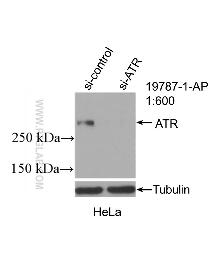
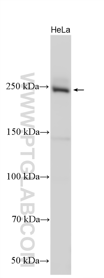
Validation Data Gallery
Tested Applications
| Positive WB detected in | HeLa cells |
Recommended dilution
| Application | Dilution |
|---|---|
| Western Blot (WB) | WB : 1:1000-1:4000 |
| It is recommended that this reagent should be titrated in each testing system to obtain optimal results. | |
| Sample-dependent, Check data in validation data gallery. | |
Published Applications
| WB | See 37 publications below |
| IHC | See 4 publications below |
| CoIP | See 1 publications below |
Product Information
19787-1-AP targets ATR in WB, IHC, CoIP, ELISA applications and shows reactivity with human samples.
| Tested Reactivity | human |
| Cited Reactivity | human, mouse, rat |
| Host / Isotype | Rabbit / IgG |
| Class | Polyclonal |
| Type | Antibody |
| Immunogen |
Peptide 相同性解析による交差性が予測される生物種 |
| Full Name | ataxia telangiectasia and Rad3 related |
| Calculated molecular weight | 301 kDa |
| Observed molecular weight | 250-290 kDa |
| GenBank accession number | NM_001184 |
| Gene Symbol | ATR |
| Gene ID (NCBI) | 545 |
| RRID | AB_10639516 |
| Conjugate | Unconjugated |
| Form | |
| Form | Liquid |
| Purification Method | Antigen affinity purification |
| UNIPROT ID | Q13535 |
| Storage Buffer | PBS with 0.02% sodium azide and 50% glycerol{{ptg:BufferTemp}}7.3 |
| Storage Conditions | Store at -20°C. Stable for one year after shipment. Aliquoting is unnecessary for -20oC storage. |
Background Information
ATR, also named as FRP1, belongs to the PI3/PI4-kinase family and ATM subfamily. ATR is a serine/threonine protein kinase which activates checkpoint signaling upon genotoxic stresses such as ionizing radiation (IR), ultraviolet light (UV), or DNA replication stalling, thereby acting as a DNA damage sensor. ATR recognizes the substrate consensus sequence [ST]-Q. ATR phosphorylates BRCA1, CHEK1, MCM2, RAD17, RPA2, SMC1 and TP53/p53, which collectively inhibit DNA replication and mitosis and promote DNA repair, recombination and apoptosis. ATR phosphorylates 'Ser-139' of histone variant H2AX/H2AFX at sites of DNA damage, thereby regulating DNA damage response mechanism. It is required for FANCD2 ubiquitination. It is critical for maintenance of fragile site stability and efficient regulation of centrosome duplication. ATR catalyze the reaction: ATP + a protein = ADP + a phosphoprotein. Defects in ATR are a cause of Seckel syndrome type 1 (SCKL1) which is a rare autosomal recessive disorder characterized by growth retardation, microcephaly with mental retardation, and a characteristic 'bird-headed' facial appearance. The antibody can recognize all the isoforms of ATR.
Protocols
| Product Specific Protocols | |
|---|---|
| WB protocol for ATR antibody 19787-1-AP | Download protocol |
| Standard Protocols | |
|---|---|
| Click here to view our Standard Protocols |
Publications
| Species | Application | Title |
|---|---|---|
Cell Extrachromosomal DNA replication and maintenance couple with DNA damage pathway in tumors | ||
Cell Res DNA damage triggers tubular endoplasmic reticulum extension to promote apoptosis by facilitating ER-mitochondria signaling. | ||
ACS Nano Graphene Oxide Causes Disordered Zonation Due to Differential Intralobular Localization in the Liver. | ||
Nat Commun ATR/Chk1 signaling induces autophagy through sumoylated RhoB-mediated lysosomal translocation of TSC2 after DNA damage. | ||
Cancer Res Combined inactivation of CTPS1 and ATR is synthetically lethal to MYC-overexpressing cancer cells. | ||
Oncogene Antifungal agent Terbinafine restrains tumor growth in preclinical models of hepatocellular carcinoma via AMPK-mTOR axis. |


