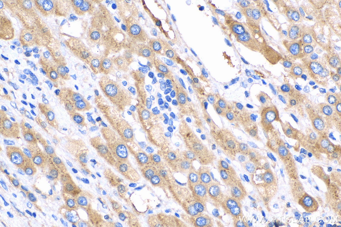
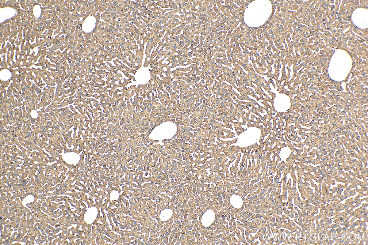
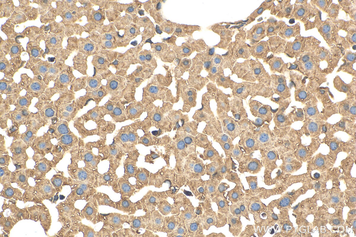
Validation Data Gallery
Tested Applications
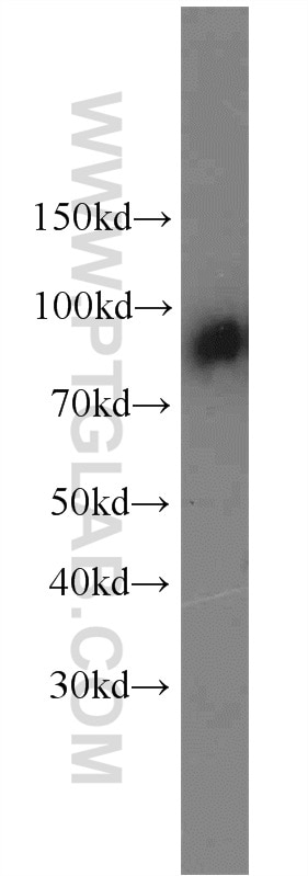
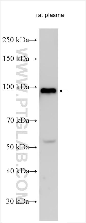
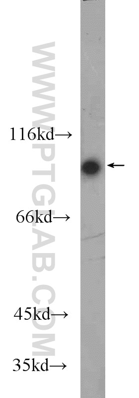
| Positive WB detected in | rat plasma, human plasma |
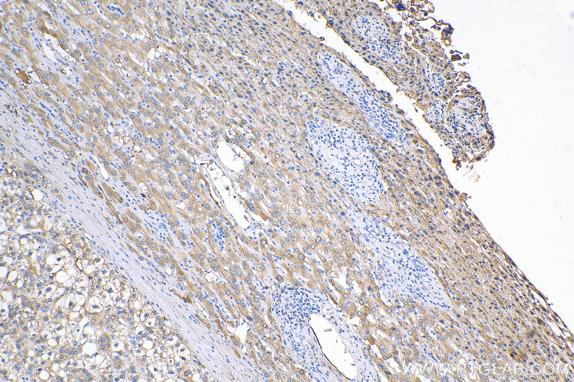
| Positive IHC detected in | human liver cancer tissue, mouse liver tissue Note: suggested antigen retrieval with TE buffer pH 9.0; (*) Alternatively, antigen retrieval may be performed with citrate buffer pH 6.0 |
Recommended dilution
| Application | Dilution |
|---|---|
| Western Blot (WB) | WB : 1:2000-1:16000 |
| Immunohistochemistry (IHC) | IHC : 1:50-1:500 |
| It is recommended that this reagent should be titrated in each testing system to obtain optimal results. | |
| Sample-dependent, Check data in validation data gallery. | |
Published Applications
| WB | See 3 publications below |
| IHC | See 1 publications below |
| IF | See 3 publications below |
Product Information
14554-1-AP targets C1S in WB, IHC, IF, ELISA applications and shows reactivity with human, mouse, rat samples.
| Tested Reactivity | human, mouse, rat |
| Cited Reactivity | human, mouse |
| Host / Isotype | Rabbit / IgG |
| Class | Polyclonal |
| Type | Antibody |
| Immunogen | C1S fusion protein Ag5977 相同性解析による交差性が予測される生物種 |
| Full Name | complement component 1, s subcomponent |
| Calculated molecular weight | 77 kDa |
| Observed molecular weight | 88 kDa |
| GenBank accession number | BC056903 |
| Gene Symbol | C1S |
| Gene ID (NCBI) | 716 |
| RRID | AB_2243610 |
| Conjugate | Unconjugated |
| Form | Liquid |
| Purification Method | Antigen affinity purification |
| UNIPROT ID | P09871 |
| Storage Buffer | PBS with 0.02% sodium azide and 50% glycerol , pH 7.3 |
| Storage Conditions | Store at -20°C. Stable for one year after shipment. Aliquoting is unnecessary for -20oC storage. |
Background Information
The first component of complement is a calcium-dependent complex of the 3 subcomponents C1q, C1r, and C1s. The serine protease subunits C1r and C1s form a Ca(2+)-dependent C1s-C1r-C1r-C1s tetramer. C1r activates C1s so that it can, in turn, activate C2 and C4. C1s is a glycoprotein of approximately 88 kDa, which is activated by cleavage into an A chain and a B chain.
Protocols
| Product Specific Protocols | |
|---|---|
| WB protocol for C1S antibody 14554-1-AP | Download protocol |
| IHC protocol for C1S antibody 14554-1-AP | Download protocol |
| Standard Protocols | |
|---|---|
| Click here to view our Standard Protocols |
Publications
| Species | Application | Title |
|---|---|---|
Int J Mol Sci Sodium Iodate-Induced Degeneration Results in Local Complement Changes and Inflammatory Processes in Murine Retina | ||
Sci Rep C-reactive protein (CRP) recognizes uric acid crystals and recruits proteases C1 and MASP1. | ||
J Transl Med Comprehensive investigation of malignant epithelial cell-related genes in clear cell renal cell carcinoma: development of a prognostic signature and exploration of tumor microenvironment interactions |
