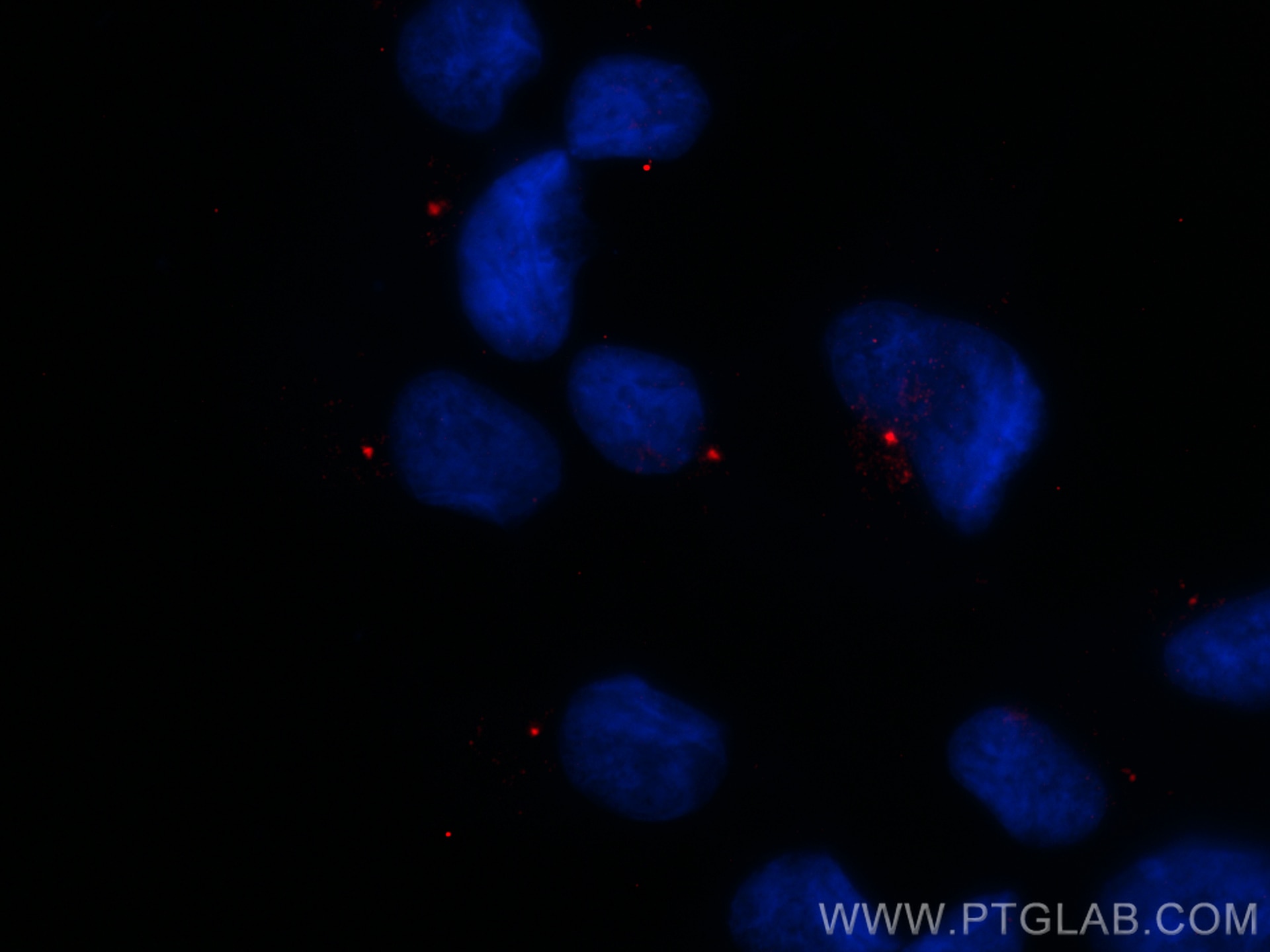
Validation Data Gallery
Tested Applications
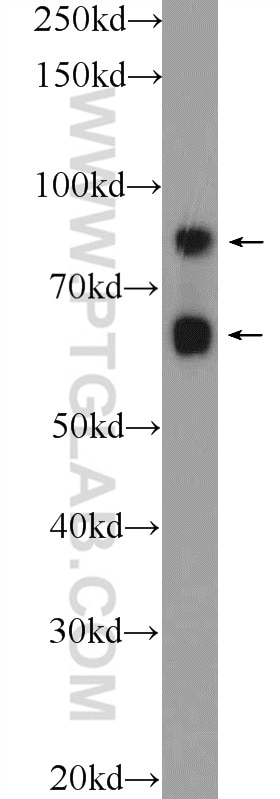
| Positive WB detected in | HEK-293 cells |
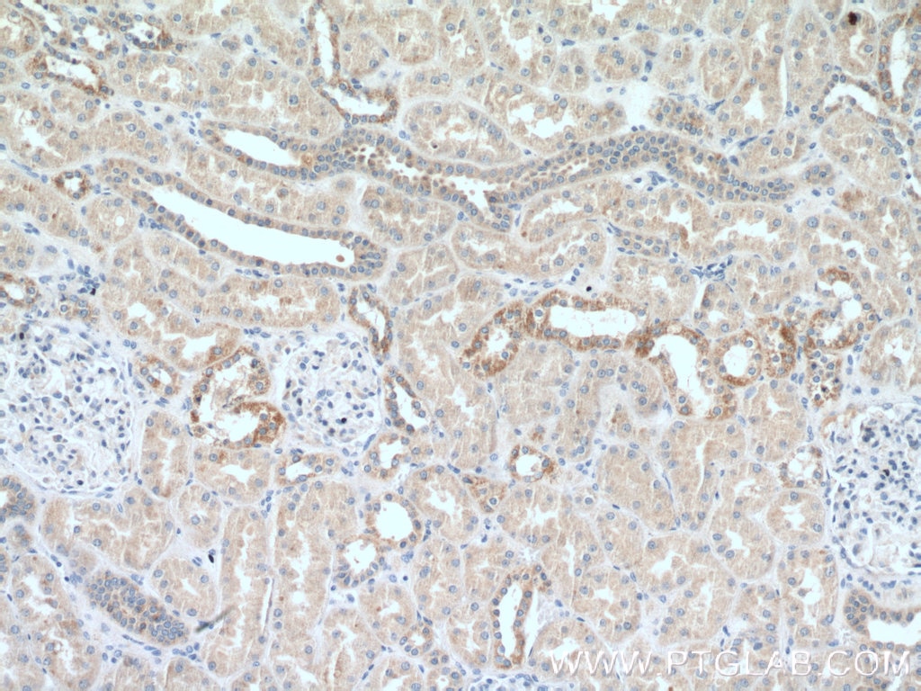
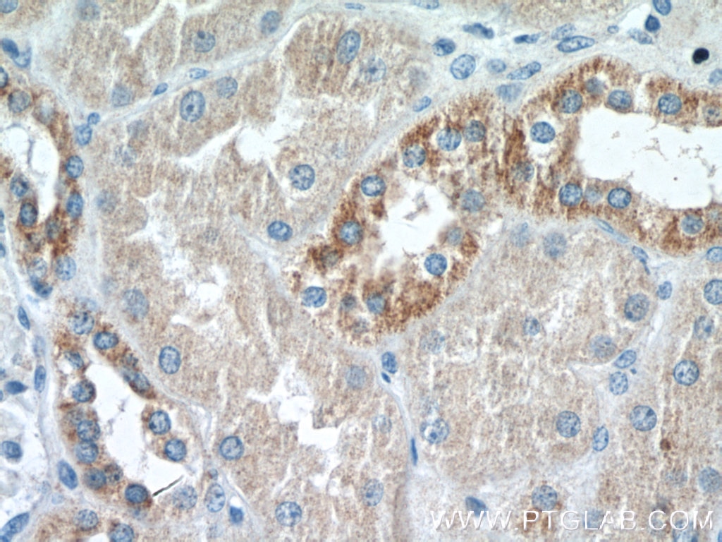
| Positive IHC detected in | human kidney tissue Note: suggested antigen retrieval with TE buffer pH 9.0; (*) Alternatively, antigen retrieval may be performed with citrate buffer pH 6.0 |
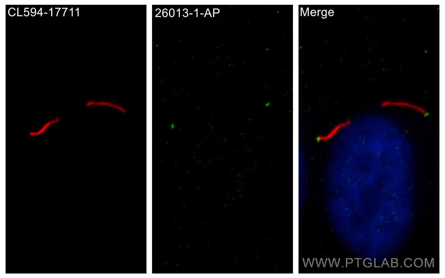
| Positive IF/ICC detected in | hTERT-RPE1 cells |
Recommended dilution
| Application | Dilution |
|---|---|
| Western Blot (WB) | WB : 1:500-1:1000 |
| Immunohistochemistry (IHC) | IHC : 1:50-1:500 |
| Immunofluorescence (IF)/ICC | IF/ICC : 1:200-1:800 |
| It is recommended that this reagent should be titrated in each testing system to obtain optimal results. | |
| Sample-dependent, Check data in validation data gallery. | |
Published Applications
| WB | See 6 publications below |
| IF | See 5 publications below |
Product Information
26013-1-AP targets CEP83 in WB, IHC, IF/ICC, ELISA applications and shows reactivity with human samples.
| Tested Reactivity | human |
| Cited Reactivity | human, mouse |
| Host / Isotype | Rabbit / IgG |
| Class | Polyclonal |
| Type | Antibody |
| Immunogen | CEP83 fusion protein Ag22826 相同性解析による交差性が予測される生物種 |
| Full Name | coiled-coil domain containing 41 |
| Calculated molecular weight | 693 aa, 82 kDa |
| Observed molecular weight | 83 kDa, 68 kDa |
| GenBank accession number | BC125087 |
| Gene Symbol | CEP83 |
| Gene ID (NCBI) | 51134 |
| RRID | AB_2880334 |
| Conjugate | Unconjugated |
| Form | Liquid |
| Purification Method | Antigen affinity purification |
| UNIPROT ID | Q9Y592 |
| Storage Buffer | PBS with 0.02% sodium azide and 50% glycerol , pH 7.3 |
| Storage Conditions | Store at -20°C. Stable for one year after shipment. Aliquoting is unnecessary for -20oC storage. |
Background Information
CEP83 (centrosomal protein, 83 kDa), also known as CCDC41 (coiled-coil domain containing 41), is required for ciliary vesicle docking to the mother centriole. It is a key component of the centriolar distal appendages. CCDC41 may collaborate with IFT20 in the trafficking of ciliary membrane proteins from the Golgi complex to the cilium during the initiation of primary cilium assembly (PMID: 23530209). Alternative splicing results in transcript variants encoding two isoforms with calculated molecular weights of 83 kDa and 68 kDa, respectively.
Protocols
| Product Specific Protocols | |
|---|---|
| WB protocol for CEP83 antibody 26013-1-AP | Download protocol |
| IHC protocol for CEP83 antibody 26013-1-AP | Download protocol |
| IF protocol for CEP83 antibody 26013-1-AP | Download protocol |
| Standard Protocols | |
|---|---|
| Click here to view our Standard Protocols |
Publications
| Species | Application | Title |
|---|---|---|
Cell Res NudCL2 is an autophagy receptor that mediates selective autophagic degradation of CP110 at mother centrioles to promote ciliogenesis. | ||
J Cell Biol The Cby3/ciBAR1 complex positions the annulus along the sperm flagellum during spermiogenesis | ||
PLoS Biol The evolutionary conserved proteins CEP90, FOPNL, and OFD1 recruit centriolar distal appendage proteins to initiate their assembly | ||
Cell Mol Life Sci Requirement of NPHP5 in the hierarchical assembly of basal feet associated with basal bodies of primary cilia. | ||
J Biol Chem The C7orf43/TRAPPC14 component links the TRAPPII complex to RABIN8 for preciliary vesicle tethering at the mother centriole during ciliogenesis. |
