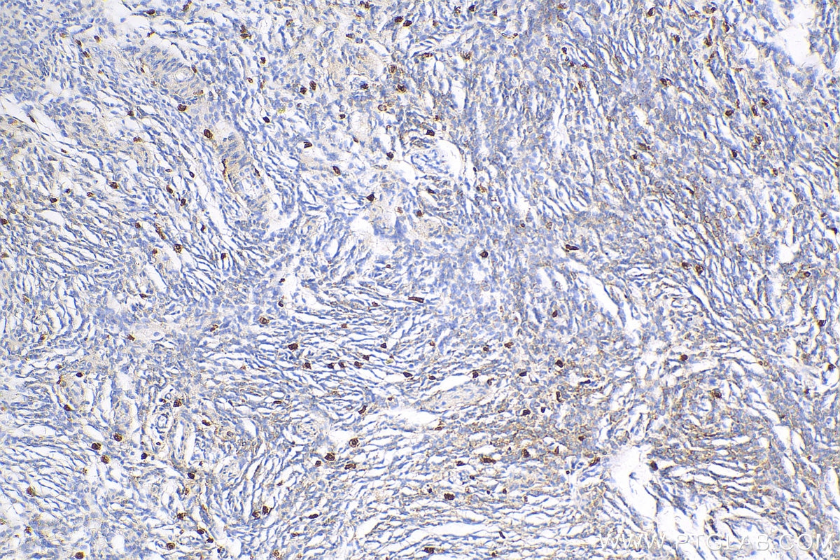
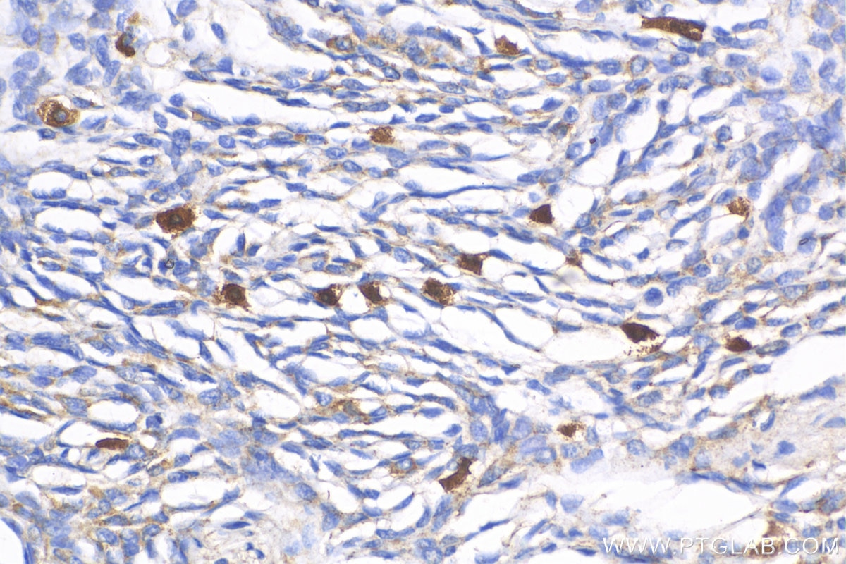
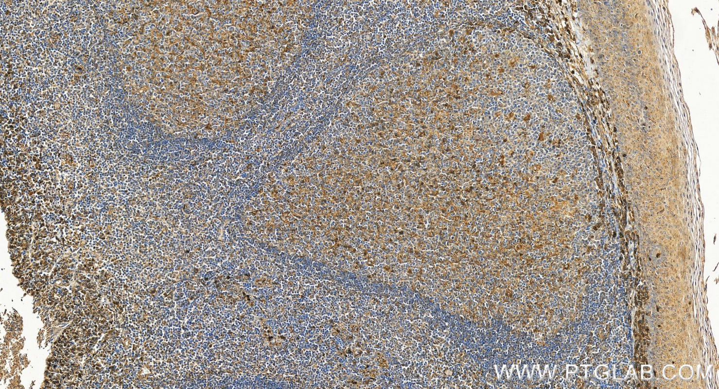
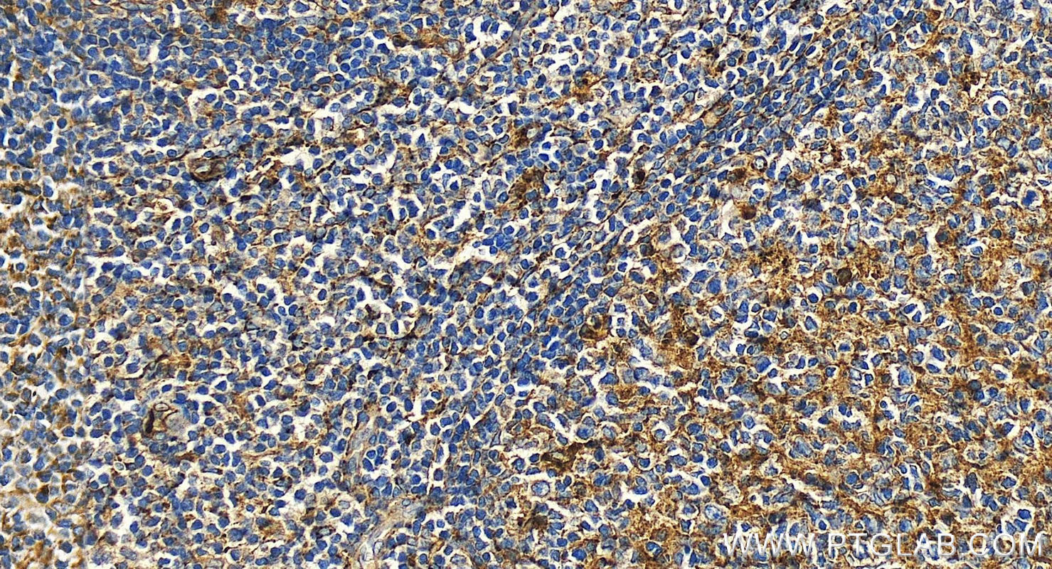
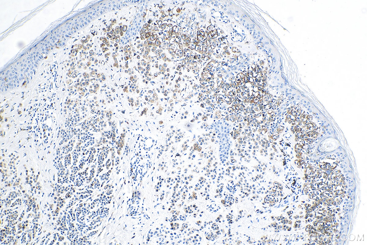
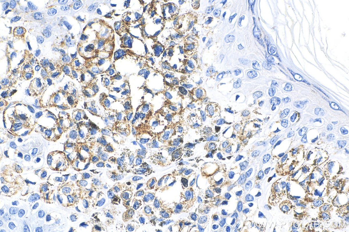
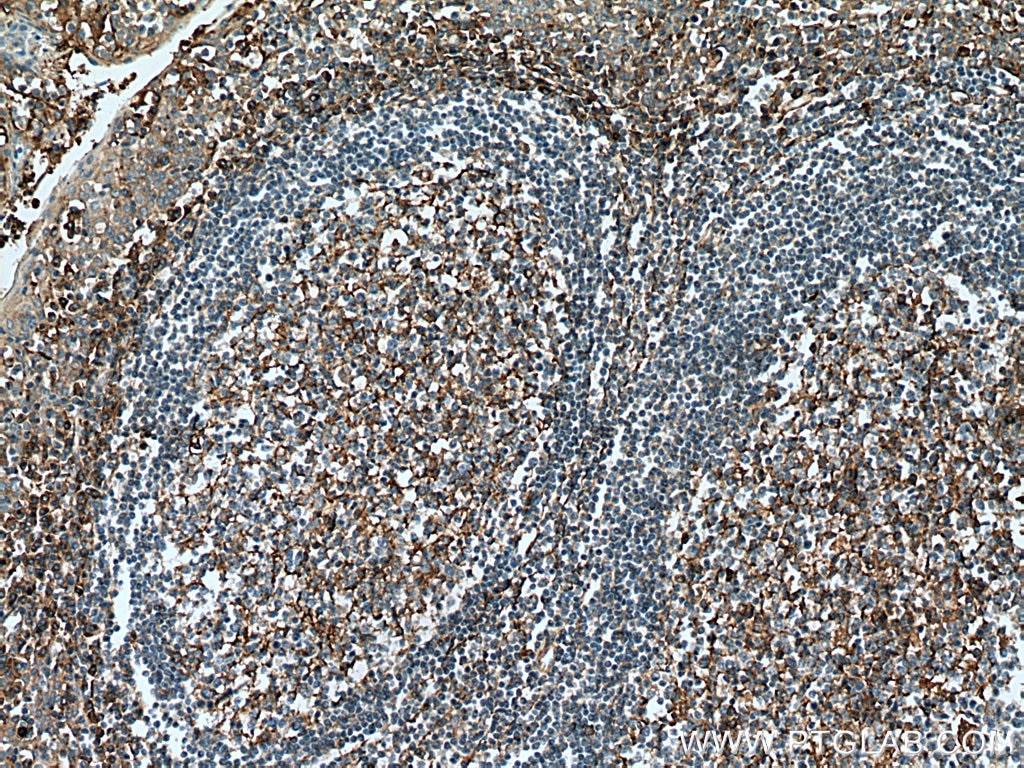
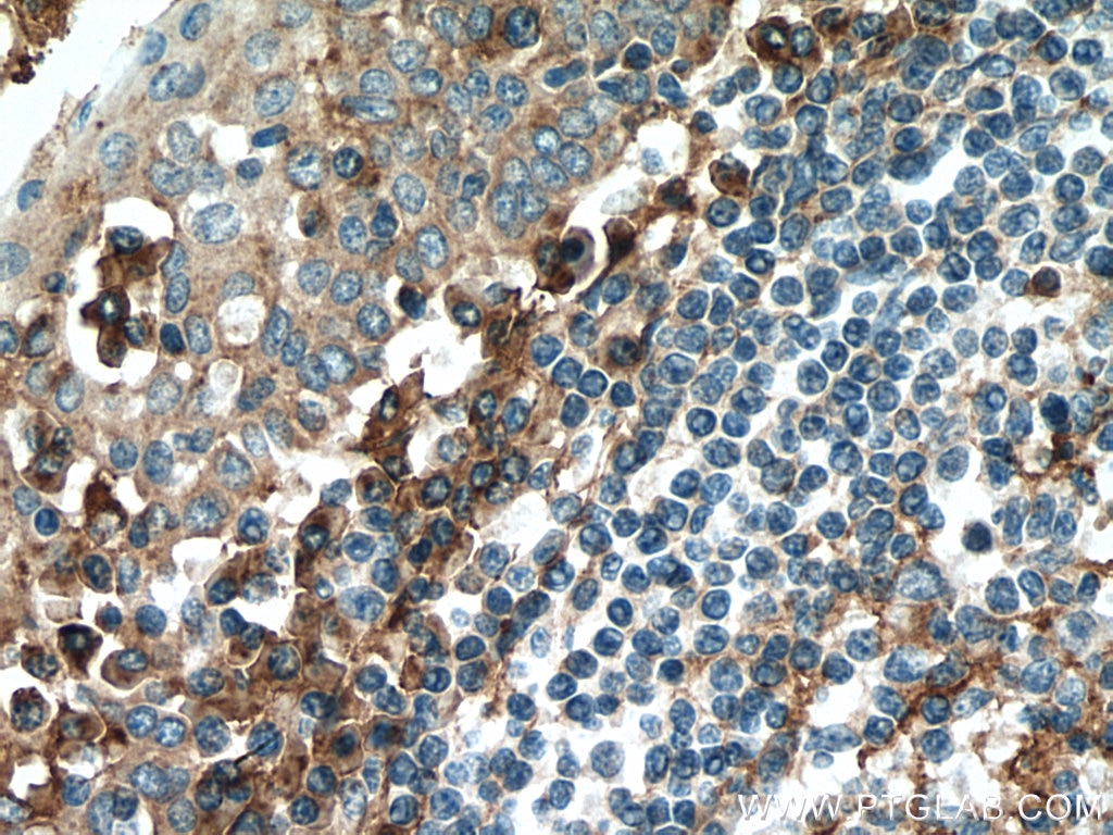
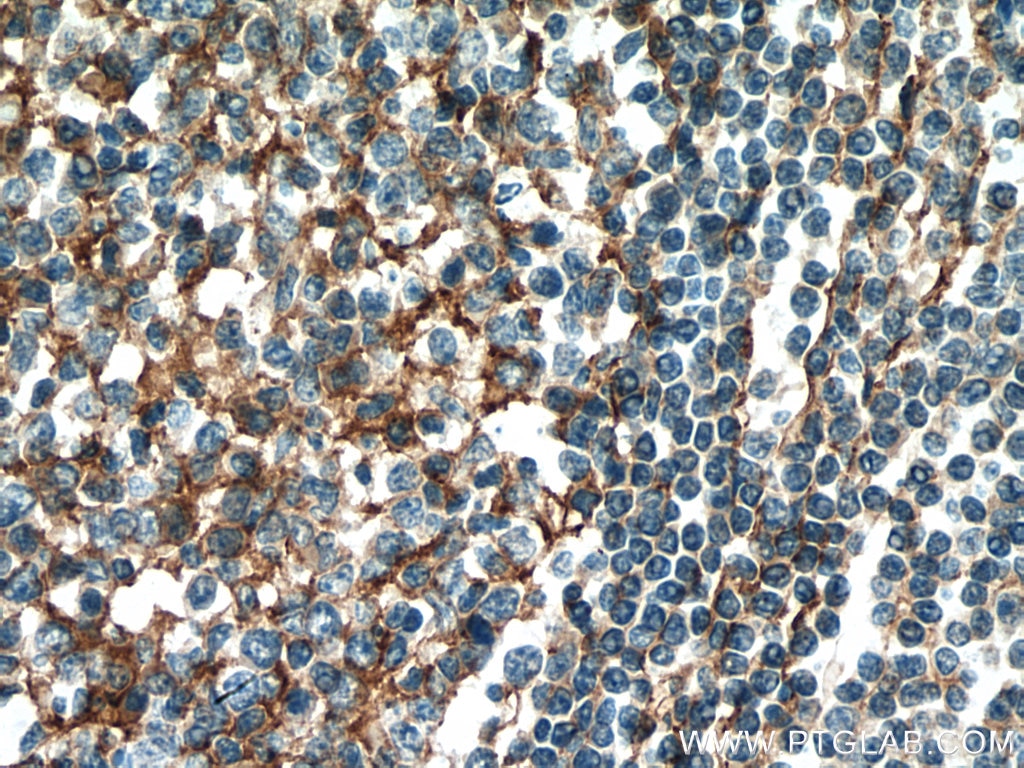
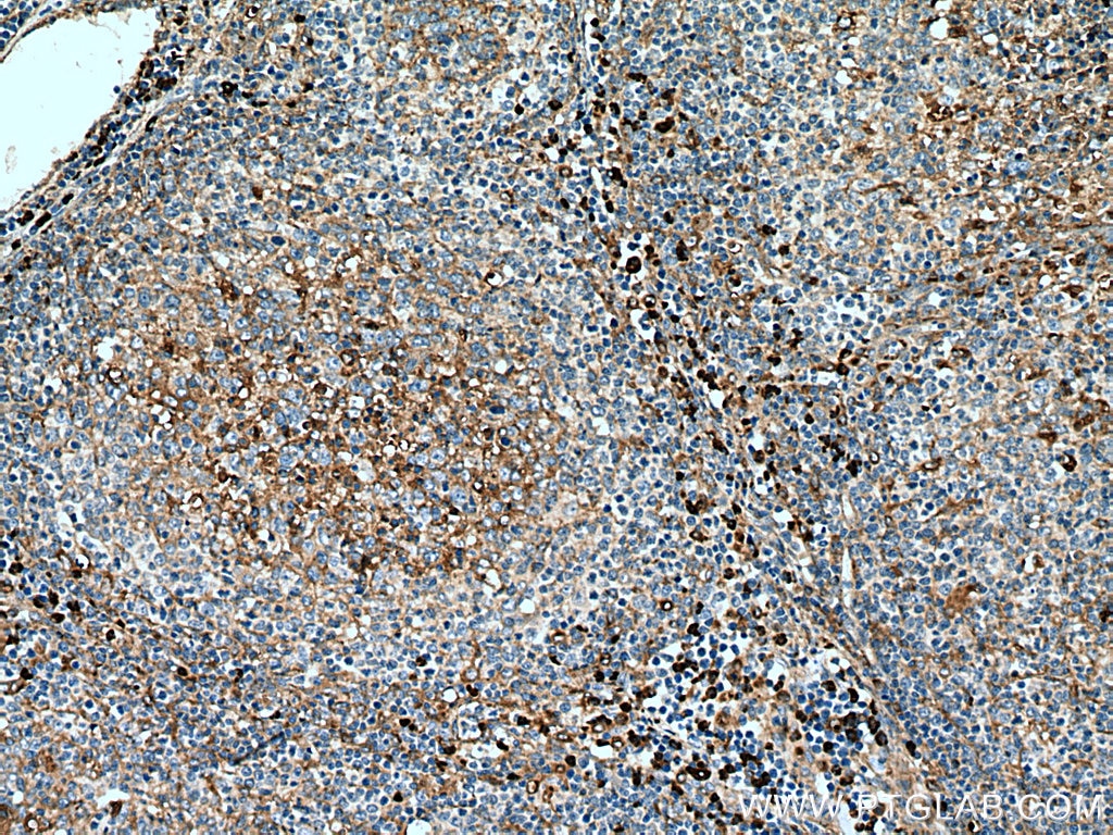
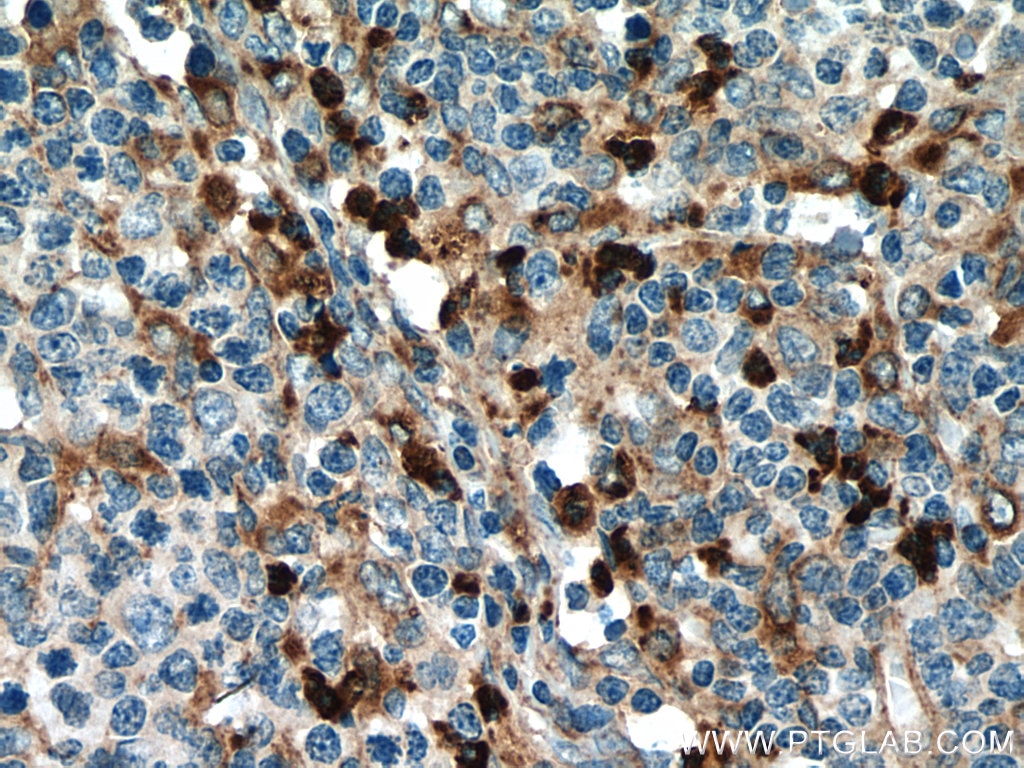
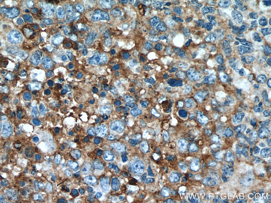
Validation Data Gallery
Tested Applications
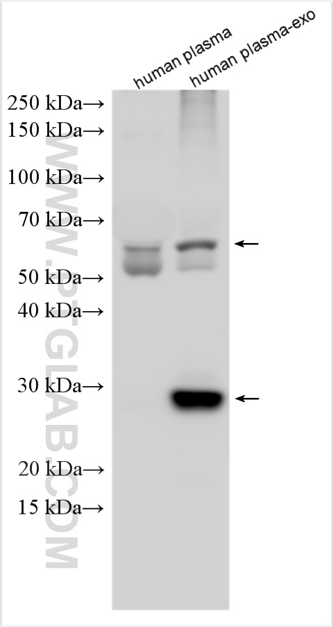
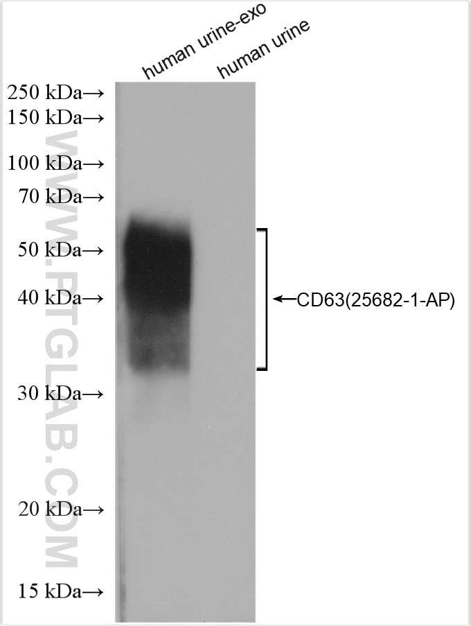
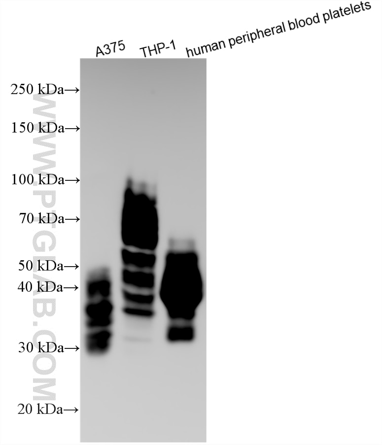
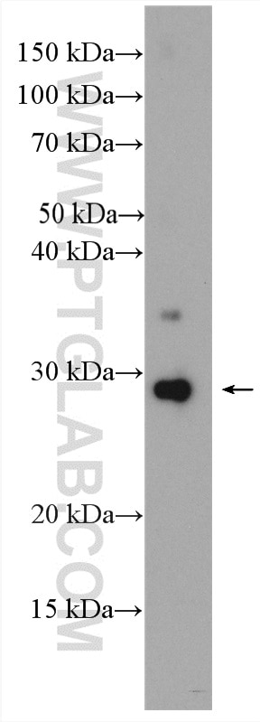
| Positive WB detected in | human urine exosomes tissue, A375 cells, human serum exosomes, THP-1 cells, human peripheral blood platelets |
| Positive IHC detected in | human malignant melanoma tissue, human lymphoma tissue, human tonsillitis tissue Note: suggested antigen retrieval with TE buffer pH 9.0; (*) Alternatively, antigen retrieval may be performed with citrate buffer pH 6.0 |
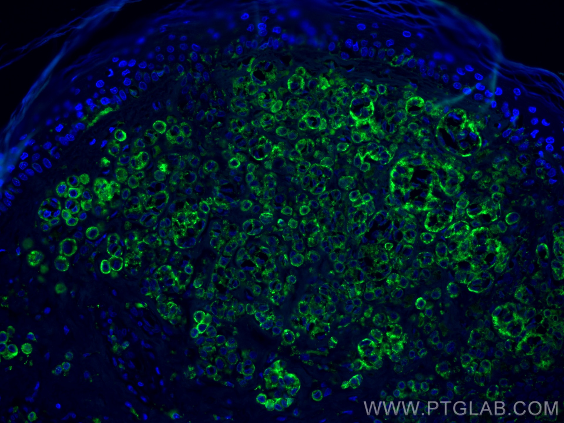
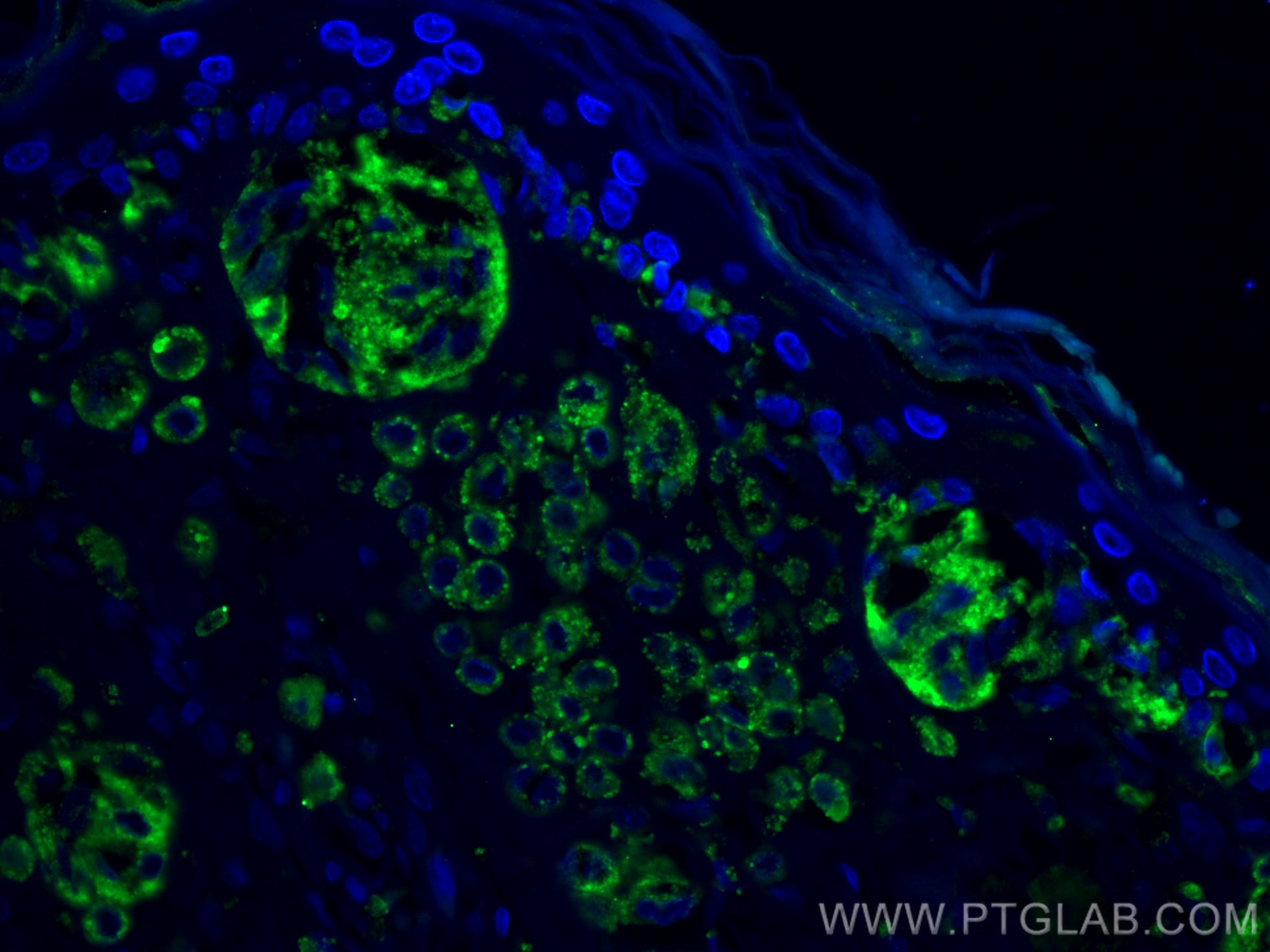
| Positive IF-P detected in | human malignant melanoma tissue |
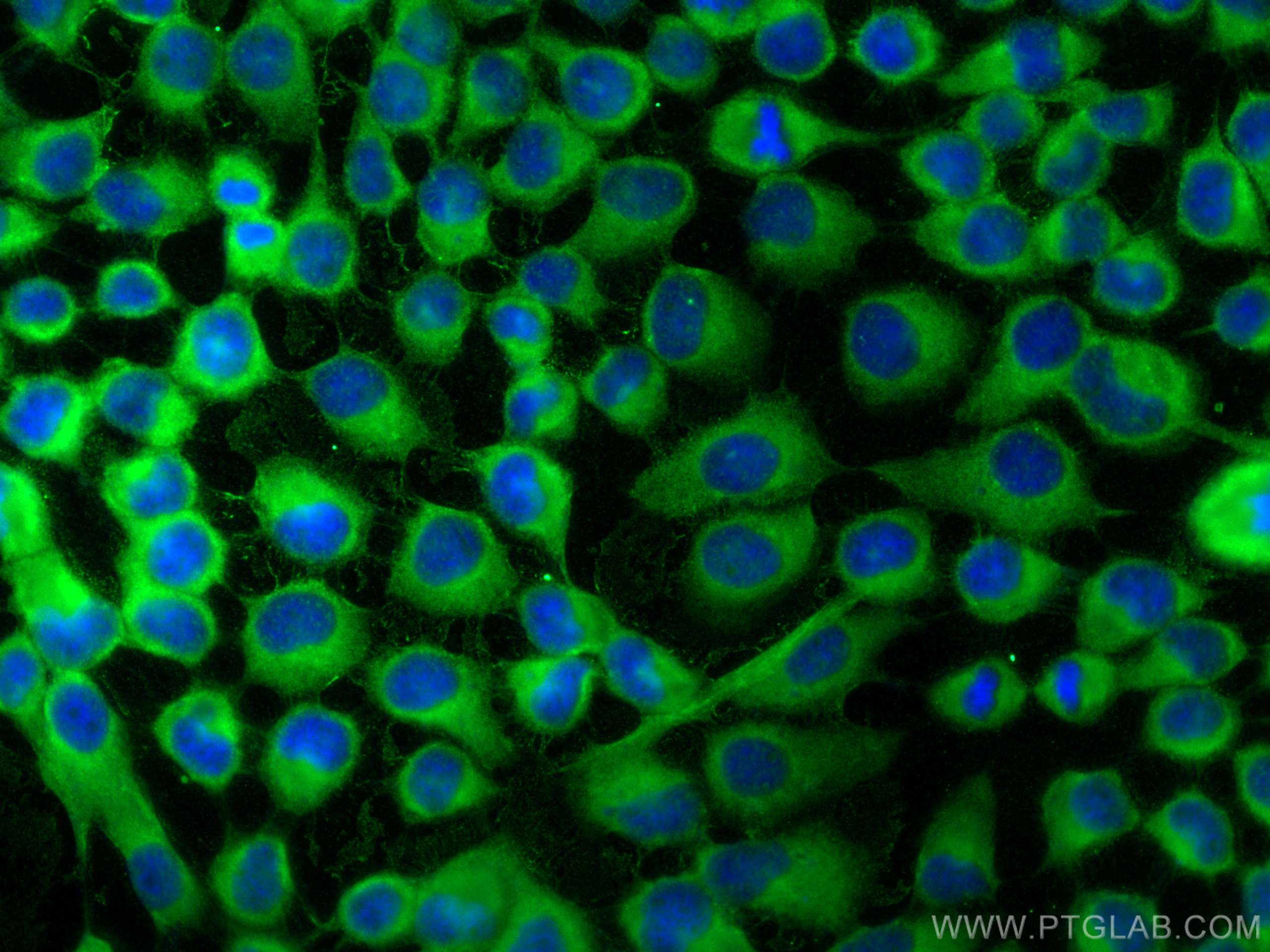
| Positive IF/ICC detected in | A431 cells |
Recommended dilution
| Application | Dilution |
|---|---|
| Western Blot (WB) | WB : 1:500-1:40000 |
| Immunohistochemistry (IHC) | IHC : 1:400-1:1600 |
| Immunofluorescence (IF)-P | IF-P : 1:50-1:500 |
| Immunofluorescence (IF)/ICC | IF/ICC : 1:200-1:800 |
| It is recommended that this reagent should be titrated in each testing system to obtain optimal results. | |
| Sample-dependent, Check data in validation data gallery. | |
Published Applications
| KD/KO | See 1 publications below |
| WB | See 453 publications below |
| IHC | See 8 publications below |
| IF | See 22 publications below |
| IP | See 2 publications below |
| ChIP | See 1 publications below |
Product Information
25682-1-AP targets CD63 in WB, IHC, IF/ICC, IF-P, IP, ChIP, ELISA applications and shows reactivity with human samples.
| Tested Reactivity | human |
| Cited Reactivity | human, pig, rabbit, monkey, hamster, goat |
| Host / Isotype | Rabbit / IgG |
| Class | Polyclonal |
| Type | Antibody |
| Immunogen |
CatNo: Ag19690 Product name: Recombinant human CD63 protein Source: e coli.-derived, PET28a Tag: 6*His Domain: 104-209 aa of BC002349 Sequence: GYVFRDKVMSEFNNNFRQQMENYPKNNHTASILDRMQADFKCCGAANYTDWEKIPSMSKNRVPDSCCINVTVGCGINFNEKAIHKEGCVEKIGGWLRKNVLVVAAA 相同性解析による交差性が予測される生物種 |
| Full Name | CD63 molecule |
| Calculated molecular weight | 26 kDa |
| Observed molecular weight | 30-60 kDa |
| GenBank accession number | BC002349 |
| Gene Symbol | CD63 |
| Gene ID (NCBI) | 967 |
| RRID | AB_2783831 |
| Conjugate | Unconjugated |
| Form | |
| Form | Liquid |
| Purification Method | Antigen affinity purification |
| UNIPROT ID | P08962 |
| Storage Buffer | PBS with 0.02% sodium azide and 50% glycerol{{ptg:BufferTemp}}7.3 |
| Storage Conditions | Store at -20°C. Stable for one year after shipment. Aliquoting is unnecessary for -20oC storage. |
Background Information
CD63 is a 30-60 kDa lysosomal membrane protein that belongs to the tetraspanin family. This protein plays many important roles in immuno-physiological functions. It mediates signal transduction events that play a role in the regulation of cell development, activation, growth, and motility. CD63 is expressed on activated platelets, thus it may function as a blood platelet activation marker. CD63 is a lysosomal membrane glycoprotein that is translocated to plasma membrane after platelet activation. The CD63 tetraspanin is highly expressed in the early stages of melanoma and decreases in advanced lesions, suggesting it as a possible suppressor of tumor progression. Deficiency of this protein is associated with Hermansky-Pudlak syndrome.
Protocols
| Product Specific Protocols | |
|---|---|
| IF protocol for CD63 antibody 25682-1-AP | Download protocol |
| IHC protocol for CD63 antibody 25682-1-AP | Download protocol |
| WB protocol for CD63 antibody 25682-1-AP | Download protocol |
| Standard Protocols | |
|---|---|
| Click here to view our Standard Protocols |
Publications
| Species | Application | Title |
|---|---|---|
ACS Nano Engineering Extracellular Vesicles Restore the Impaired Cellular Uptake and Attenuate Intervertebral Disc Degeneration. | ||
Nat Commun Injectable ECM-mimetic dynamic hydrogels abolish ferroptosis-induced post-discectomy herniation through delivering nucleus pulposus progenitor cell-derived exosomes | ||
Transl Neurodegener Abnormal α-synuclein binds to synaptotagmin 13, impairing extracellular vesicle release in synucleinopathies | ||
Adv Sci (Weinh) Tβ4-Engineered ADSC Extracellular Vesicles Rescue Cell Senescence Through Separable Microneedle Patches for Diabetic Wound Healing | ||
Adv Sci (Weinh) Astrocyte-Derived Extracellular Vesicular miR-143-3p Dampens Autophagic Degradation of Endothelial Adhesion Molecules and Promotes Neutrophil Transendothelial Migration after Acute Brain Injury |

