Validation Data Gallery
Tested Applications
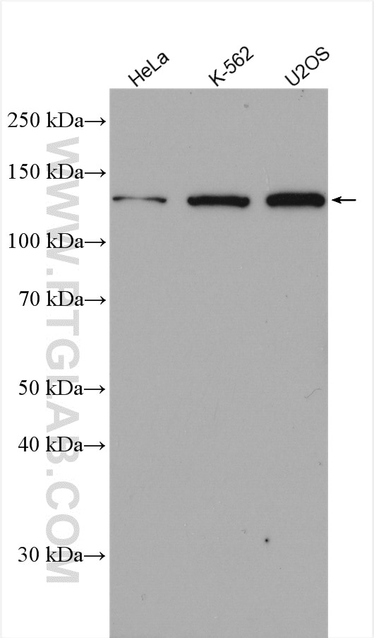
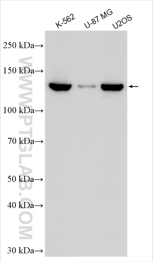
| Positive WB detected in | HeLa cells, K-562 cells, U2OS cells, U-87 MG cells |
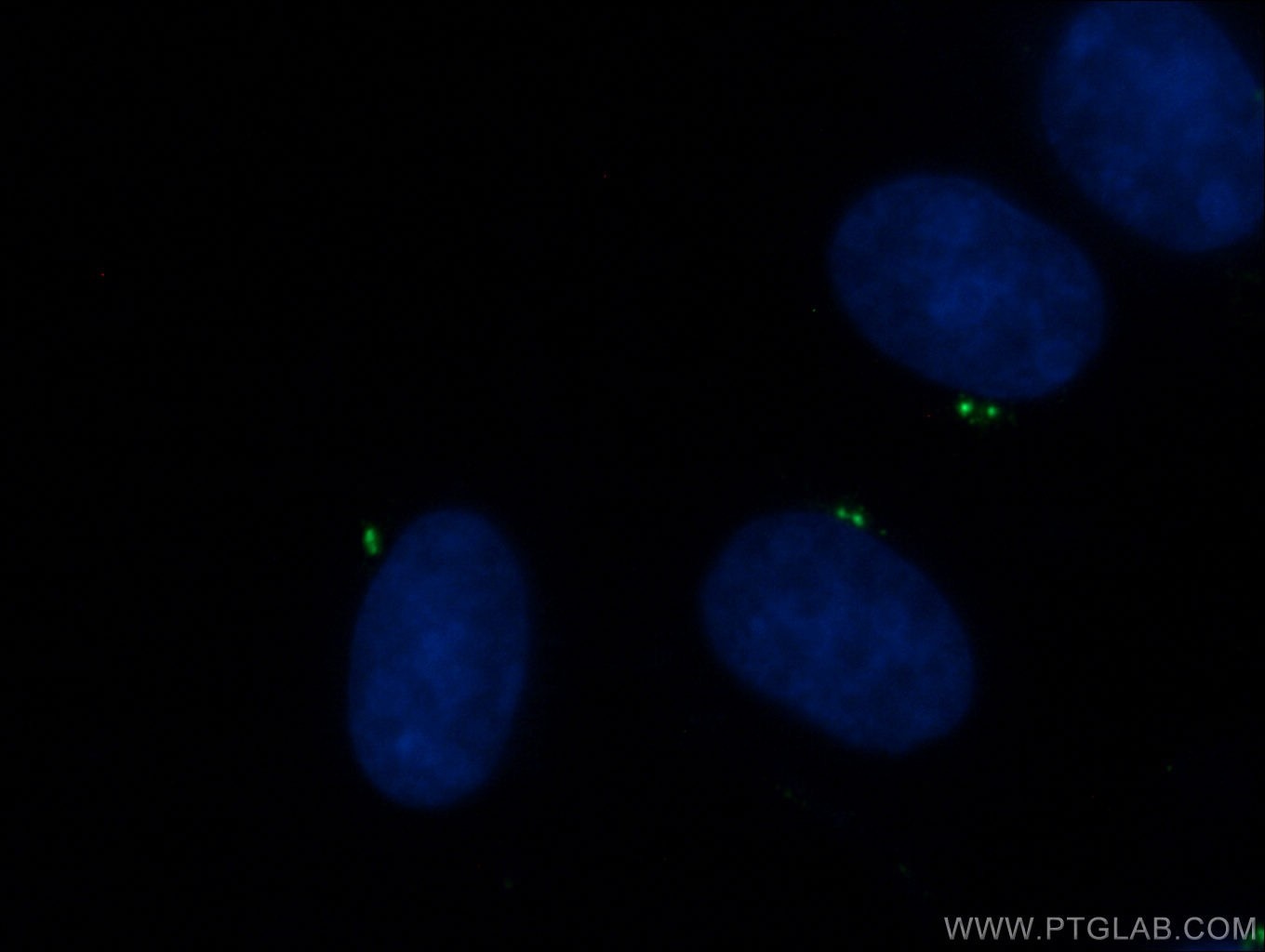
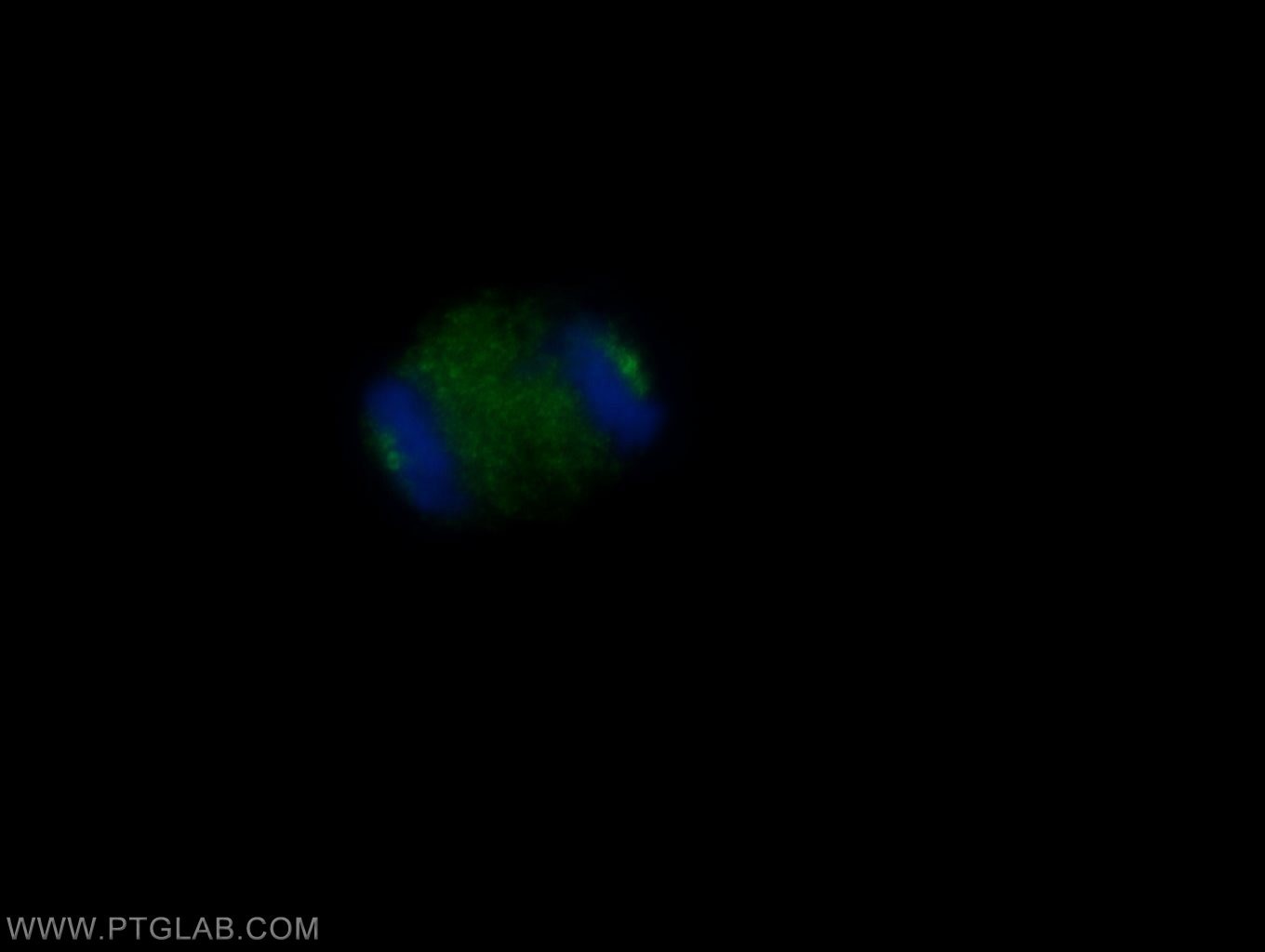
| Positive IF/ICC detected in | MDCK cells |
Recommended dilution
| Application | Dilution |
|---|---|
| Western Blot (WB) | WB : 1:1000-1:3000 |
| Immunofluorescence (IF)/ICC | IF/ICC : 1:50-1:500 |
| It is recommended that this reagent should be titrated in each testing system to obtain optimal results. | |
| Sample-dependent, Check data in validation data gallery. | |
Published Applications
| WB | See 2 publications below |
| IF | See 8 publications below |
Product Information
24428-1-AP targets CEP135 in WB, IF/ICC, ELISA applications and shows reactivity with human, Canine samples.
| Tested Reactivity | human, Canine |
| Cited Reactivity | human, mouse |
| Host / Isotype | Rabbit / IgG |
| Class | Polyclonal |
| Type | Antibody |
| Immunogen | CEP135 fusion protein Ag19122 相同性解析による交差性が予測される生物種 |
| Full Name | centrosomal protein 135kDa |
| Calculated molecular weight | 1140 aa, 134 kDa |
| Observed molecular weight | 135 kDa |
| GenBank accession number | BC136535 |
| Gene Symbol | CEP135 |
| Gene ID (NCBI) | 9662 |
| RRID | AB_2879543 |
| Conjugate | Unconjugated |
| Form | Liquid |
| Purification Method | Antigen affinity purification |
| UNIPROT ID | Q66GS9 |
| Storage Buffer | PBS with 0.02% sodium azide and 50% glycerol , pH 7.3 |
| Storage Conditions | Store at -20°C. Stable for one year after shipment. Aliquoting is unnecessary for -20oC storage. |
Background Information
CEP135 is a 135 kDa centrosomal protein that is present in a wide range of organisms. CEP135 is located at the centrosome throughout the cell cycle, and localization is independent of the microtubule network. It distributes throughout the centrosomal area in association with the electron-dense material surrounding centrioles.
Protocols
| Product Specific Protocols | |
|---|---|
| WB protocol for CEP135 antibody 24428-1-AP | Download protocol |
| IF protocol for CEP135 antibody 24428-1-AP | Download protocol |
| Standard Protocols | |
|---|---|
| Click here to view our Standard Protocols |
Publications
| Species | Application | Title |
|---|---|---|
Curr Biol SAS-6 Association with γ-Tubulin Ring Complex Is Required for Centriole Duplication in Human Cells. | ||
EMBO Rep ENKD1 promotes CP110 removal through competing with CEP97 to initiate ciliogenesis. | ||
Elife WDR90 is a centriolar microtubule wall protein important for centriole architecture integrity. | ||
Cells Mitotic Maturation Compensates for Premature Centrosome Splitting and PCM Loss in Human cep135 Knockout Cells. | ||
J Cell Sci Separation-of-function MCPH-associated mutations in CPAP affect centriole number and length |


