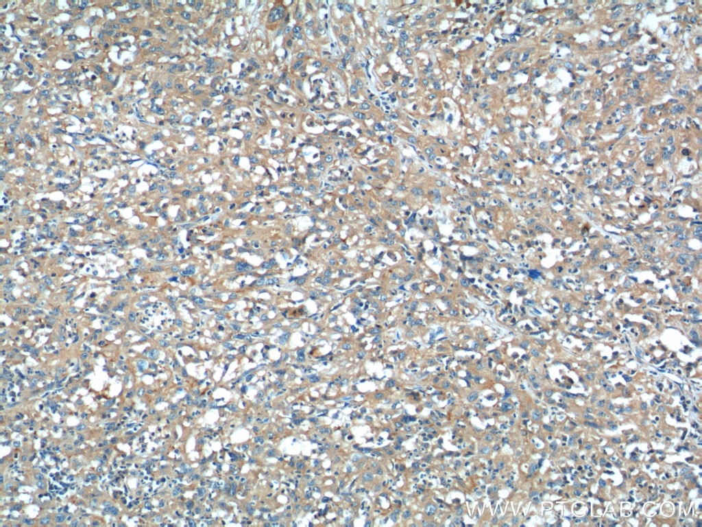
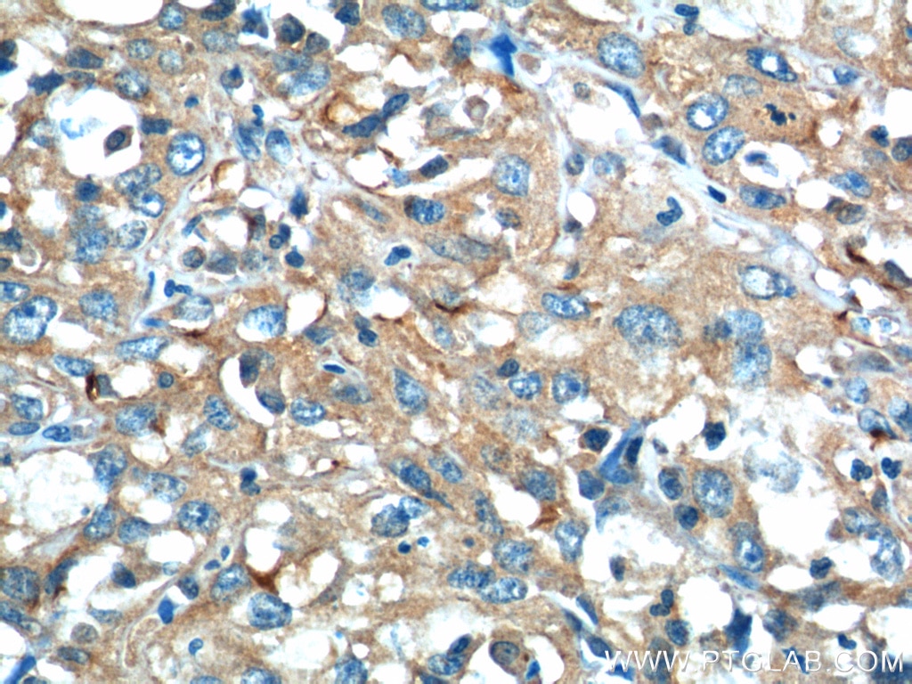
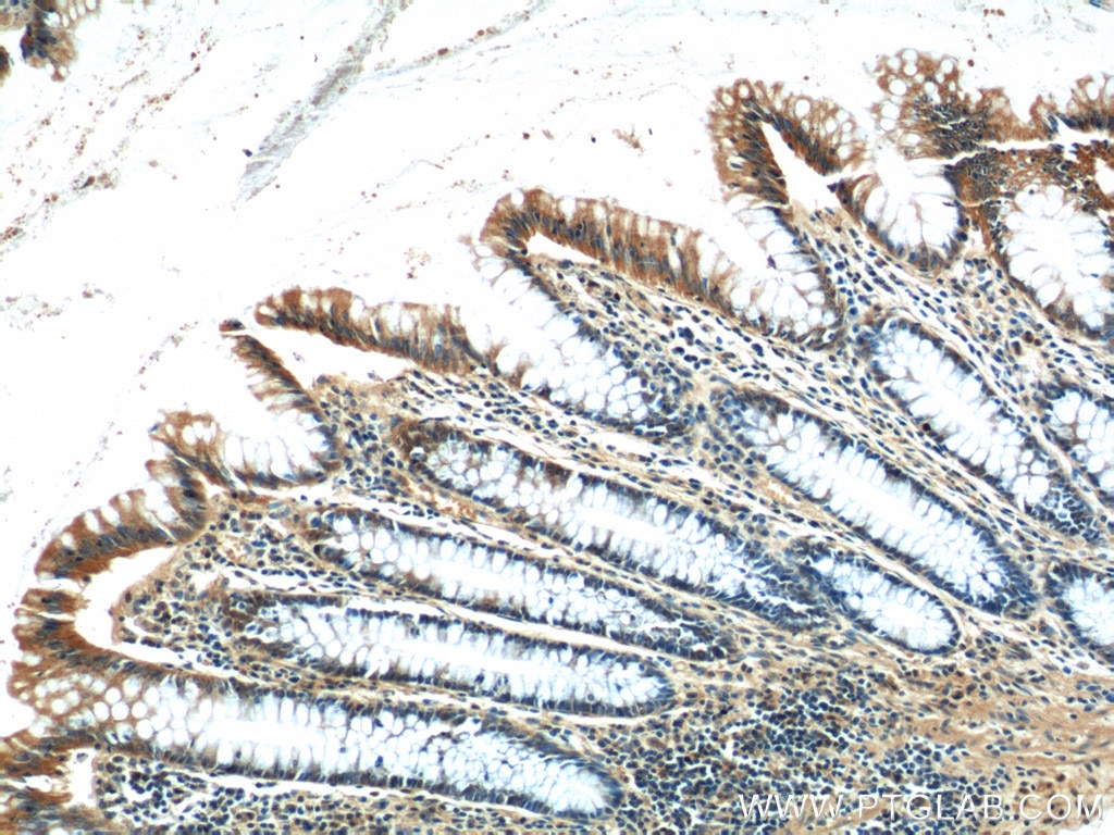
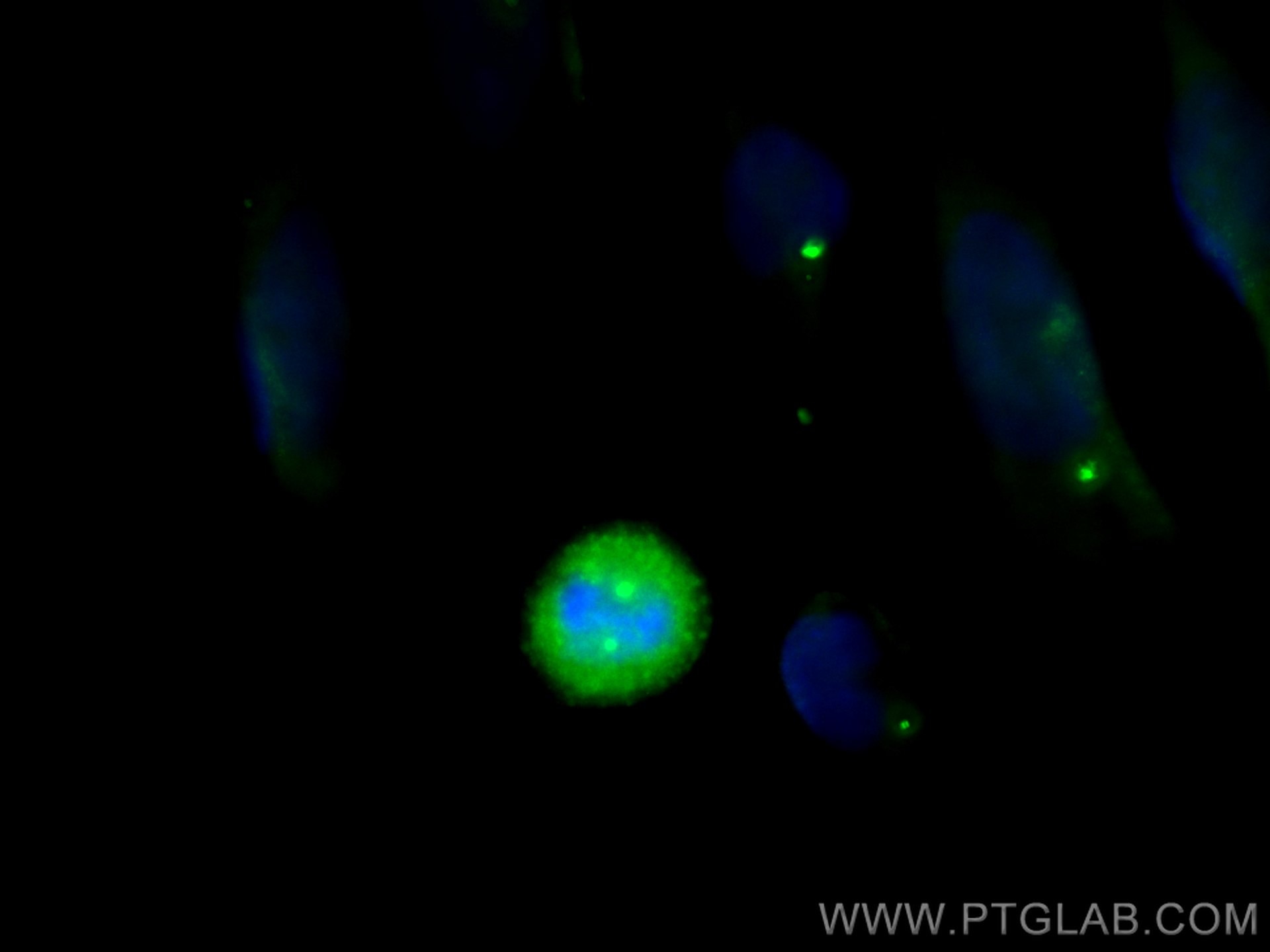
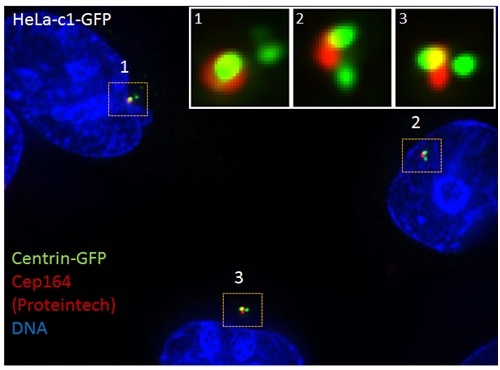
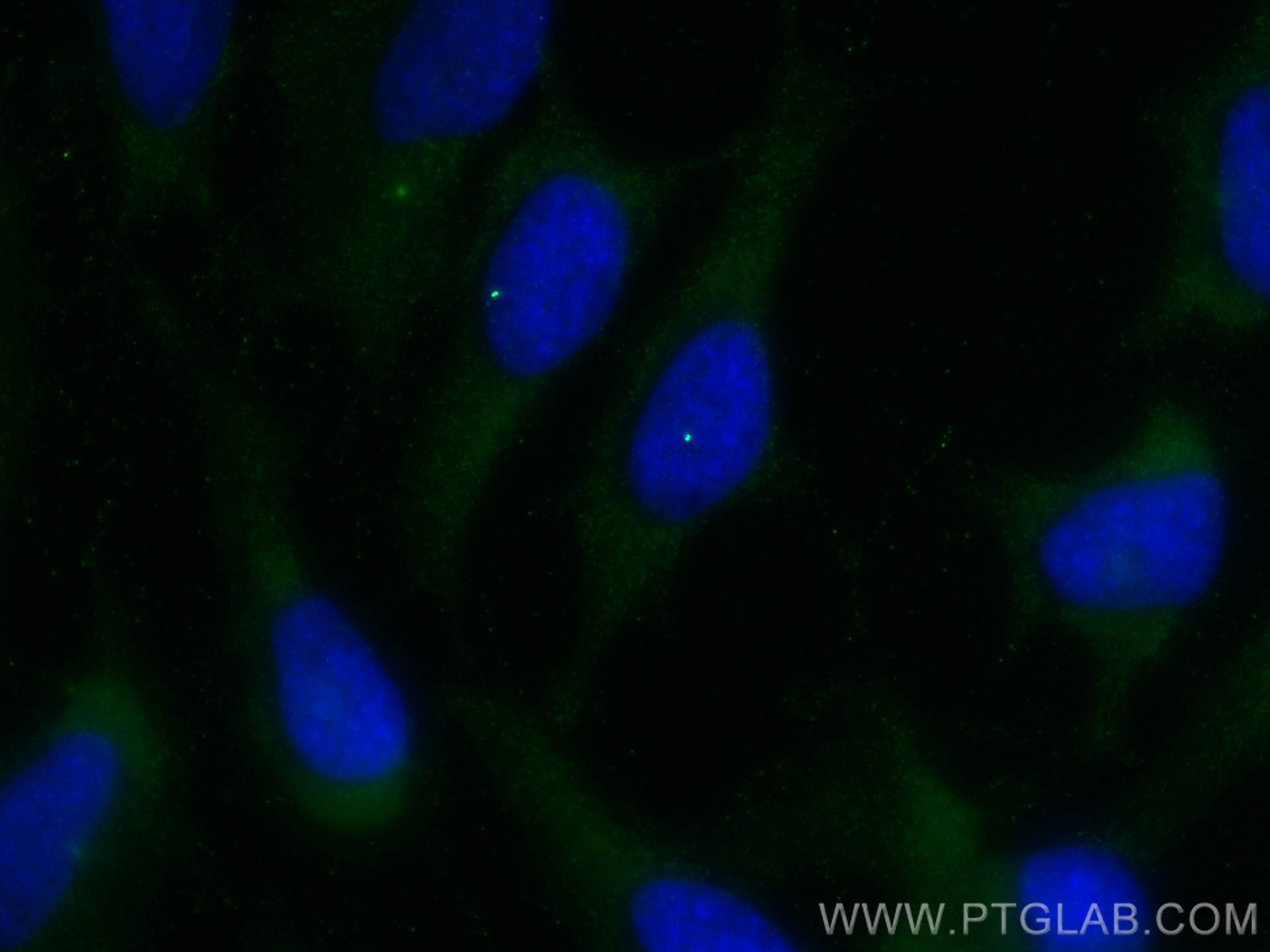
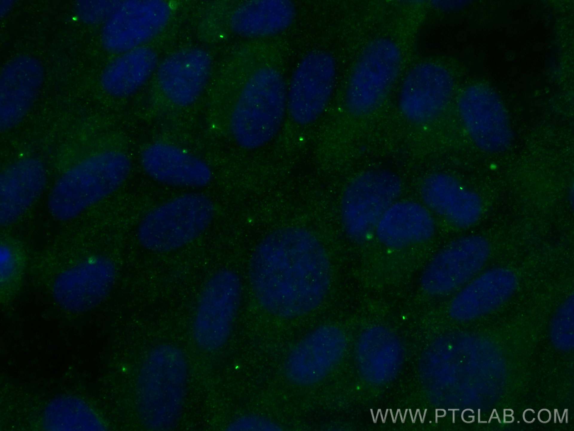
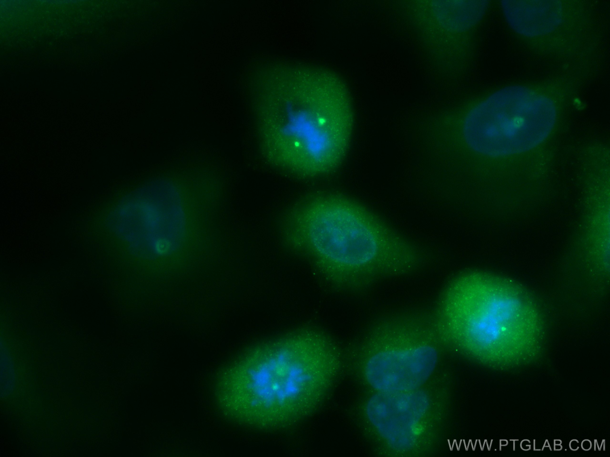
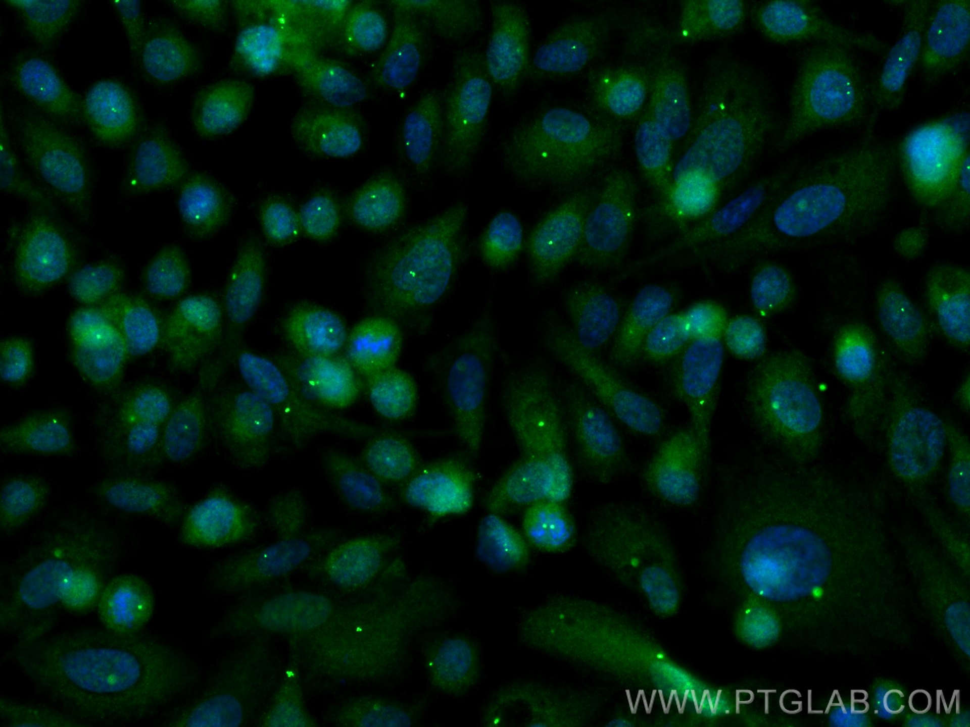
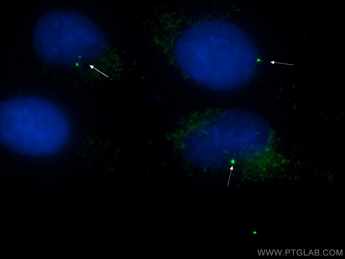
Validation Data Gallery
Tested Applications
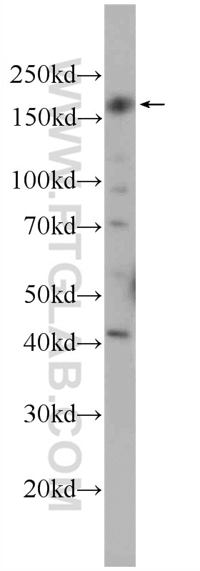
| Positive WB detected in | HEK-293 cells |
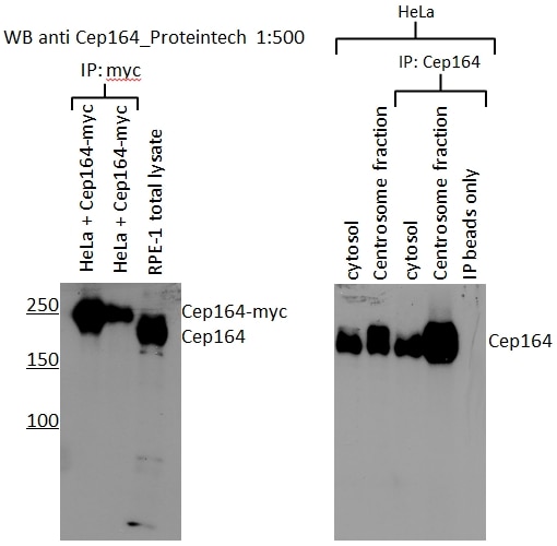
| Positive IP detected in | RPE1 cells |
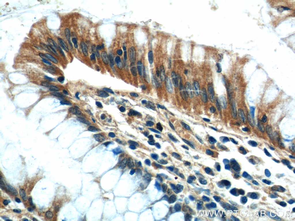
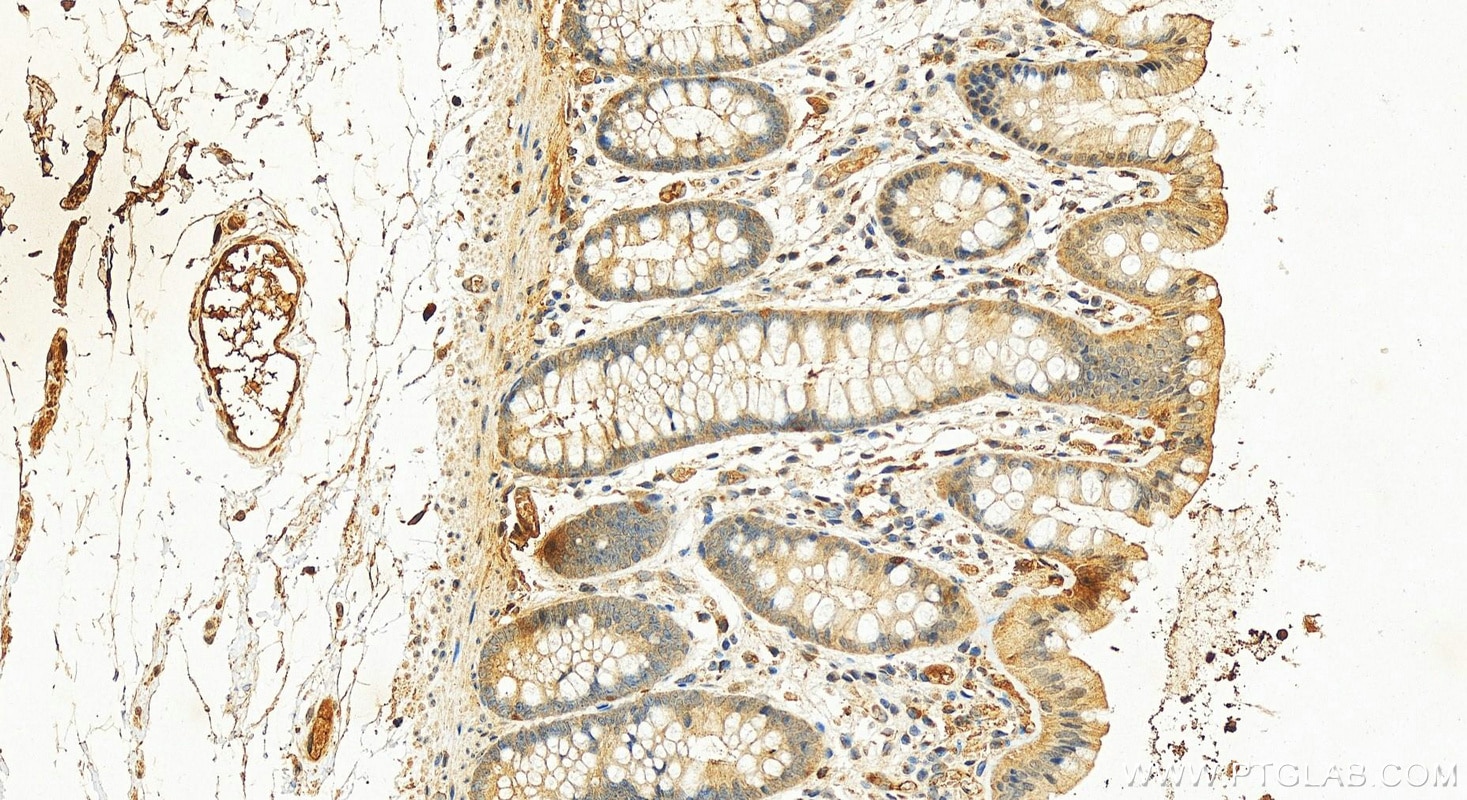
| Positive IHC detected in | human colon tissue, human cervical cancer tissue Note: suggested antigen retrieval with TE buffer pH 9.0; (*) Alternatively, antigen retrieval may be performed with citrate buffer pH 6.0 |
| Positive IF/ICC detected in | HeLa cells, A549 cells, PC-3 cells, MDCK cells, hTERT-RPE1 cells |
Recommended dilution
| Application | Dilution |
|---|---|
| Western Blot (WB) | WB : 1:200-1:1000 |
| Immunoprecipitation (IP) | IP : 0.5-4.0 ug for 1.0-3.0 mg of total protein lysate |
| Immunohistochemistry (IHC) | IHC : 1:100-1:1000 |
| Immunofluorescence (IF)/ICC | IF/ICC : 1:200-1:800 |
| It is recommended that this reagent should be titrated in each testing system to obtain optimal results. | |
| Sample-dependent, Check data in validation data gallery. | |
Published Applications
| KD/KO | See 2 publications below |
| WB | See 10 publications below |
| IHC | See 1 publications below |
| IF | See 53 publications below |
| IP | See 1 publications below |
Product Information
22227-1-AP targets CEP164 in WB, IHC, IF/ICC, IP, ELISA applications and shows reactivity with human, canine samples.
| Tested Reactivity | human, canine |
| Cited Reactivity | human, mouse |
| Host / Isotype | Rabbit / IgG |
| Class | Polyclonal |
| Type | Antibody |
| Immunogen | CEP164 fusion protein Ag17570 相同性解析による交差性が予測される生物種 |
| Full Name | centrosomal protein 164kDa |
| Calculated molecular weight | 1460 aa, 164 kDa |
| Observed molecular weight | 164 kDa |
| GenBank accession number | BC000602 |
| Gene Symbol | CEP164 |
| Gene ID (NCBI) | 22897 |
| RRID | AB_2651175 |
| Conjugate | Unconjugated |
| Form | Liquid |
| Purification Method | Antigen affinity purification |
| UNIPROT ID | Q9UPV0 |
| Storage Buffer | PBS with 0.02% sodium azide and 50% glycerol , pH 7.3 |
| Storage Conditions | Store at -20°C. Stable for one year after shipment. Aliquoting is unnecessary for -20oC storage. |
Background Information
CEP164, also called KIAA1052 or NPHP15, is a 1460 amino acid protein containing 1 WW domain. CEP164 localizes in the microtubule organizing center and is expressed in several cell lines. CEP164 plays a role in microtubule organization and/or maintenance for the formation of primary cilia, a microtubule-based structure that protrudes from the surface of epithelial cells. CEP164 plays a critical role in the G2/M checkpoint and nuclear divisions. The expression of CEP164 is normally limited to the mother centriole, and CEP164 can be used as a useful marker for mother centriole.
Protocols
| Product Specific Protocols | |
|---|---|
| WB protocol for CEP164 antibody 22227-1-AP | Download protocol |
| IHC protocol for CEP164 antibody 22227-1-AP | Download protocol |
| IF protocol for CEP164 antibody 22227-1-AP | Download protocol |
| Standard Protocols | |
|---|---|
| Click here to view our Standard Protocols |
Publications
| Species | Application | Title |
|---|---|---|
Cell Res NudCL2 is an autophagy receptor that mediates selective autophagic degradation of CP110 at mother centrioles to promote ciliogenesis. | ||
Nat Commun M-Phase Phosphoprotein 9 regulates ciliogenesis by modulating CP110-CEP97 complex localization at the mother centriole. | ||
Nat Commun DNA replication licensing factor Cdc6 and Plk4 kinase antagonistically regulate centrosome duplication via Sas-6. | ||
EMBO J ANKRD26 recruits PIDD1 to centriolar distal appendages to activate the PIDDosome following centrosome amplification. | ||
