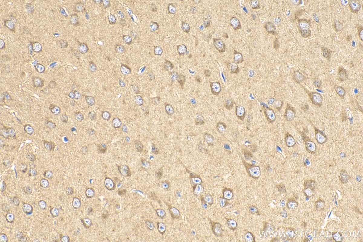
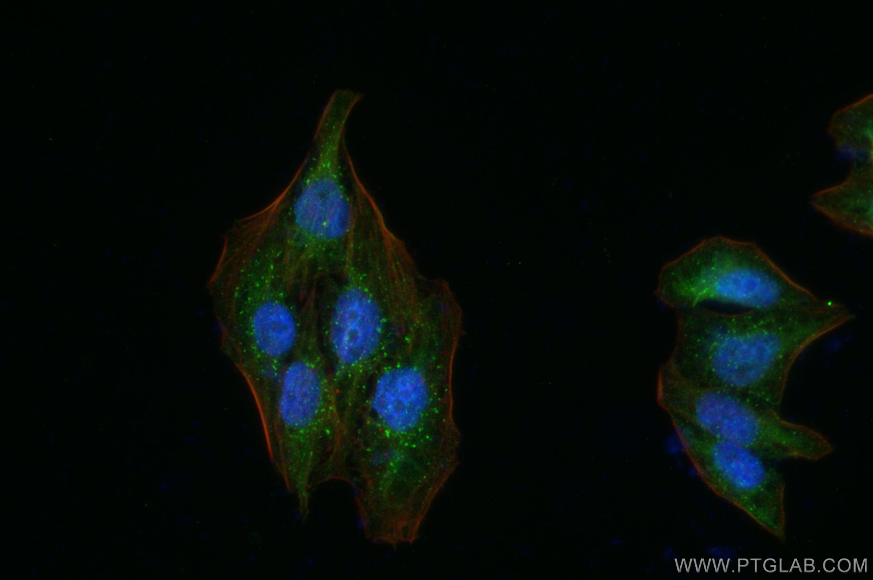
Validation Data Gallery
Tested Applications
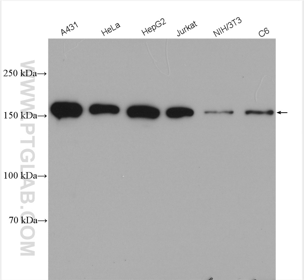
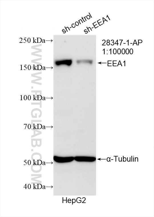
| Positive WB detected in | A431 cells, HepG2 cells, HeLa cells, Jurkat cells, NIH/3T3 cells, C6 cells |
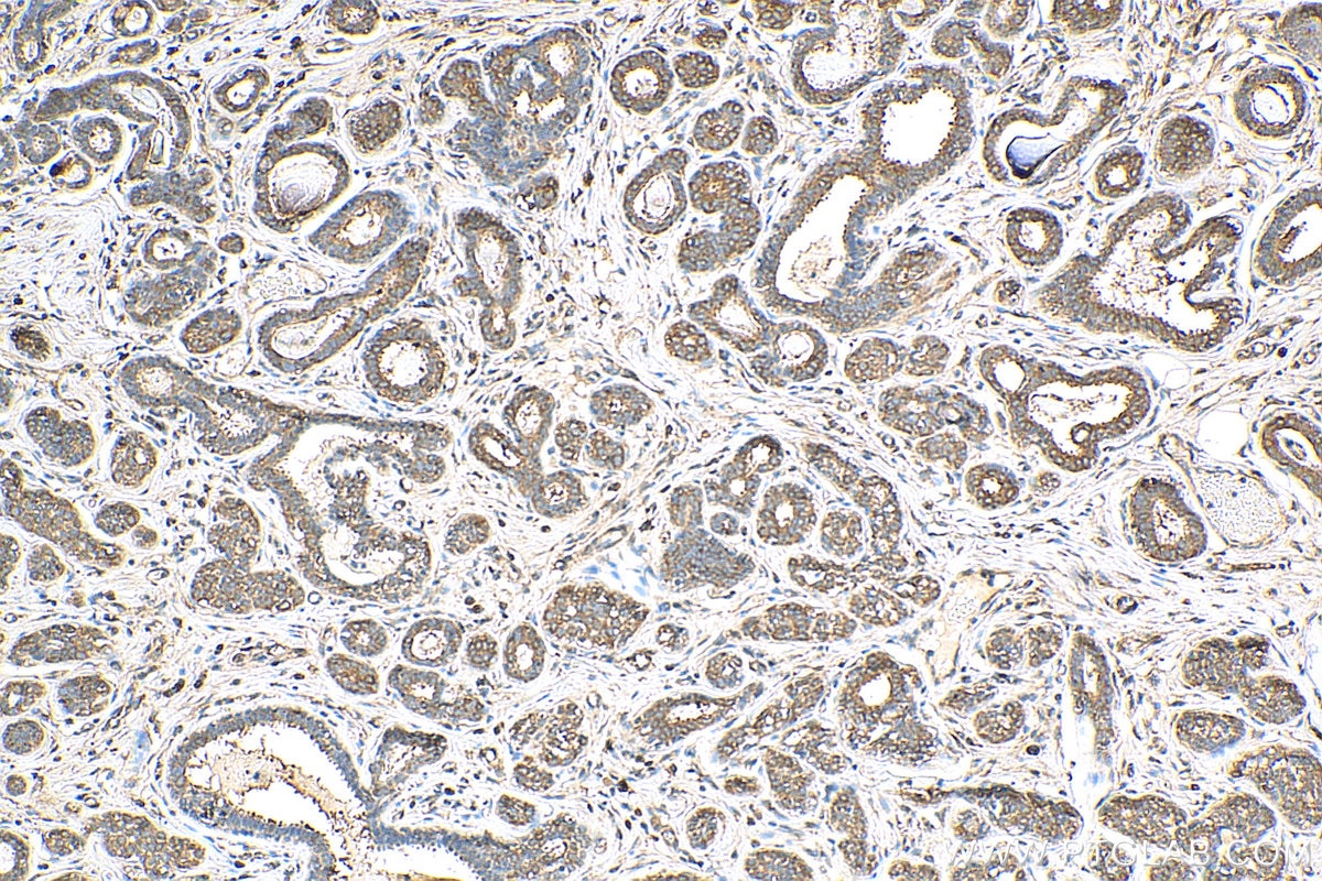
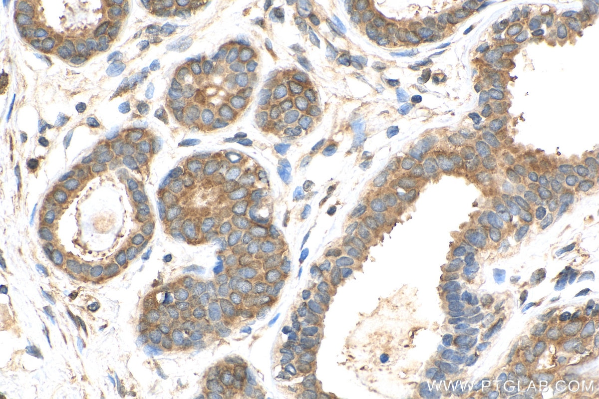
| Positive IHC detected in | human breast cancer tissue, mouse brain tissue Note: suggested antigen retrieval with TE buffer pH 9.0; (*) Alternatively, antigen retrieval may be performed with citrate buffer pH 6.0 |
| Positive IF/ICC detected in | HepG2 cells |
Recommended dilution
| Application | Dilution |
|---|---|
| Western Blot (WB) | WB : 1:2000-1:16000 |
| Immunohistochemistry (IHC) | IHC : 1:50-1:500 |
| Immunofluorescence (IF)/ICC | IF/ICC : 1:200-1:800 |
| It is recommended that this reagent should be titrated in each testing system to obtain optimal results. | |
| Sample-dependent, Check data in validation data gallery. | |
Published Applications
| IF | See 7 publications below |
Product Information
28347-1-AP targets EEA1 in WB, IHC, IF/ICC, ELISA applications and shows reactivity with Human, Mouse, Rat samples.
| Tested Reactivity | Human, Mouse, Rat |
| Cited Reactivity | human, mouse, monkey |
| Host / Isotype | Rabbit / IgG |
| Class | Polyclonal |
| Type | Antibody |
| Immunogen | EEA1 fusion protein Ag28814 相同性解析による交差性が予測される生物種 |
| Full Name | early endosome antigen 1 |
| Calculated molecular weight | 162 kDa |
| Observed molecular weight | 170 kDa |
| GenBank accession number | NM_003566 |
| Gene Symbol | EEA1 |
| Gene ID (NCBI) | 8411 |
| RRID | AB_2881117 |
| Conjugate | Unconjugated |
| Form | Liquid |
| Purification Method | Antigen affinity purification |
| UNIPROT ID | Q15075 |
| Storage Buffer | PBS with 0.02% sodium azide and 50% glycerol , pH 7.3 |
| Storage Conditions | Store at -20°C. Stable for one year after shipment. Aliquoting is unnecessary for -20oC storage. |
Background Information
Early endosome antigen 1 (EEA1) is a marker of early endosomes and has an important role in endosomal trafficking. EEA1 contains coiled-coil regions and an FYVE-type zinc finger which interacts with phosphatidylinositol-3-phosphate. EEA1 functions as a Rab5 effector, mediates endosome docking and, together with SNAREs, leads to membrane fusion.
Protocols
| Product Specific Protocols | |
|---|---|
| WB protocol for EEA1 antibody 28347-1-AP | Download protocol |
| IHC protocol for EEA1 antibody 28347-1-AP | Download protocol |
| IF protocol for EEA1 antibody 28347-1-AP | Download protocol |
| Standard Protocols | |
|---|---|
| Click here to view our Standard Protocols |
Publications
| Species | Application | Title |
|---|---|---|
Nucleic Acids Res Nucleosomes enter cells by clathrin- and caveolin-dependent endocytosis. | ||
Front Immunol Host MKRN1-Mediated Mycobacterial PPE Protein Ubiquitination Suppresses Innate Immune Response. | ||
Chemistry Investigation of Glycosylphosphatidylinositol (GPI)-Plasma Membrane Interaction in Live Cells and the Influence of GPI Glycan Structure on the Interaction | ||
Toxicol Lett Anti-ricin toxin human neutralizing antibodies and DMAbs protection against ricin toxin poisoning | ||
Nat Commun Sortilin-mediated translocation of mitochondrial ACSL1 impairs adipocyte thermogenesis and energy expenditure in male mice |
