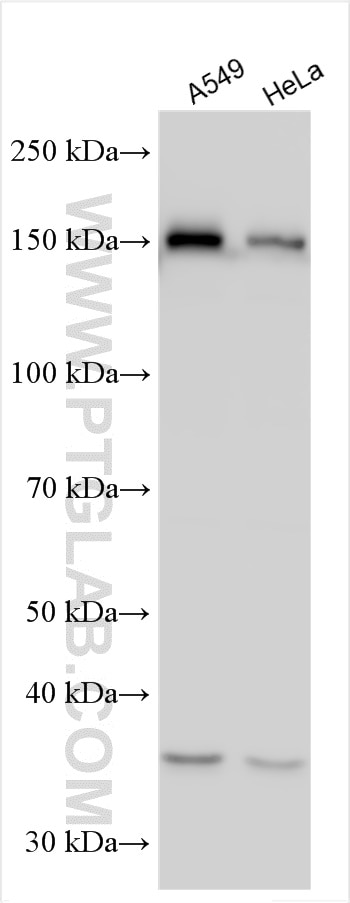
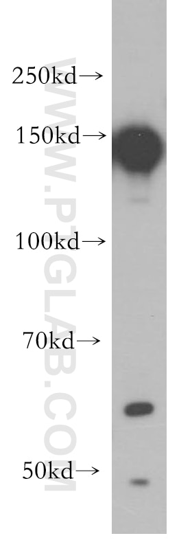
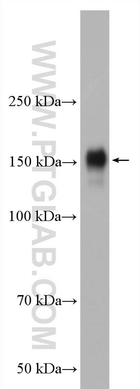
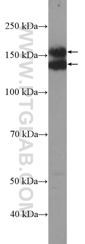
Validation Data Gallery
Tested Applications
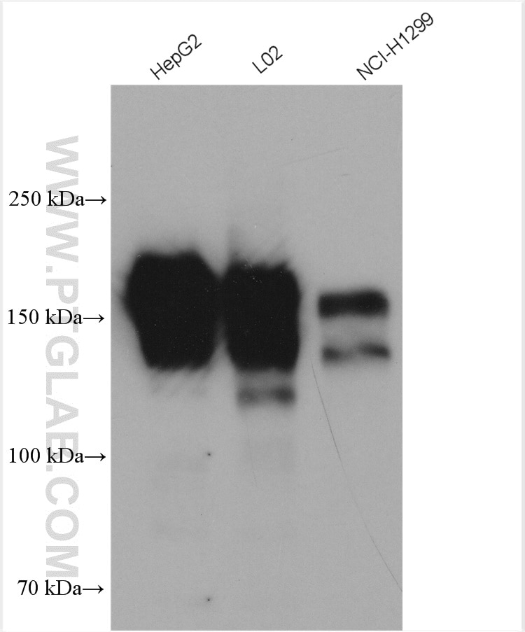
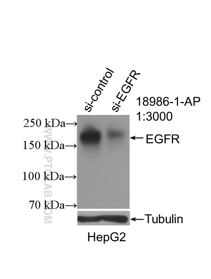
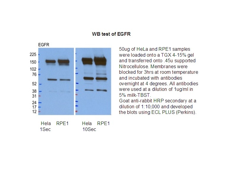
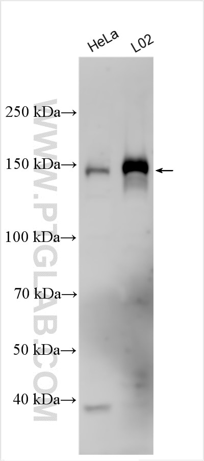
| Positive WB detected in | HepG2 cells, MCF-7 cells, A431 cells, HeLa/RPE1 cells, NCI-H1299 cells, HeLa cells, A549 cells, L02 cells, NCL-H1299 cells |
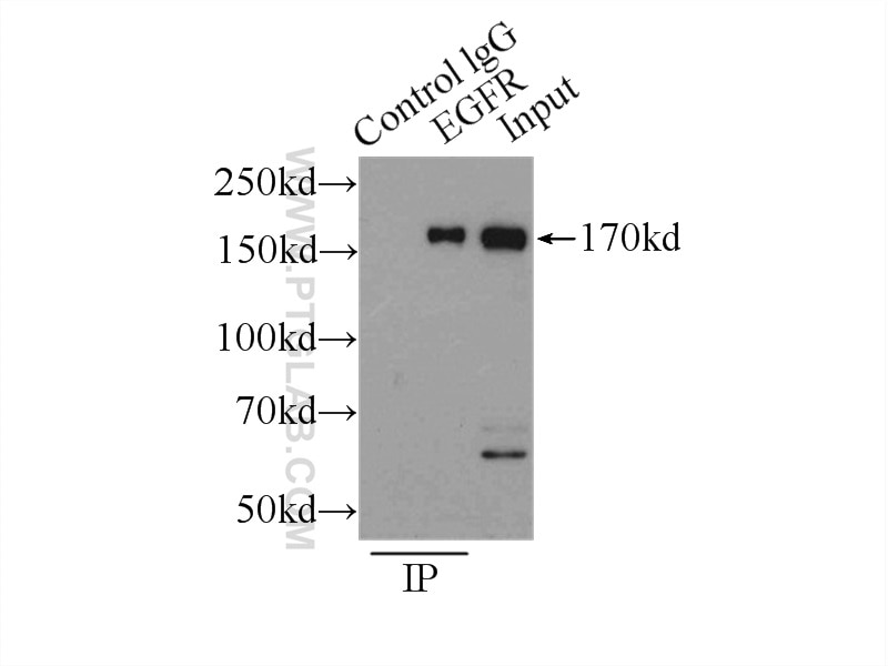
| Positive IP detected in | MCF-7 cells |
Recommended dilution
| Application | Dilution |
|---|---|
| Western Blot (WB) | WB : 1:1000-1:6000 |
| Immunoprecipitation (IP) | IP : 0.5-4.0 ug for 1.0-3.0 mg of total protein lysate |
| It is recommended that this reagent should be titrated in each testing system to obtain optimal results. | |
| Sample-dependent, Check data in validation data gallery. | |
Product Information
18986-1-AP targets EGFR-Specific in WB, IHC, IF, IP, CoIP, ChIP, ELISA applications and shows reactivity with human samples.
| Tested Reactivity | human |
| Cited Reactivity | human, mouse, rat, pig, sheep, goat, sus scrofa (pig) |
| Host / Isotype | Rabbit / IgG |
| Class | Polyclonal |
| Type | Antibody |
| Immunogen |
Peptide 相同性解析による交差性が予測される生物種 |
| Full Name | epidermal growth factor receptor (erythroblastic leukemia viral (v-erb-b) oncogene homolog, avian) |
| Calculated molecular weight | 134 kDa |
| Observed molecular weight | 145-165 kDa |
| GenBank accession number | NM_005228 |
| Gene Symbol | EGFR |
| Gene ID (NCBI) | 1956 |
| RRID | AB_10596476 |
| Conjugate | Unconjugated |
| Form | |
| Form | Liquid |
| Purification Method | Antigen affinity purification |
| UNIPROT ID | P00533 |
| Storage Buffer | PBS with 0.02% sodium azide and 50% glycerol{{ptg:BufferTemp}}7.3 |
| Storage Conditions | Store at -20°C. Stable for one year after shipment. Aliquoting is unnecessary for -20oC storage. |
Background Information
ERBB1, also named as EGFR, is a receptor for EGF, but also for other members of the EGF family, as TGF-alpha, amphiregulin, betacellulin, heparin-binding EGF-like growth factor, GP30 and vaccinia virus growth factor. ERBB1 is involved in the control of cell growth and differentiation. Phosphorylates MUC1 in breast cancer cells and increases the interaction of MUC1 with C-SRC and CTNNB1/beta-catenin. Isoform 2/truncated isoform of ERBB1 may act as an antagonist. EGFR is associated with lung cancer. The antibody is specific to isoform1.
Protocols
| Product Specific Protocols | |
|---|---|
| IP protocol for EGFR-Specific antibody 18986-1-AP | Download protocol |
| WB protocol for EGFR-Specific antibody 18986-1-AP | Download protocol |
| Standard Protocols | |
|---|---|
| Click here to view our Standard Protocols |
Publications
| Species | Application | Title |
|---|---|---|
Nat Microbiol Interferon-induced transmembrane protein-1 competitively blocks Ephrin receptor A2-mediated Epstein-Barr virus entry into epithelial cells | ||
Cell Res In vivo self-assembled small RNAs as a new generation of RNAi therapeutics.
| ||
Nat Commun Inhalable liposomal delivery of osimertinib and DNA for treating primary and metastasis lung cancer | ||
Cancer Commun (Lond) Blockage of EGFR/AKT and mevalonate pathways synergize the antitumor effect of temozolomide by reprogramming energy metabolism in glioblastoma | ||
Adv Sci (Weinh) Splicing Shift of RAC1 Accelerates Tumorigenesis and Defines a Potent Therapeutic Target in Lung Cancer | ||

