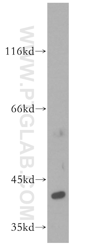
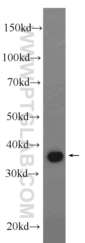
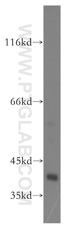
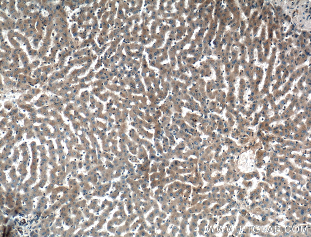
Validation Data Gallery
Tested Applications
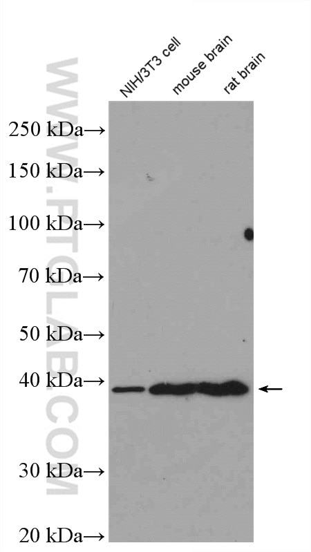
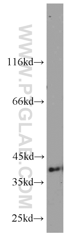
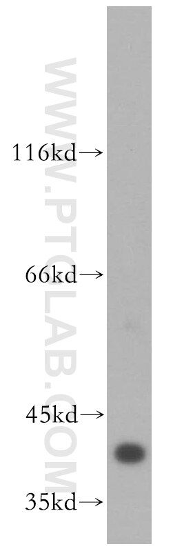
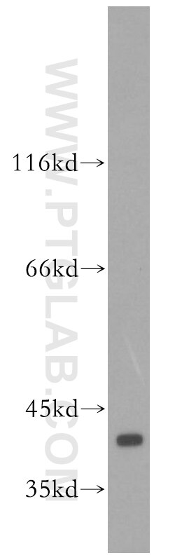
| Positive WB detected in | NIH/3T3 cells, A431 cells, mouse kidney tissue, HEK-293 cells, HeLa cells, PC-3 cells, rat kidney tissue, mouse brain tissue, rat brain tissue |
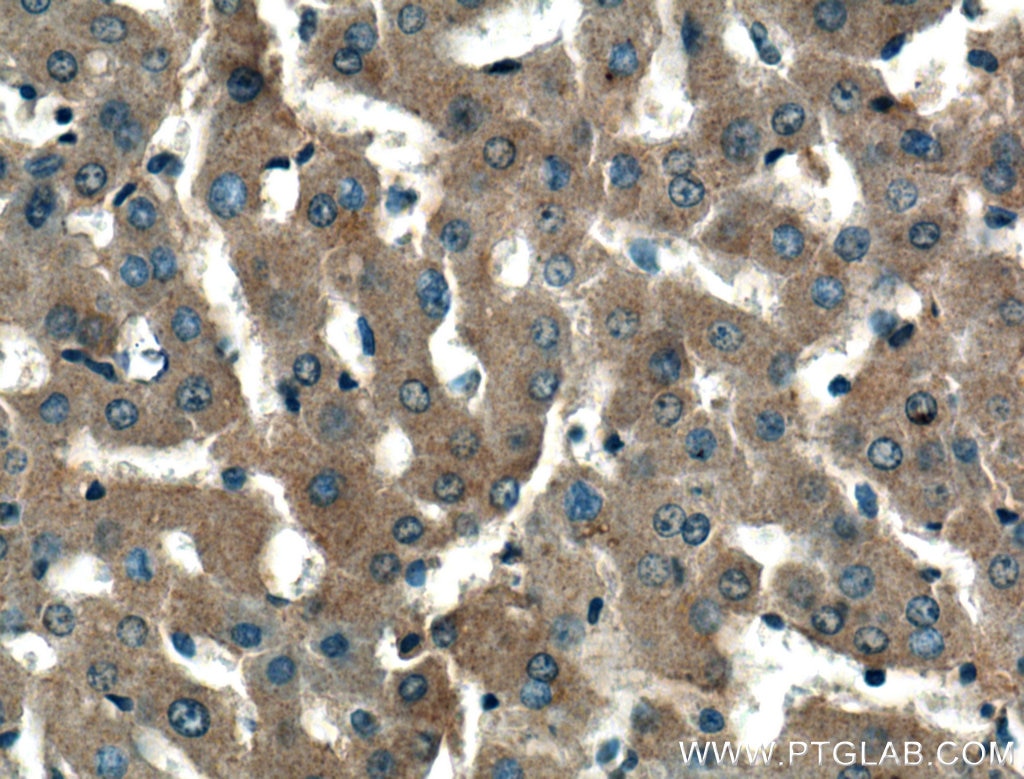
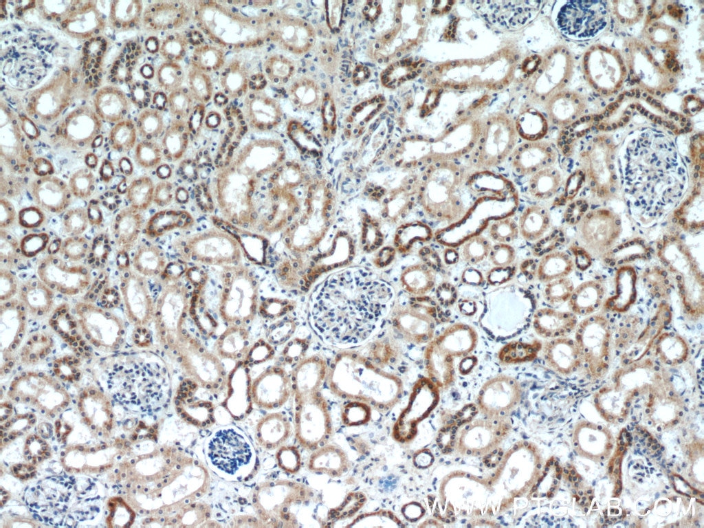
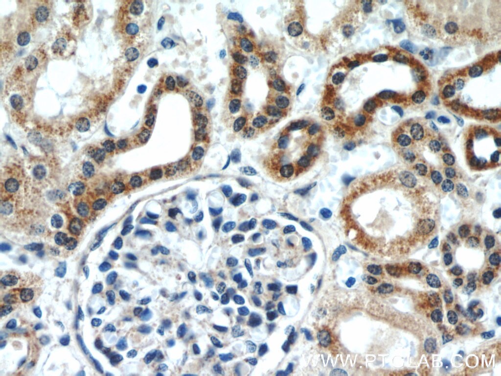
| Positive IHC detected in | human liver tissue, human kidney tissue Note: suggested antigen retrieval with TE buffer pH 9.0; (*) Alternatively, antigen retrieval may be performed with citrate buffer pH 6.0 |
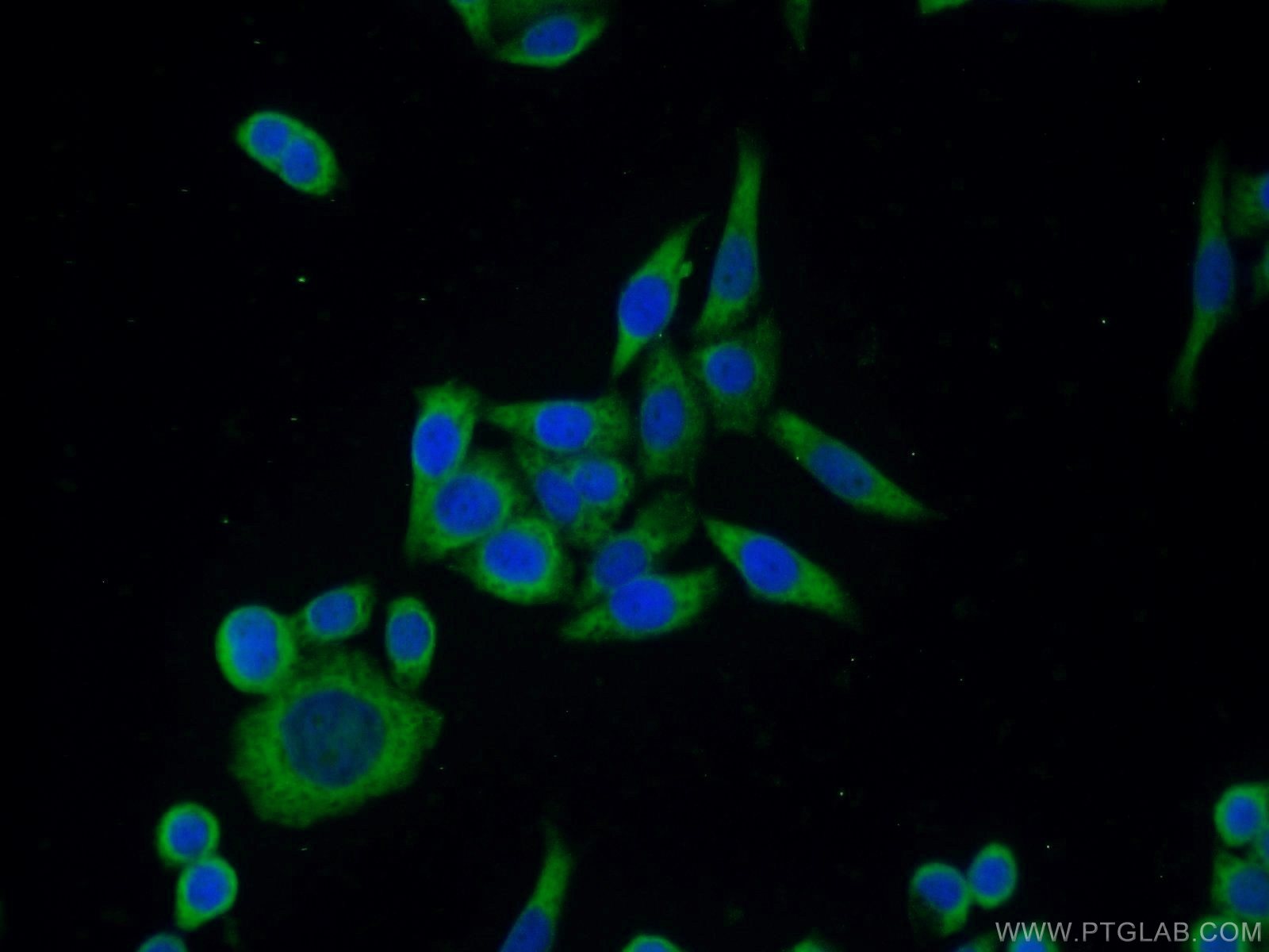
| Positive IF/ICC detected in | HeLa cells |
Recommended dilution
| Application | Dilution |
|---|---|
| Western Blot (WB) | WB : 1:1000-1:4000 |
| Immunohistochemistry (IHC) | IHC : 1:50-1:500 |
| Immunofluorescence (IF)/ICC | IF/ICC : 1:10-1:100 |
| It is recommended that this reagent should be titrated in each testing system to obtain optimal results. | |
| Sample-dependent, Check data in validation data gallery. | |
Published Applications
| KD/KO | See 1 publications below |
| WB | See 414 publications below |
| IHC | See 6 publications below |
| IF | See 4 publications below |
| IP | See 1 publications below |
Product Information
16443-1-AP targets ERK1/2 in WB, IHC, IF/ICC, IP, ELISA applications and shows reactivity with human, mouse, rat samples.
| Tested Reactivity | human, mouse, rat |
| Cited Reactivity | human, mouse, rat, pig, chicken, sheep, eriocheir sinensis |
| Host / Isotype | Rabbit / IgG |
| Class | Polyclonal |
| Type | Antibody |
| Immunogen |
CatNo: Ag9772 Product name: Recombinant human ERK1/2 protein Source: e coli.-derived, PGEX-4T Tag: GST Domain: 25-360 aa of BC017832 Sequence: YTNLSYIGEGAYGMVCSAYDNVNKVRVAIKKISPFEHQTYCQRTLREIKILLRFRHENIIGINDIIRAPTIEQMKDVYIVQDLMETDLYKLLKTQHLSNDHICYFLYQILRGLKYIHSANVLHRDLKPSNLLLNTTCDLKICDFGLARVADPDHDHTGFLTEYVATRWYRAPEIMLNSKGYTKSIDIWSVGCILAEMLSNRPIFPGKHYLDQLNHILGILGSPSQEDLNCIINLKARNYLLSLPHKNKVPWNRLFPNADSKALDLLDKMLTFNPHKRIEVEQALAHPYLEQYYDPSDEPIAEAPFKFDMELDDLPKEKLKELIFEETARFQPGYRS 相同性解析による交差性が予測される生物種 |
| Full Name | mitogen-activated protein kinase 1 |
| Calculated molecular weight | 360 aa, 41 kDa |
| Observed molecular weight | 38-44 kDa |
| GenBank accession number | BC017832 |
| Gene Symbol | ERK2 |
| Gene ID (NCBI) | 5594 |
| RRID | AB_10603369 |
| Conjugate | Unconjugated |
| Form | |
| Form | Liquid |
| Purification Method | Antigen affinity purification |
| UNIPROT ID | P28482 |
| Storage Buffer | PBS with 0.02% sodium azide and 50% glycerol{{ptg:BufferTemp}}7.3 |
| Storage Conditions | Store at -20°C. Stable for one year after shipment. Aliquoting is unnecessary for -20oC storage. |
Background Information
ERK1 and ERK2 belongs to the protein kinase superfamily. It is involved in both the initiation and regulation of meiosis, mitosis, and postmitotic functions in differentiated cells by phosphorylating a number of transcription factors such as ELK-1. ERK1/2 catalized the reaction: ATP + a protein = ADP + a phosphoprotein. It is activated by tyrosine phosphorylation in response to INS and NGF. This antibody can recognize both ERK1 and ERK2 with the molecular mass of 38-44 kDa.
Protocols
| Product Specific Protocols | |
|---|---|
| IF protocol for ERK1/2 antibody 16443-1-AP | Download protocol |
| IHC protocol for ERK1/2 antibody 16443-1-AP | Download protocol |
| WB protocol for ERK1/2 antibody 16443-1-AP | Download protocol |
| Standard Protocols | |
|---|---|
| Click here to view our Standard Protocols |
Publications
| Species | Application | Title |
|---|---|---|
Dev Cell Lactylation of LSD1 is an acquired epigenetic vulnerability of BRAFi/MEKi-resistant melanoma | ||
Cancer Lett Direct contact between tumor cells and platelets initiates a FAK-dependent F3/TGF-β positive feedback loop that promotes tumor progression and EMT in osteosarcoma | ||
Adv Sci (Weinh) An Important Function of Petrosiol E in Inducing the Differentiation of Neuronal Progenitors and in Protecting Them against Oxidative Stress. | ||
J Hazard Mater ZnO NPs delay the recovery of psoriasis-like skin lesions through promoting nuclear translocation of p-NFκB p65 and cysteine deficiency in keratinocytes. | ||
Cell Death Dis 4EBP1 senses extracellular glucose deprivation and initiates cell death signaling in lung cancer |

