Validation Data Gallery
Tested Applications
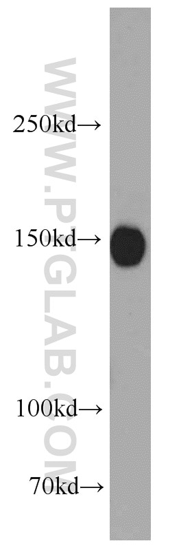
| Positive WB detected in | mouse brain tissue |
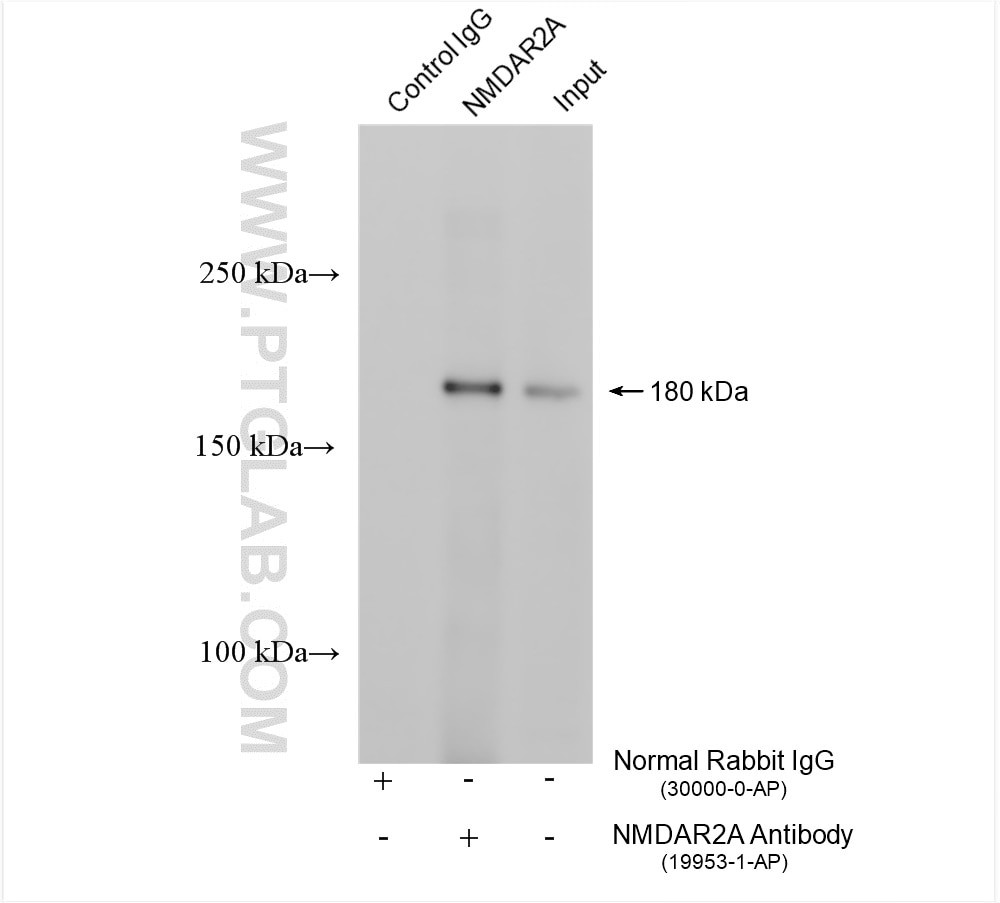
| Positive IP detected in | mouse brain tissue |
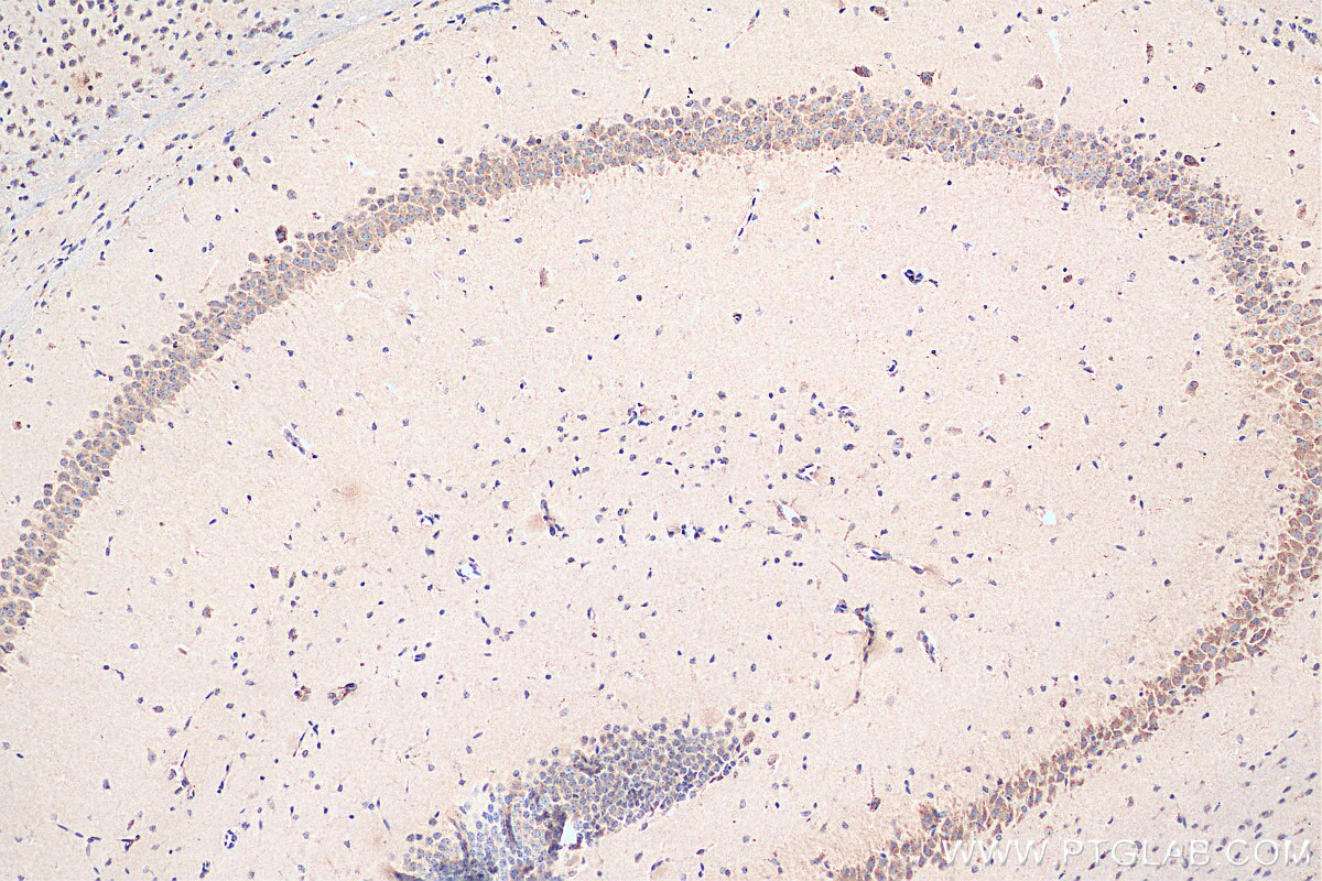
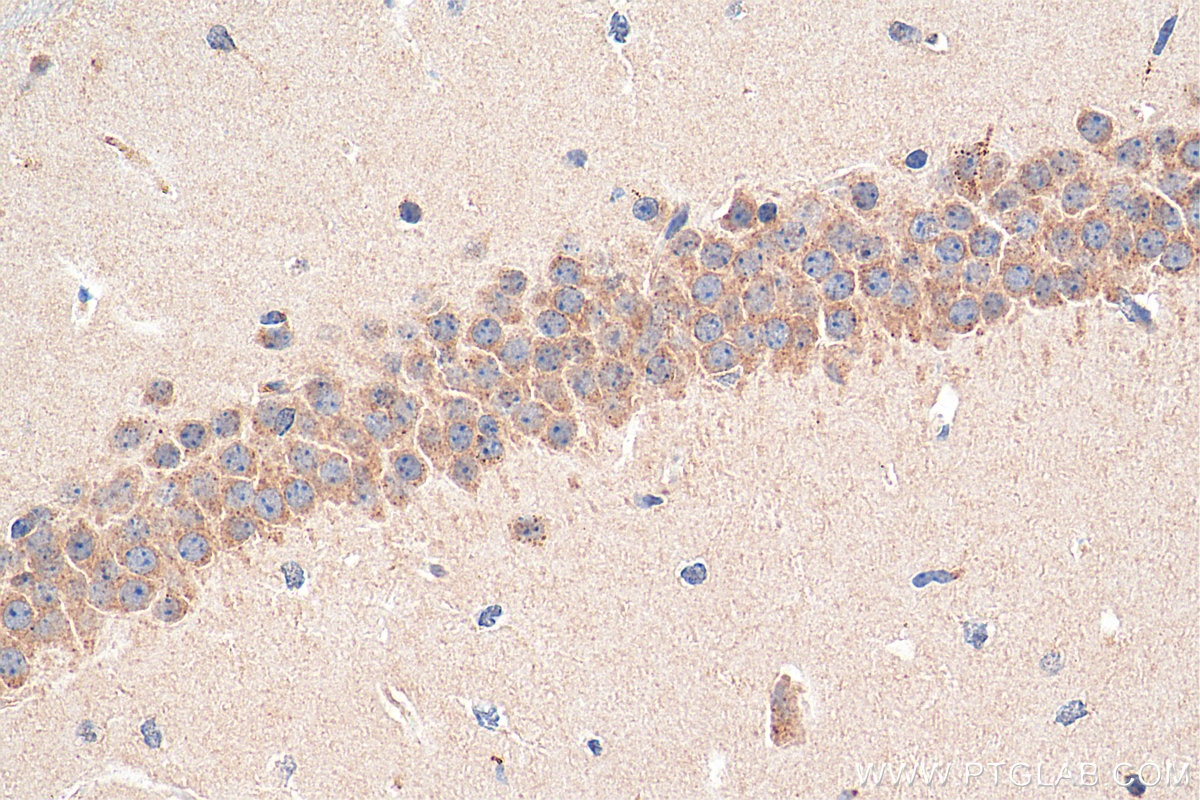
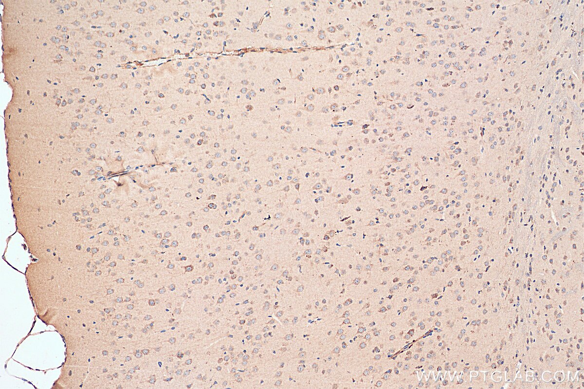
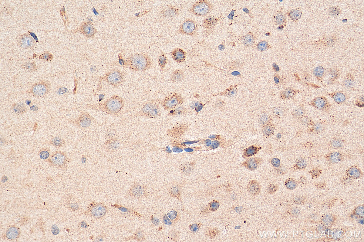
| Positive IHC detected in | mouse brain tissue Note: suggested antigen retrieval with TE buffer pH 9.0; (*) Alternatively, antigen retrieval may be performed with citrate buffer pH 6.0 |
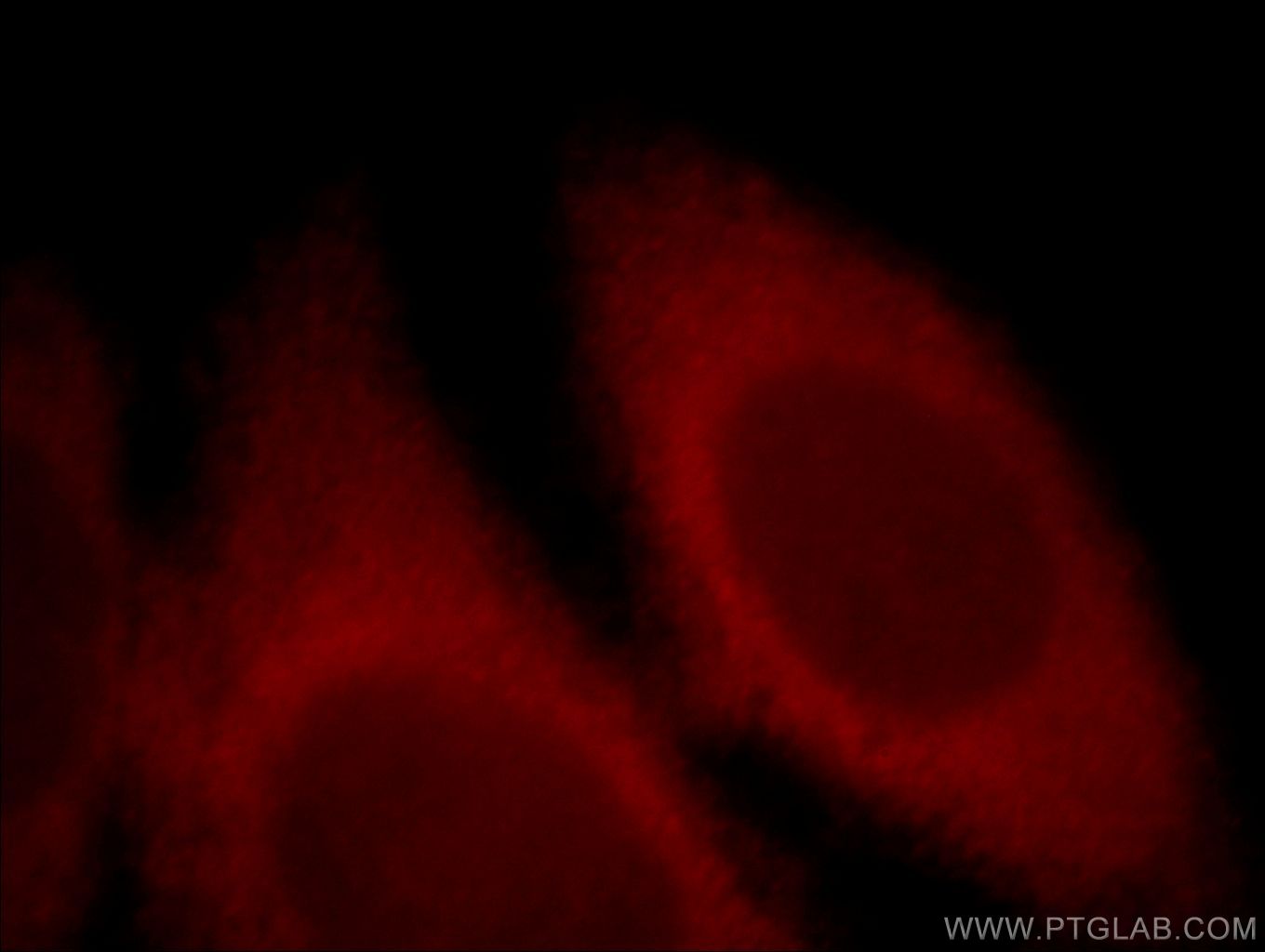
| Positive IF/ICC detected in | Hela cells |
Recommended dilution
| Application | Dilution |
|---|---|
| Western Blot (WB) | WB : 1:500-1:1000 |
| Immunoprecipitation (IP) | IP : 0.5-4.0 ug for 1.0-3.0 mg of total protein lysate |
| Immunohistochemistry (IHC) | IHC : 1:200-1:800 |
| Immunofluorescence (IF)/ICC | IF/ICC : 1:10-1:100 |
| It is recommended that this reagent should be titrated in each testing system to obtain optimal results. | |
| Sample-dependent, Check data in validation data gallery. | |
Published Applications
| WB | See 29 publications below |
| IF | See 2 publications below |
| IP | See 1 publications below |
Product Information
19953-1-AP targets NMDAR2A/GRIN2A in WB, IHC, IF/ICC, IP, ELISA applications and shows reactivity with human, mouse samples.
| Tested Reactivity | human, mouse |
| Cited Reactivity | human, mouse, rat |
| Host / Isotype | Rabbit / IgG |
| Class | Polyclonal |
| Type | Antibody |
| Immunogen | Peptide 相同性解析による交差性が予測される生物種 |
| Full Name | glutamate receptor, ionotropic, N-methyl D-aspartate 2A |
| Calculated molecular weight | 165 kDa |
| Observed molecular weight | 150-160 kDa |
| GenBank accession number | NM_000833 |
| Gene Symbol | GRIN2A |
| Gene ID (NCBI) | 2903 |
| RRID | AB_10640434 |
| Conjugate | Unconjugated |
| Form | Liquid |
| Purification Method | Antigen affinity purification |
| UNIPROT ID | Q12879 |
| Storage Buffer | PBS with 0.02% sodium azide and 50% glycerol , pH 7.3 |
| Storage Conditions | Store at -20°C. Stable for one year after shipment. Aliquoting is unnecessary for -20oC storage. |
Background Information
GRIN2A, also named as NMDAR2A and NR2A, belongs to the glutamate-gated ion channel (TC 1.A.10) family. GRIN2A is NMDA receptor subtype of glutamate-gated ion channels possesses high calcium permeability and voltage-dependent sensitivity to magnesium. Activation of GRIN2A requires binding of agonist to both types of subunits. The antibody recognize the C-term of GRIN2A.
Protocols
| Product Specific Protocols | |
|---|---|
| WB protocol for NMDAR2A/GRIN2A antibody 19953-1-AP | Download protocol |
| IHC protocol for NMDAR2A/GRIN2A antibody 19953-1-AP | Download protocol |
| IF protocol for NMDAR2A/GRIN2A antibody 19953-1-AP | Download protocol |
| IP protocol for NMDAR2A/GRIN2A antibody 19953-1-AP | Download protocol |
| Standard Protocols | |
|---|---|
| Click here to view our Standard Protocols |
Publications
| Species | Application | Title |
|---|---|---|
Sci Adv Loss of schizophrenia-related miR-501-3p in mice impairs sociability and memory by enhancing mGluR5-mediated glutamatergic transmission | ||
Sci Adv RNF220 is an E3 ubiquitin ligase for AMPA receptors to regulate synaptic transmission | ||
Mol Psychiatry A recurrent SHANK1 mutation implicated in autism spectrum disorder causes autistic-like core behaviors in mice via downregulation of mGluR1-IP3R1-calcium signaling. | ||
J Clin Invest TMEM25 modulates neuronal excitability and NMDA receptor subunit NR2B degradation. | ||
EBioMedicine Inhibition of Nwd1 activity attenuates neuronal hyperexcitability and GluN2B phosphorylation in the hippocampus. | ||
Aging (Albany NY) Enriched environment remedies cognitive dysfunctions and synaptic plasticity through NMDAR-Ca2+-Activin A circuit in chronic cerebral hypoperfusion rats. |
