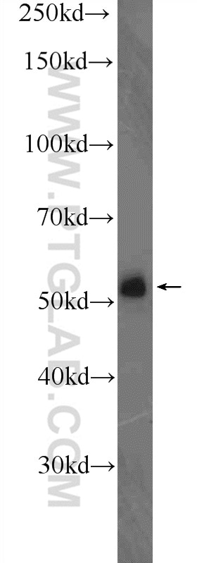
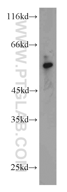
Validation Data Gallery
Tested Applications
| Positive WB detected in | mouse lung tissue |
Recommended dilution
| Application | Dilution |
|---|---|
| Western Blot (WB) | WB : 1:200-1:1000 |
| It is recommended that this reagent should be titrated in each testing system to obtain optimal results. | |
| Sample-dependent, Check data in validation data gallery. | |
Published Applications
| WB | See 1 publications below |
Product Information
21904-1-AP targets DELE1 in WB, ELISA applications and shows reactivity with human, mouse samples.
| Tested Reactivity | human, mouse |
| Cited Reactivity | mouse |
| Host / Isotype | Rabbit / IgG |
| Class | Polyclonal |
| Type | Antibody |
| Immunogen | DELE1 fusion protein Ag14615 相同性解析による交差性が予測される生物種 |
| Full Name | KIAA0141 |
| Calculated molecular weight | 515 aa, 56 kDa |
| Observed molecular weight | 56 kDa |
| GenBank accession number | BC007855 |
| Gene Symbol | DELE1 |
| Gene ID (NCBI) | 9812 |
| RRID | AB_2878942 |
| Conjugate | Unconjugated |
| Form | Liquid |
| Purification Method | Antigen affinity purification |
| UNIPROT ID | Q14154 |
| Storage Buffer | PBS with 0.02% sodium azide and 50% glycerol , pH 7.3 |
| Storage Conditions | Store at -20°C. Stable for one year after shipment. Aliquoting is unnecessary for -20oC storage. |
Background Information
DELE1, also known as KIAA0141, is a 515 amino acid protein, which localizes in Mitochondrion and is detected in liver, skeletal muscle, kidney, pancreas, spleen, thyroid, testis, ovary, small intestine and colon. DELE1 is essential for the induction of death receptor-mediated apoptosis through the regulation of caspase activation. Mitochondrial malfunction needs to be relayed to the cytosol, where an integrated stress response is triggered by the phosphorylation of eukaryotic translation initiation factor 2α(eIF2α) in mammalian cells. eIF2αphosphorylation is mediated by the four eIF2α kinases GCN2, HRI, PERK and PKR, which are activated by diverse types of cellular stress. OMA1, a mitochondrial stress-activated protease; and DELE1, a little-characterized protein that we found was associated with the inner mitochondrial membrane. Mitochondrial stress stimulates OMA1-dependent cleavage of DELE1 and leads to the accumulation of DELE1 in the cytosol, where it interacts with HRI and activates the eIF2α kinase activity of HRI. In addition, DELE1 is required for ATF4 translation downstream of eIF2α phosphorylation.
Protocols
| Product Specific Protocols | |
|---|---|
| WB protocol for DELE1 antibody 21904-1-AP | Download protocol |
| Standard Protocols | |
|---|---|
| Click here to view our Standard Protocols |
Publications
| Species | Application | Title |
|---|---|---|
Antioxidants (Basel) Increased Mobile Zinc Regulates Retinal Ganglion Cell Survival via Activating Mitochondrial OMA1 and Integrated Stress Response |

