MYC tag Monoclonal antibody

MYC tag Monoclonal Antibody for WB, IF/ICC, FC (Intra), IP, ELISA

Host / Isotype

Mouse / IgG1

Reactivity

recombinant protein and More (1)

Applications

WB, IF/ICC, FC (Intra), IP, CoIP, ChIP, ELISA

Conjugate

Unconjugated

CloneNo.

1A5A2

Cat no : 60003-2-Ig

Synonyms

myc, MYC-tag, EQKLISEEDL


"MYC tag Antibodies" Comparison

View side-by-side comparison of MYC tag antibodies from other vendors to find the one that best suits your research needs.
Filter:
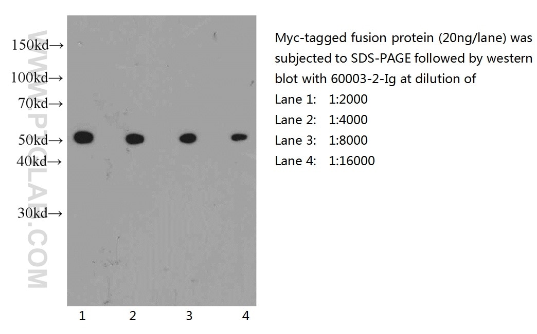
  • Western Blot (WB) analysis of recombinant proein using MYC tag Monoclonal antibody (60003-2-Ig)

    WB analysis of recombinant proein using 60003-2-Ig

    Western blot of Myc-tagged fusion protein with anti-Myc-tag (60003-2-Ig) at various dilutions.

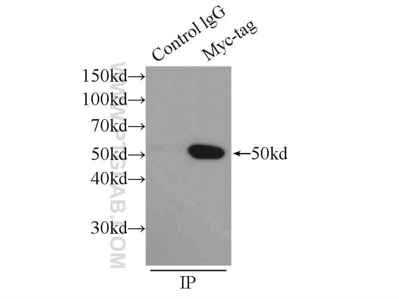
  • Immunoprecipitation (IP) experiment of Transfected HEK-293 cells using MYC tag Monoclonal antibody (60003-2-Ig)

    IP experiment of Transfected HEK-293 using 60003-2-Ig

    IP result of anti-MYC tag (IP:60003-2-Ig, 4ug; Detection:60003-2-Ig 1:10000) with Transfected HEK-293 cells lysate 300ug.

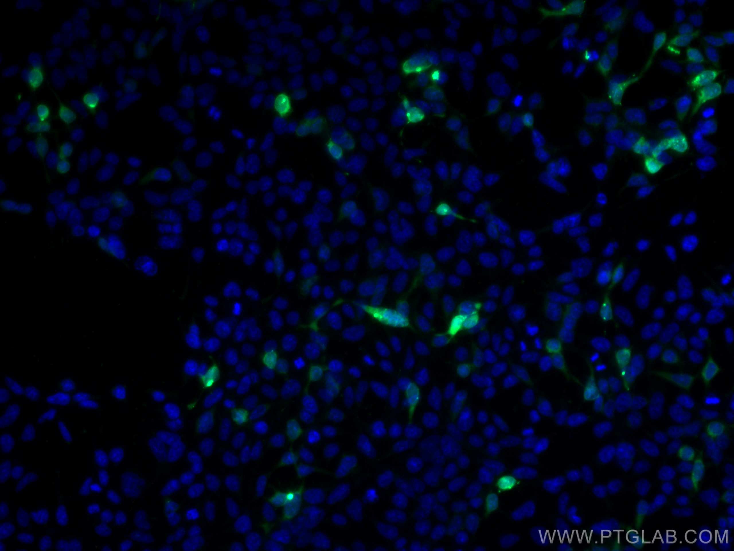
  • Immunofluorescence (IF) / fluorescent staining of Transfected HEK-293 cells using MYC tag Monoclonal antibody (60003-2-Ig)

    IF Staining of Transfected HEK-293 using 60003-2-Ig

    Immunofluorescent analysis of (-20°C Ethanol) fixed Transfected HEK-293 cells using MYC tag antibody (60003-2-Ig, Clone: 1A5A2 ) at dilution of 1:1000 and CoraLite®488-Conjugated AffiniPure Goat Anti-Mouse IgG(H+L).

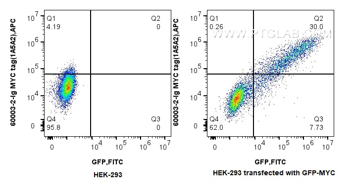
  • Flow cytometry (FC) experiment of HEK-293  transfected with GFP-MYC using MYC tag Monoclonal antibody (60003-2-Ig)

    FC experiment of HEK-293 transfected with GFP-MYC using 60003-2-Ig

    1x10^6 HEK-293 transfected with GFP-MYC were intracellularly stained with 0.25 ug MYC tag Monoclonal antibody (60003-2-Ig, Clone:1A5A2) and Multi-rAb CoraLite® Plus 647-Goat Anti-Mouse Recombinant Secondary Antibody (H+L)(RGAM005). Cells were fixed with 4% PFA and permeabilized with Flow Cytometry Perm Buffer (PF00011-C).

☆1/31(金)までお得に買える! >> IHC・ELISA・IPキット最大30%OFFキャンペーン


Tested Applications

Positive WB detected inrecombinant protein
Positive IP detected inTransfected HEK-293 cells
Positive IF/ICC detected inTransfected HEK-293 cells
Positive FC (Intra) detected inTransfected HEK-293 cells

Recommended dilution

ApplicationDilution
Western Blot (WB)WB : 1:2000-1:10000
Immunoprecipitation (IP)IP : 0.5-4.0 ug for 1.0-3.0 mg of total protein lysate
Immunofluorescence (IF)/ICCIF/ICC : 1:500-1:2000
Flow Cytometry (FC) (INTRA)FC (INTRA) : 0.50 ug per 10^6 cells in a 100 µl suspension
It is recommended that this reagent should be titrated in each testing system to obtain optimal results.
Sample-dependent, Check data in validation data gallery.

Product Information

60003-2-Ig targets MYC tag in WB, IF/ICC, FC (Intra), IP, CoIP, ChIP, ELISA applications and shows reactivity with recombinant protein samples.

Tested Reactivity recombinant protein
Cited Reactivityhuman
Host / Isotype Mouse / IgG1
Class Monoclonal
Type Antibody
Immunogen Peptide 相同性解析による交差性が予測される生物種
Full Name MYC tag
Gene symbol
Gene ID (NCBI)
RRIDAB_2734122
Conjugate Unconjugated
Form Liquid
Purification MethodProtein A purification
Storage Buffer PBS with 0.02% sodium azide and 50% glycerol pH 7.3.
Storage ConditionsStore at -20°C. Stable for one year after shipment. Aliquoting is unnecessary for -20oC storage.

Background Information

Protein tags are protein or peptide sequences located either on the C- or N- terminal of the target protein, which facilitates one or several of the following characteristics: solubility, detection, purification, localization and expression. The c-Myc tag corresponds to amino acid residues(EQKLISEEDL) of the human c-Myc protein. It can be used for affinity chromatography, then used to separate recombinant, overexpressed protein from wild type protein expressed by the host organism. It can also be used in the isolation of protein complexes with multiple subunits. Myc-Tag mouse mAb detects recombinant proteins containing the Myc tag. The antibody recognizes the Myc-tag EQKLISEEDL fused to either the amino- or carboxy- terminus of targeted proteins.

Protocols

Product Specific Protocols
IF protocol for MYC tag antibody 60003-2-IgDownload protocol
IP protocol for MYC tag antibody 60003-2-IgDownload protocol
FC protocol for MYC tag antibody 60003-2-IgDownload protocol
Standard Protocols
Click here to view our Standard Protocols

Publications

SpeciesApplicationTitle
WB

Nature

Promotion of DNA end resection by BRCA1-BARD1 in homologous recombination

Authors - Sameer Salunkhe
WB

Cell Discov

Glc7/PP1 dephosphorylates histone H3T11 to regulate autophagy and telomere silencing in response to nutrient availability

Authors - Xinyu Zhang
WB

Nat Cancer

Lymphatic endothelial-like cells promote glioblastoma stem cell growth through cytokine-driven cholesterol metabolism

Authors - Linjie Zhao
CoIP

Nat Commun

Phosphoglycerate dehydrogenase activates PKM2 to phosphorylate histone H3T11 and attenuate cellular senescence

Authors - Yinsheng Wu
WB

Nat Commun

UV-B irradiation-activated E3 ligase GmILPA1 modulates gibberellin catabolism to increase plant height in soybean

Authors - Jiaqi Sun
IP, WB

Am J Respir Crit Care Med

Loss of DP1 Aggravates Vascular Remodeling in Pulmonary Arterial Hypertension via mTORC1 Signaling.

Authors - Yuhu He

Reviews

The reviews below have been submitted by verified Proteintech customers who received an incentive forproviding their feedback.


FH

RASHMI (Verified Customer) (07-26-2024)

Used this product, highly recommended

  • Applications: Western Blot
  • Primary Antibody Dilution: 1:2000
  • Cell Tissue Type: HEK-293
FH

Sarah (Verified Customer) (10-27-2023)

works well at low dilution for WB and worked for immunoprecipitation

  • Applications: Western Blot, Immunoprecipitation
  • Primary Antibody Dilution: 1:10,000
  • Cell Tissue Type: 293T
FH

manohar (Verified Customer) (07-10-2023)

Used Nitrocellulose membrane with 5% milk as blocking, antibody diluted in 1% milk.

  • Applications: Western Blot, Immunofluorescence
  • Primary Antibody Dilution: 1:2000
  • Cell Tissue Type: HEK293, Neuronal stem cells
FH

Amy (Verified Customer) (03-05-2023)

Clean bands for detecting over expressed Myc-tagged proteins in HEK293T cell lysates.

  • Applications: Western Blot
  • Primary Antibody Dilution: 1:500
  • Cell Tissue Type: HEK293T
MYC tag Antibody Western Blot validation (1:500 dilution) in HEK293T (Cat no:60003-2-Ig)
FH

Isabelle Cristine (Verified Customer) (01-14-2022)

The antibody works generally well for the detection of MYC-tagged proteins.

  • Applications: Western Blot
  • Primary Antibody Dilution: 1:500
  • Cell Tissue Type: HEK293 cells
MYC tag Antibody Western Blot validation (1:500 dilution) in HEK293 cells (Cat no:60003-2-Ig)
FH

Tom (Verified Customer) (02-10-2021)

HEK293T cells were transfected with a Myc-tagged construct. The 60003-2-Ig antibody (1:2000) resulted in a lot of background. An alternative antibody was used alongside this one, with a stronger band and less background being achieved.

  • Applications: Western Blot
  • Primary Antibody Dilution: 1 in 2000
  • Cell Tissue Type: HEK293
MYC tag Antibody Western Blot validation (1 in 2000 dilution) in HEK293 (Cat no:60003-2-Ig)
FH

Alinda (Verified Customer) (10-09-2019)

This antibody was used at 1:1000 in HEK cells transfected with a myc-tagged insert cloned into an overxpression. Cells were blocked for 1 hr at room temp in 5%goat serum PBST.This antibody has a lot of background; another antibody from cell signalling was tested in parallel and performed much better

  • Applications: Immunofluorescence,
  • Primary Antibody Dilution: 1:1000
  • Cell Tissue Type: HEK cells