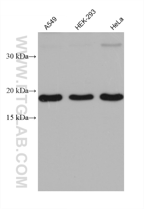
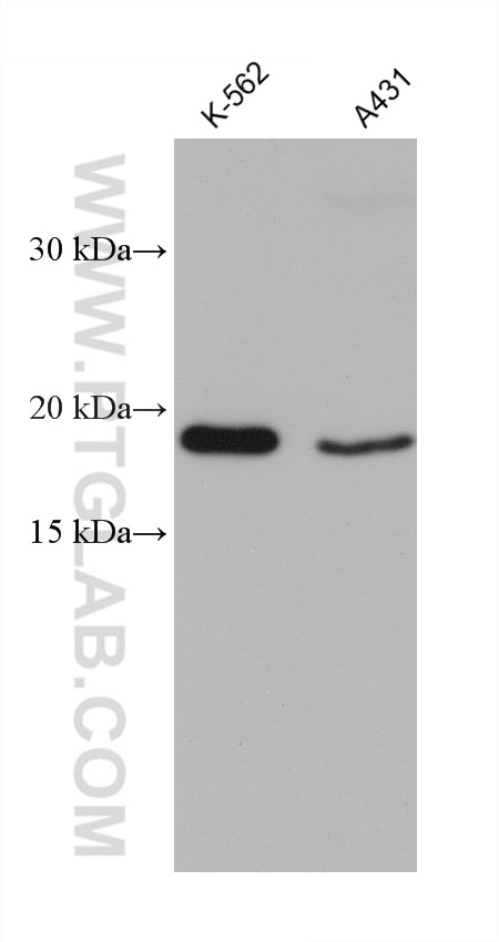
Validation Data Gallery
Tested Applications
| Positive WB detected in | A549 cells, K-562 cells, A431 cells, HEK-293 cells, HeLa cells |
Recommended dilution
| Application | Dilution |
|---|---|
| Western Blot (WB) | WB : 1:1000-1:6000 |
| It is recommended that this reagent should be titrated in each testing system to obtain optimal results. | |
| Sample-dependent, Check data in validation data gallery. | |
Product Information
68241-1-Ig targets OPA3 in WB, ELISA applications and shows reactivity with Human samples.
| Tested Reactivity | Human |
| Host / Isotype | Mouse / IgG1 |
| Class | Monoclonal |
| Type | Antibody |
| Immunogen | OPA3 fusion protein Ag8173 相同性解析による交差性が予測される生物種 |
| Full Name | optic atrophy 3 (autosomal recessive, with chorea and spastic paraplegia) |
| Calculated molecular weight | 179 aa, 20 kDa |
| Observed molecular weight | 20 kDa |
| GenBank accession number | BC005059 |
| Gene Symbol | OPA3 |
| Gene ID (NCBI) | 80207 |
| RRID | AB_2935328 |
| Conjugate | Unconjugated |
| Form | Liquid |
| Purification Method | Protein G purification |
| UNIPROT ID | Q9H6K4 |
| Storage Buffer | PBS with 0.02% sodium azide and 50% glycerol , pH 7.3 |
| Storage Conditions | Store at -20°C. Stable for one year after shipment. Aliquoting is unnecessary for -20oC storage. |
Background Information
The OPA3 cDNA encodes a deduced 179-amino acid protein. Northern blot analysis demonstrated a primary transcript of approximately 5.0 kb that was ubiquitously expressed, most prominently in skeletal muscle and kidney. Mutations in this gene have been shown to result in 3-methylglutaconic aciduria type III and autosomal dominant optic atrophy and cataract.
Protocols
| Product Specific Protocols | |
|---|---|
| WB protocol for OPA3 antibody 68241-1-Ig | Download protocol |
| Standard Protocols | |
|---|---|
| Click here to view our Standard Protocols |

