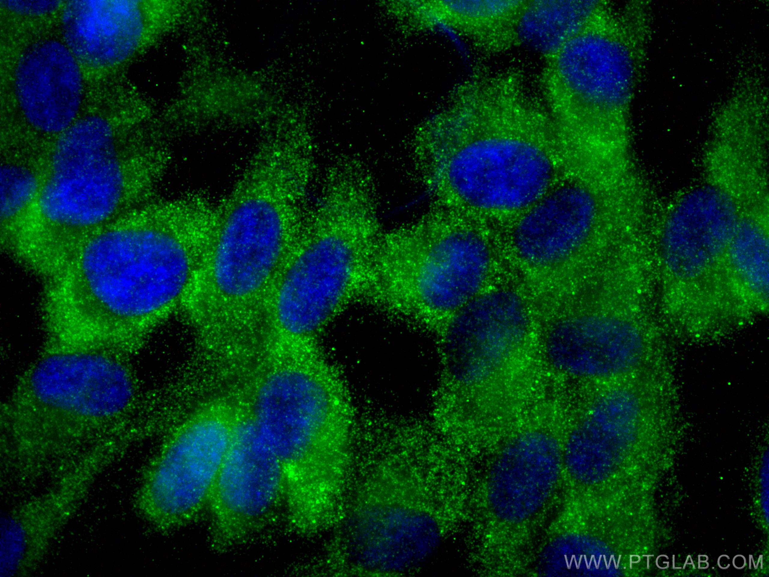
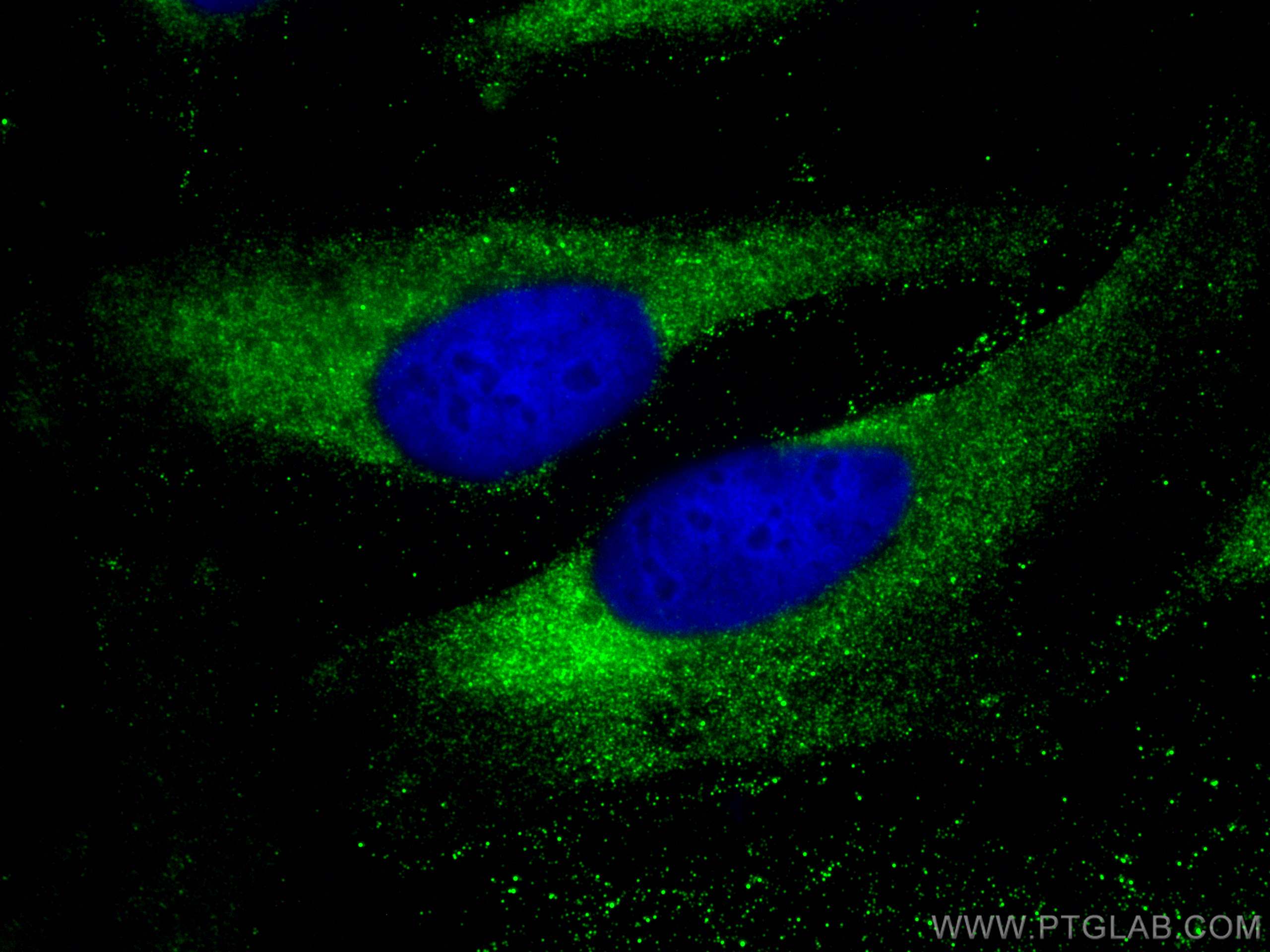
Validation Data Gallery
Tested Applications
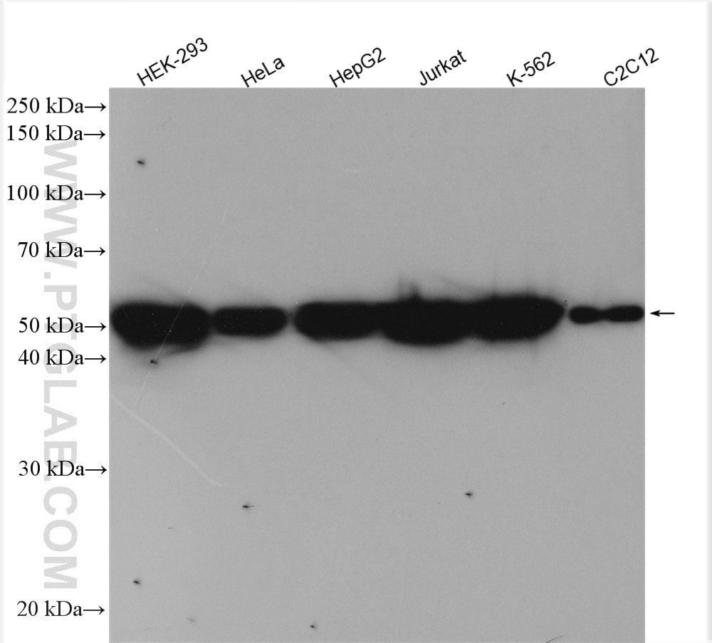
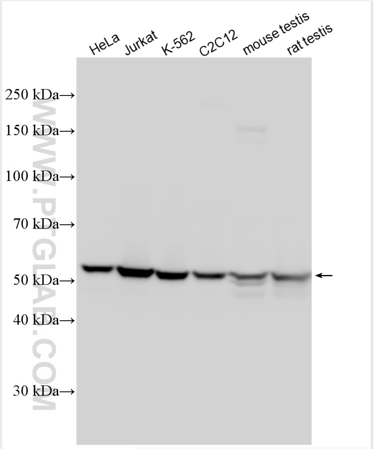
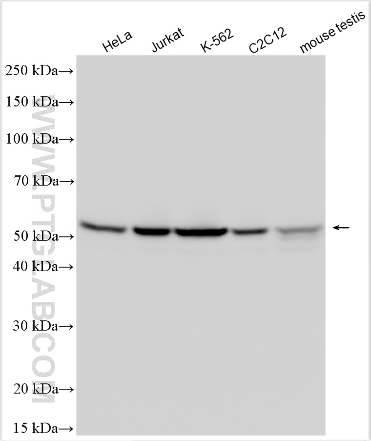
| Positive WB detected in | HeLa cells, HEK-293 cells, HepG2 cells, Jurkat cells, K-562 cells, C2C12 cells, mouse testis tissue, rat testis tissue |
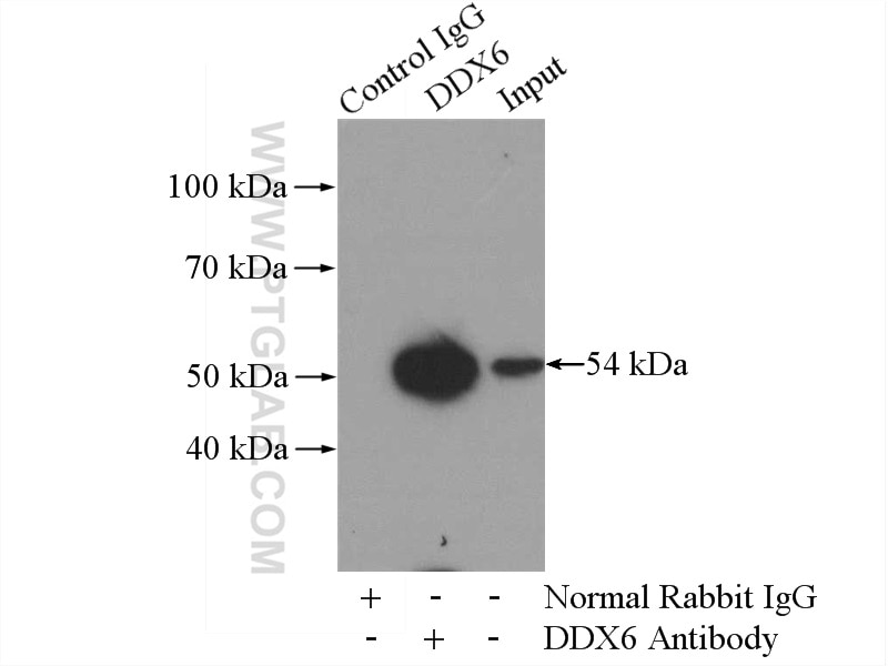
| Positive IP detected in | mouse testis tissue |
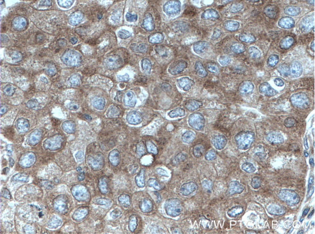
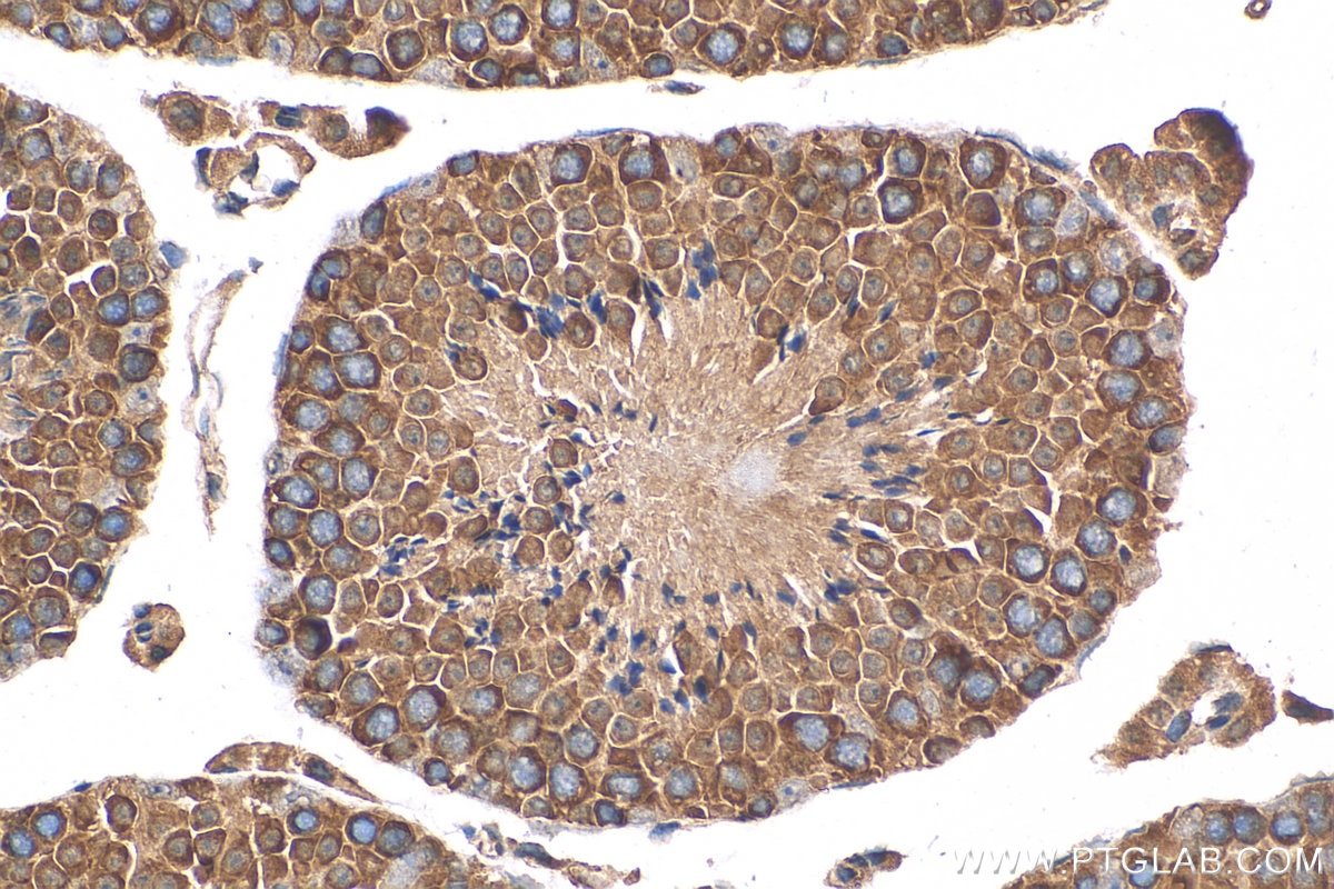
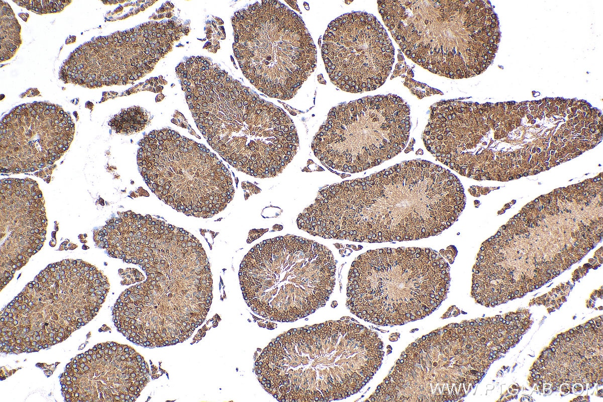
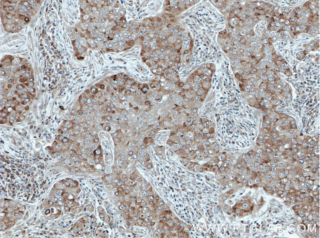
| Positive IHC detected in | human breast cancer tissue, mouse testis tissue Note: suggested antigen retrieval with TE buffer pH 9.0; (*) Alternatively, antigen retrieval may be performed with citrate buffer pH 6.0 |
| Positive IF/ICC detected in | HeLa cells, hTERT-RPE1 cells |
Recommended dilution
| Application | Dilution |
|---|---|
| Western Blot (WB) | WB : 1:2000-1:16000 |
| Immunoprecipitation (IP) | IP : 0.5-4.0 ug for 1.0-3.0 mg of total protein lysate |
| Immunohistochemistry (IHC) | IHC : 1:50-1:500 |
| Immunofluorescence (IF)/ICC | IF/ICC : 1:200-1:800 |
| It is recommended that this reagent should be titrated in each testing system to obtain optimal results. | |
| Sample-dependent, Check data in validation data gallery. | |
Published Applications
| KD/KO | See 1 publications below |
| WB | See 10 publications below |
| IF | See 5 publications below |
Product Information
14632-1-AP targets DDX6 in WB, IHC, IF/ICC, IP, ELISA applications and shows reactivity with human, mouse, rat samples.
| Tested Reactivity | human, mouse, rat |
| Cited Reactivity | human, mouse |
| Host / Isotype | Rabbit / IgG |
| Class | Polyclonal |
| Type | Antibody |
| Immunogen | DDX6 fusion protein Ag6211 相同性解析による交差性が予測される生物種 |
| Full Name | DEAD (Asp-Glu-Ala-Asp) box polypeptide 6 |
| Calculated molecular weight | 54 kDa |
| Observed molecular weight | 54 kDa |
| GenBank accession number | BC065007 |
| Gene Symbol | DDX6 |
| Gene ID (NCBI) | 1656 |
| RRID | AB_2091264 |
| Conjugate | Unconjugated |
| Form | Liquid |
| Purification Method | Antigen affinity purification |
| UNIPROT ID | P26196 |
| Storage Buffer | PBS with 0.02% sodium azide and 50% glycerol , pH 7.3 |
| Storage Conditions | Store at -20°C. Stable for one year after shipment. Aliquoting is unnecessary for -20oC storage. |
Background Information
DDX6, also named as P54, HLR2, and RCK, is a 483 amino acid protein, which contains one helicase C-terminal domain, one helicase ATP-binding domain, and belongs to the DEAD box helicase family. DDX6/DHH1 subfamily. P54 is abundantly expressed in most tissues. In the process of mRNA degradation, P54 may play a role in mRNA decapping. P54 is involved in the replication of hepatitis C virus genomes in hepatocytes and in tumourigenesis of hepatocellular carcinomas.
Protocols
| Product Specific Protocols | |
|---|---|
| WB protocol for DDX6 antibody 14632-1-AP | Download protocol |
| IHC protocol for DDX6 antibody 14632-1-AP | Download protocol |
| IF protocol for DDX6 antibody 14632-1-AP | Download protocol |
| IP protocol for DDX6 antibody 14632-1-AP | Download protocol |
| Standard Protocols | |
|---|---|
| Click here to view our Standard Protocols |
Publications
| Species | Application | Title |
|---|---|---|
J Genet Genomics LSM14B coordinates protein component expression in the P-body and controls oocyte maturation | ||
iScience The SARS-CoV-2 protein NSP2 impairs the silencing capacity of the human 4EHP-GIGYF2 complex. | ||
Biol Reprod Translational Activation of Developmental Messenger RNAs During Neonatal Mouse Testis Development. | ||
Biochem Biophys Res Commun DDX6 is involved in the pathogenesis of inflammatory diseases via NF-κB activation
| ||
EMBO J Energy stress promotes P-bodies formation via lysine-63-linked polyubiquitination of HAX1 | ||
BMC Bioinformatics SpotitPy: a semi-automated tool for object-based co-localization of fluorescent labels in microscopy images |
