Validation Data Gallery
Tested Applications
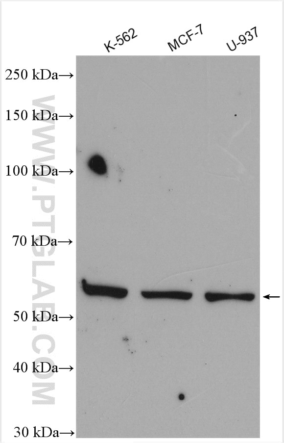
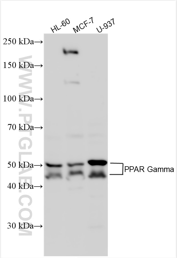
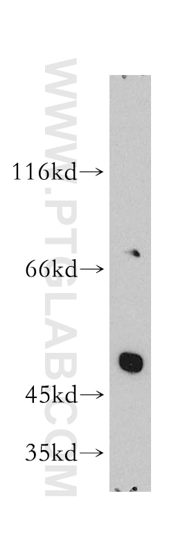
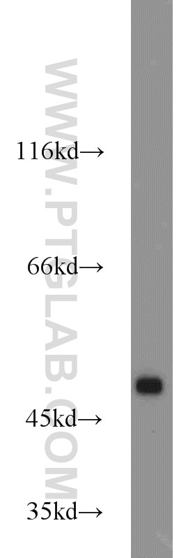
| Positive WB detected in | K-562 cells, HL-60 cells, mouse heart tissue, human heart tissue, MCF-7 cells, U-937 cells |
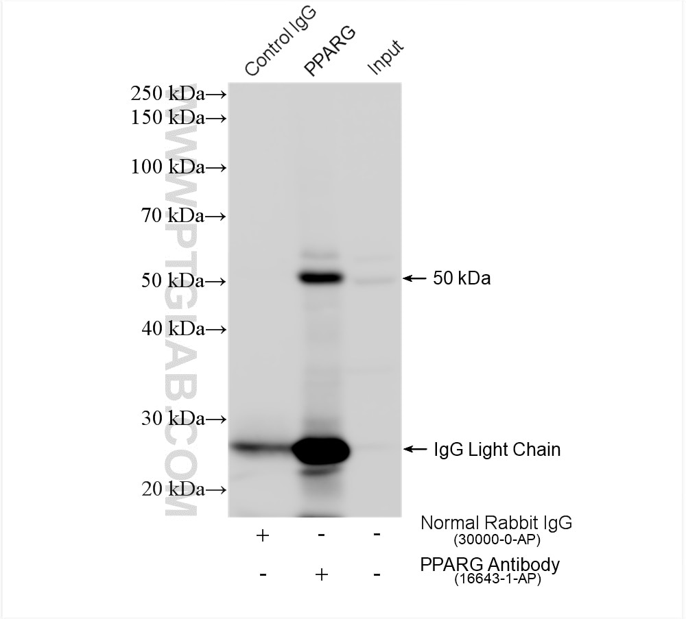
| Positive IP detected in | HL-60 cells |
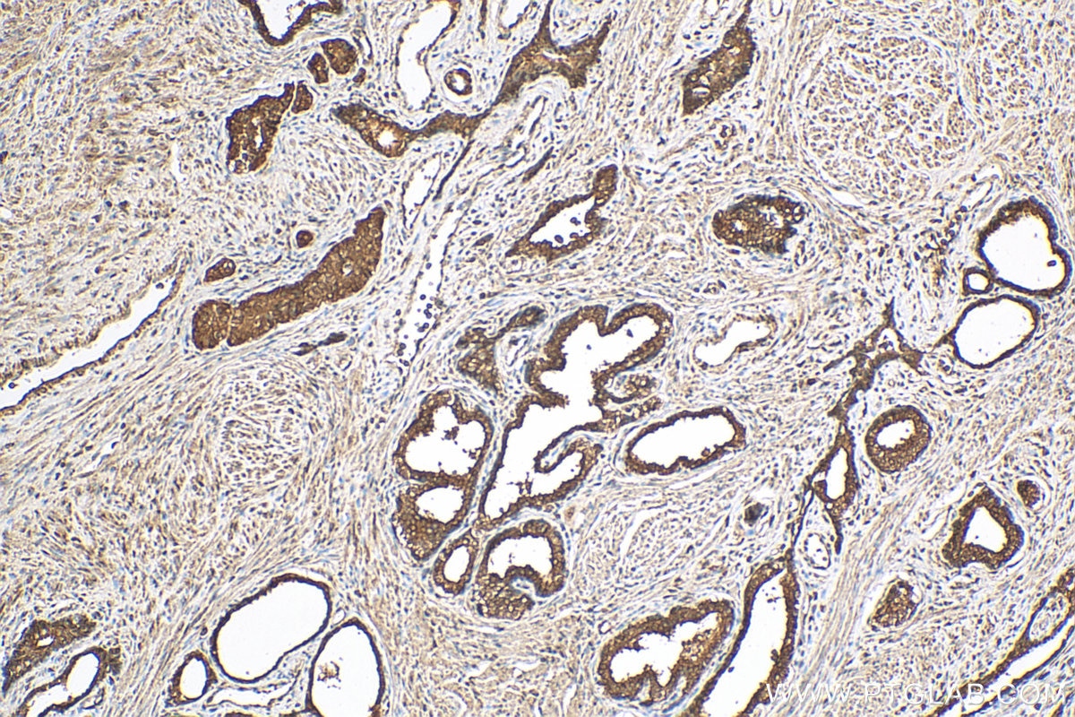
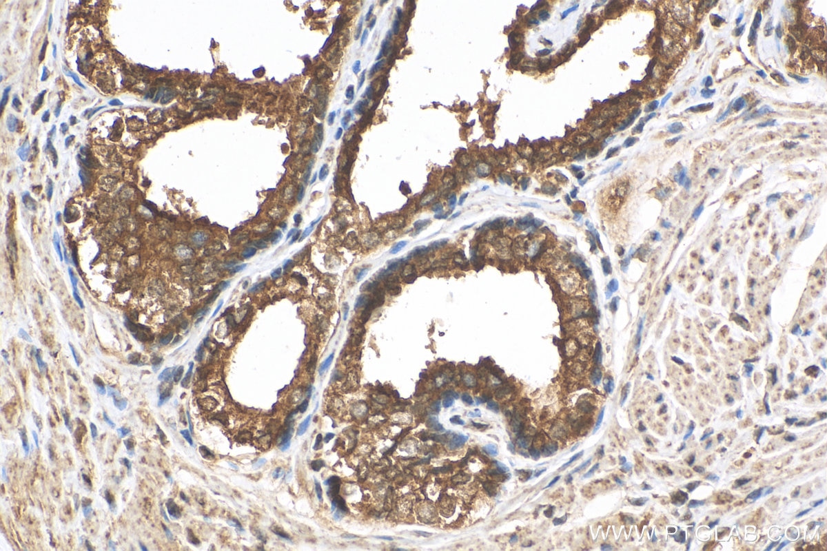
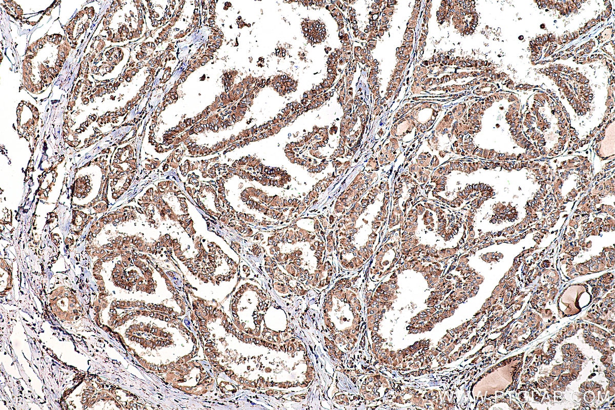
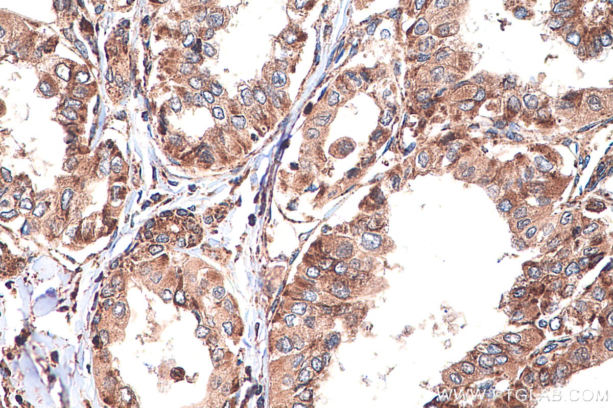
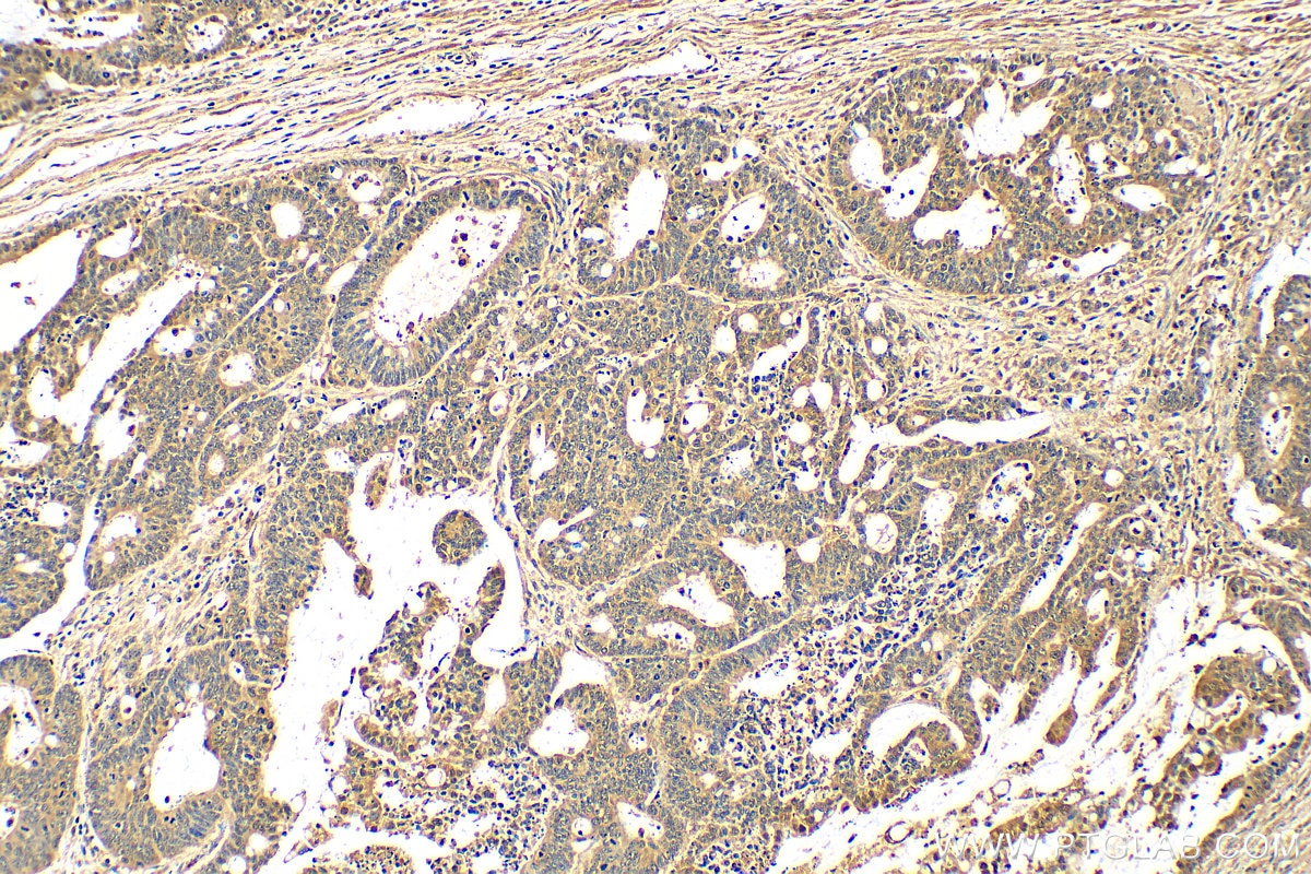
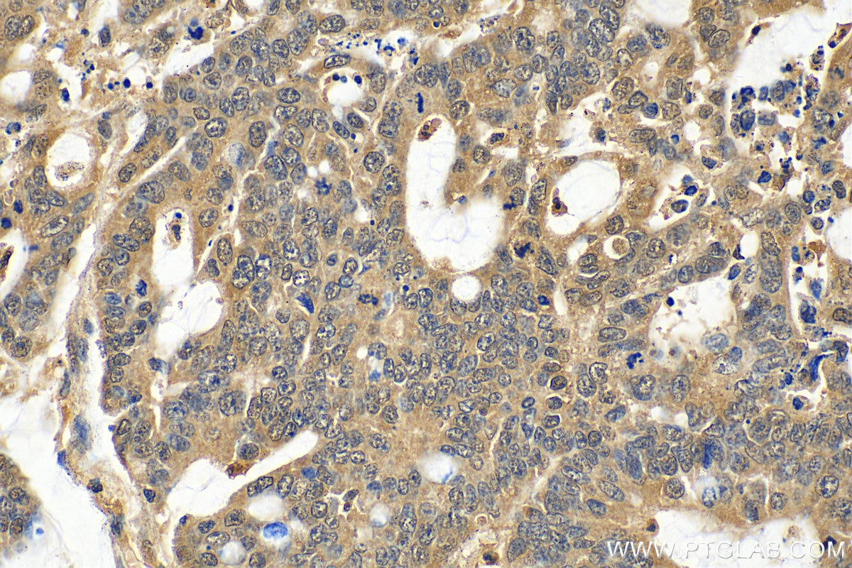
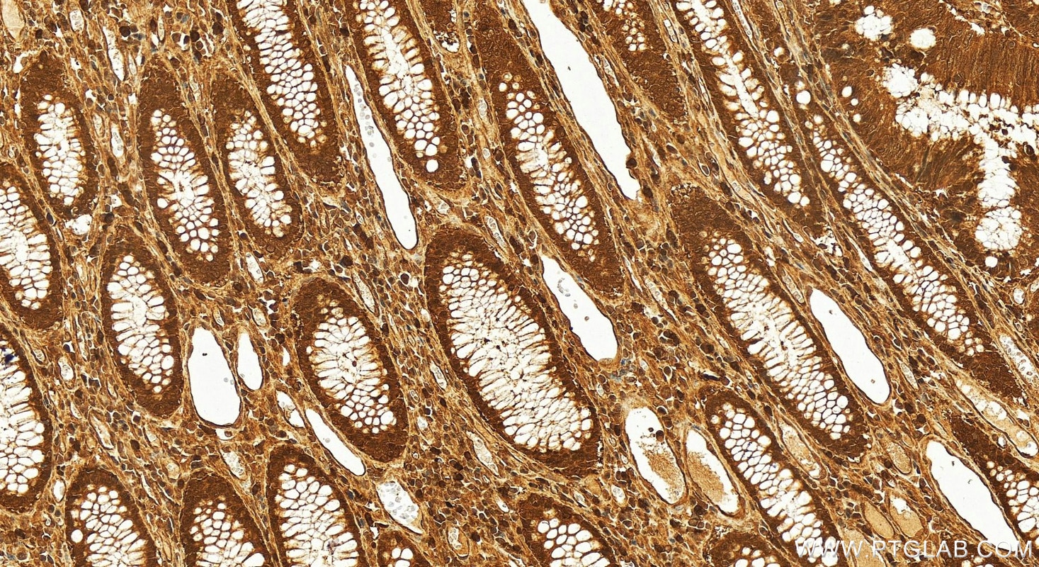
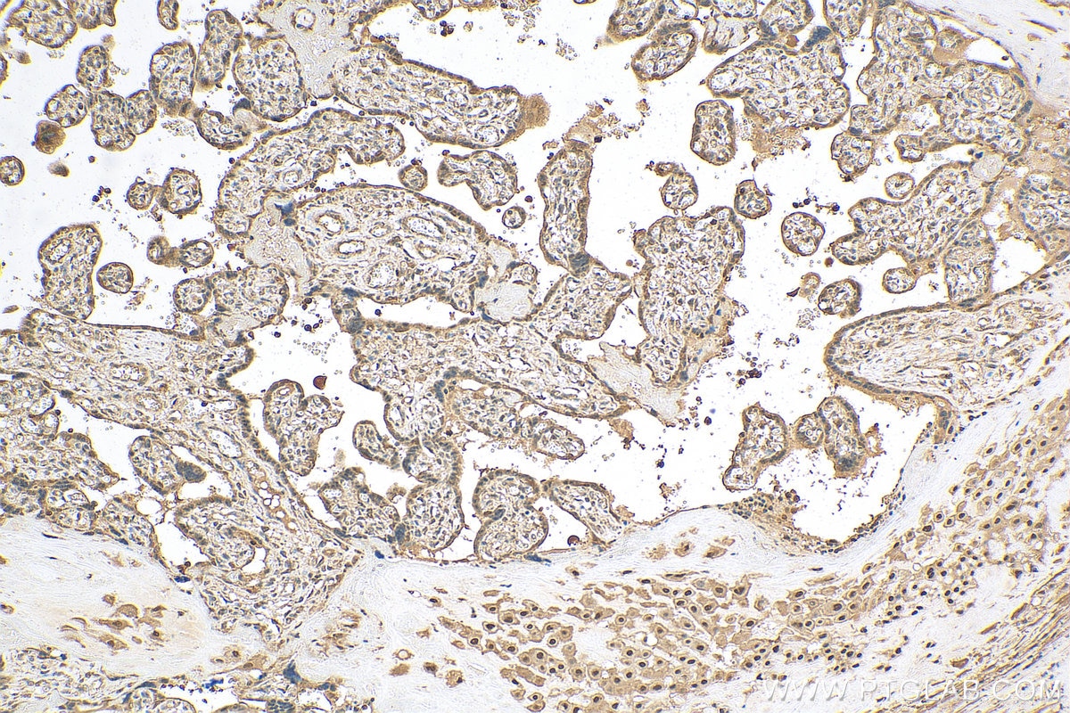
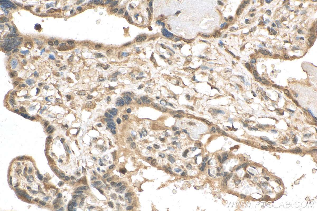
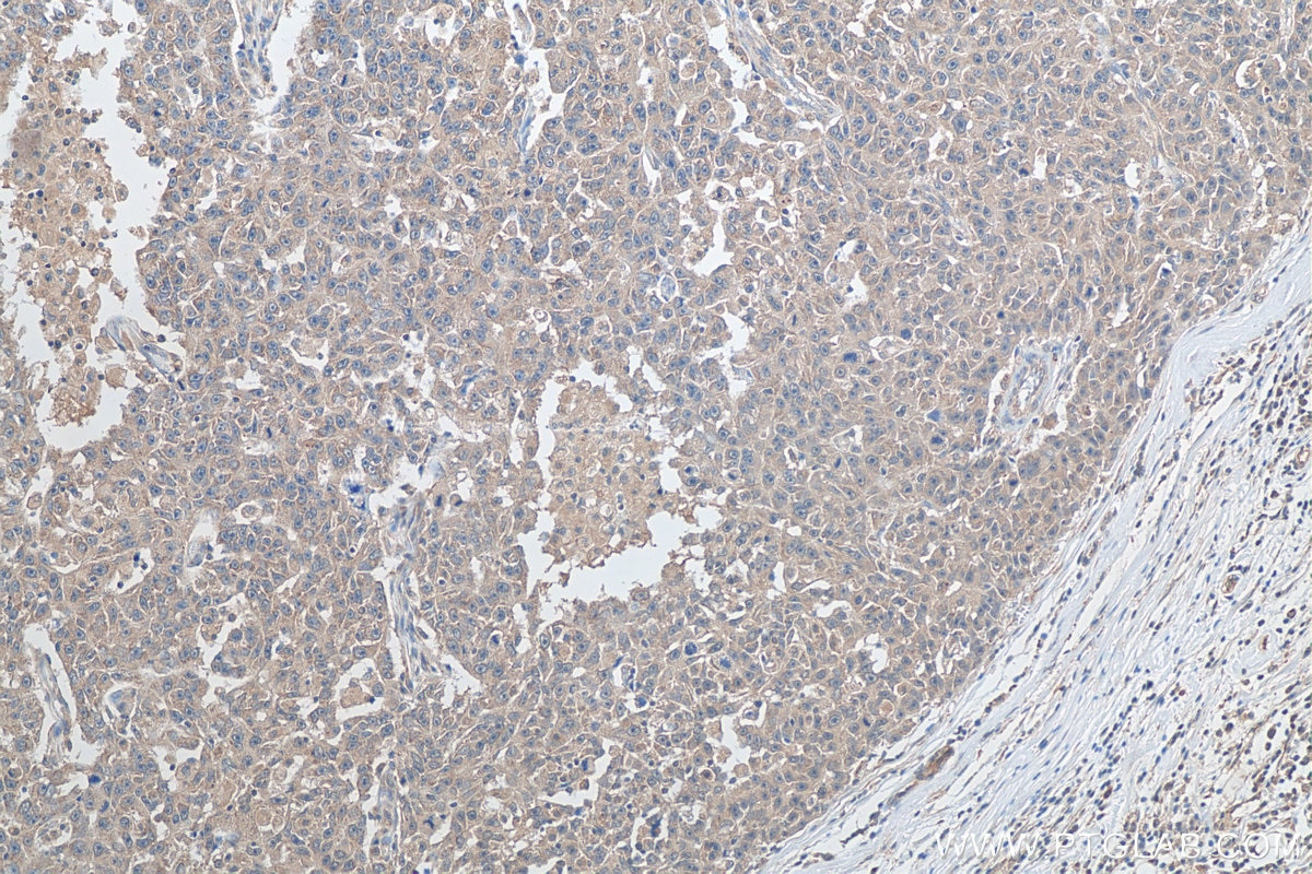
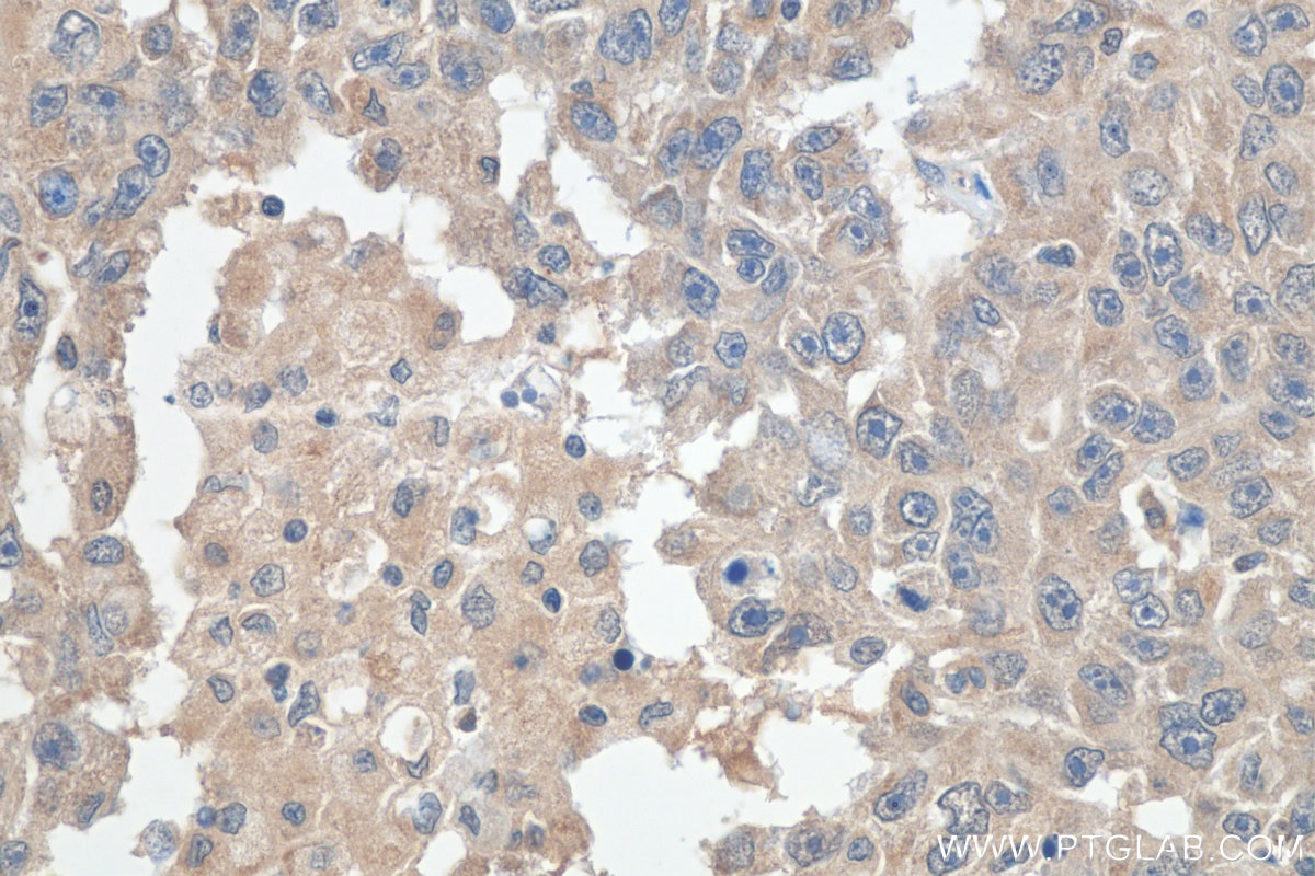
| Positive IHC detected in | human prostate cancer tissue, human breast cancer tissue, human colon cancer tissue, human placenta tissue, human thyroid cancer tissue Note: suggested antigen retrieval with TE buffer pH 9.0; (*) Alternatively, antigen retrieval may be performed with citrate buffer pH 6.0 |
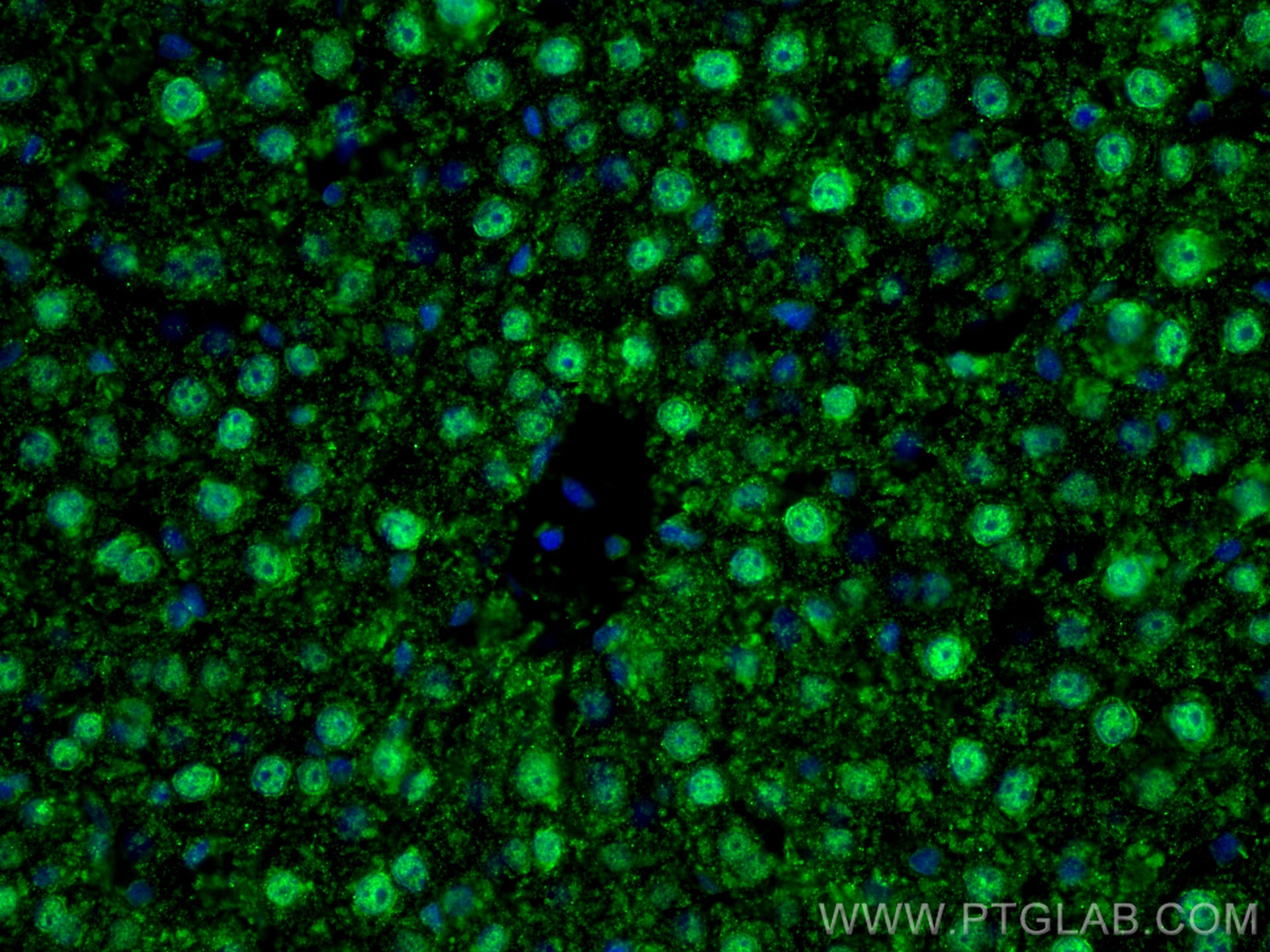
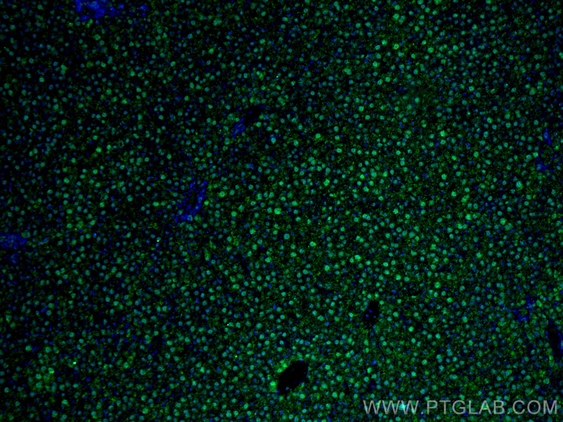
| Positive IF-P detected in | rat liver tissue |
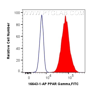
| Positive FC (Intra) detected in | HeLa cells |
This antibody is not recommended for immunocytofluorescent assays.
Recommended dilution
| Application | Dilution |
|---|---|
| Western Blot (WB) | WB : 1:1000-1:5000 |
| Immunoprecipitation (IP) | IP : 0.5-4.0 ug for 1.0-3.0 mg of total protein lysate |
| Immunohistochemistry (IHC) | IHC : 1:200-1:800 |
| Immunofluorescence (IF)-P | IF-P : 1:50-1:500 |
| Flow Cytometry (FC) (INTRA) | FC (INTRA) : 0.40 ug per 10^6 cells in a 100 µl suspension |
| It is recommended that this reagent should be titrated in each testing system to obtain optimal results. | |
| Sample-dependent, Check data in validation data gallery. | |
Published Applications
| KD/KO | See 17 publications below |
| WB | See 480 publications below |
| IHC | See 50 publications below |
| IF | See 48 publications below |
| IP | See 9 publications below |
| CoIP | See 10 publications below |
| ChIP | See 8 publications below |
Product Information
16643-1-AP targets PPAR Gamma in WB, IHC, IF-P, FC (Intra), IP, CoIP, CHIP, ELISA applications and shows reactivity with human, mouse, rat samples.
| Tested Reactivity | human, mouse, rat |
| Cited Reactivity | human, mouse, rat, pig, rabbit, chicken, zebrafish, hamster, sheep, goat |
| Host / Isotype | Rabbit / IgG |
| Class | Polyclonal |
| Type | Antibody |
| Immunogen | PPAR Gamma fusion protein Ag10005 相同性解析による交差性が予測される生物種 |
| Full Name | peroxisome proliferator-activated receptor gamma |
| Calculated molecular weight | 58 kDa |
| Observed molecular weight | 50-60 kDa |
| GenBank accession number | BC006811 |
| Gene Symbol | PPARG |
| Gene ID (NCBI) | 5468 |
| RRID | AB_10596794 |
| Conjugate | Unconjugated |
| Form | Liquid |
| Purification Method | Antigen affinity purification |
| UNIPROT ID | P37231 |
| Storage Buffer | PBS with 0.02% sodium azide and 50% glycerol , pH 7.3 |
| Storage Conditions | Store at -20°C. Stable for one year after shipment. Aliquoting is unnecessary for -20oC storage. |
Background Information
Peroxisome Proliferator-Activated Receptors (PPARs) are ligand-activated intracellular transcription factors, members of the nuclear hormone receptor superfamily (NR), that includes estrogen, thyroid hormone receptors, retinoic acid, Vitamin D3 as well as retinoid X receptors (RXRs). The PPAR subfamily consists of three subtypes encoded by distinct genes denoted PPARα (NR1C1), PPARβ/δ (NR1C2) and PPARγ (NR1C3), which are activated by selective ligands. PPARγ, also named as PPARG, contains one nuclear receptor DNA-binding domain and is a receptor that binds peroxisome proliferators such as hypolipidemic drugs and fatty acids. It plays an important role in the regulation of lipid homeostasis, adipogenesis, ins resistance, and development of various organs. Defects in PPARG are the cause of familial partial lipodystrophy type 3 (FPLD3) and may be associated with susceptibility to obesity. Defects in PPARG can lead to type 2 ins-resistant diabetes and hypertension. PPARG mutations may be associated with colon cancer. Genetic variations in PPARG are associated with susceptibility to glioma type 1 (GLM1). PPARG has two isoforms with molecular weight 57 kDa and 54 kDa (PMID: 9831621), but modified PPARG is about 67 KDa (PMID: 16809887). PPARG2 is a splice variant and has an additional 30 amino acids at the N-terminus (PMID: 15689403). Experimental data indicate that a 45 kDa protein displaying three different sequences immunologically related to the nuclear receptor PPARG2 is located in mitochondria (mt-PPAR). However, the molecular weight of this protein is clearly less when compared to that of PPARG2 (57 kDa) (PMID: 10922459). PPARG has been reported to be localized mainly (but not always) in the nucleus. PPARG can also be detected in the cytoplasm and was reported to possess extra-nuclear/non-genomic actions (PMID: 17611413; 19432669; 14681322).
Protocols
| Product Specific Protocols | |
|---|---|
| WB protocol for PPAR Gamma antibody 16643-1-AP | Download protocol |
| IHC protocol for PPAR Gamma antibody 16643-1-AP | Download protocol |
| IF protocol for PPAR Gamma antibody 16643-1-AP | Download protocol |
| IP protocol for PPAR Gamma antibody 16643-1-AP | Download protocol |
| FC protocol for PPAR Gamma antibody 16643-1-AP | Download protocol |
| Standard Protocols | |
|---|---|
| Click here to view our Standard Protocols |
Publications
| Species | Application | Title |
|---|---|---|
Nat Nanotechnol Photoacoustic molecular imaging-escorted adipose photodynamic-browning synergy for fighting obesity with virus-like complexes. | ||
ACS Nano Dual Inhibition of Endoplasmic Reticulum Stress and Oxidation Stress Manipulates the Polarization of Macrophages under Hypoxia to Sensitize Immunotherapy. | ||
J Pineal Res Single-cell RNA sequencing of preadipocytes reveals the cell fate heterogeneity induced by melatonin. | ||
Nat Commun N1-methyladenosine methylation in tRNA drives liver tumourigenesis by regulating cholesterol metabolism. | ||
Nat Commun METTL3 is essential for postnatal development of brown adipose tissue and energy expenditure in mice. | ||
Research (Wash D C) Herpetrione, a New Type of PPARα Ligand as a Therapeutic Strategy Against Nonalcoholic Steatohepatitis |
