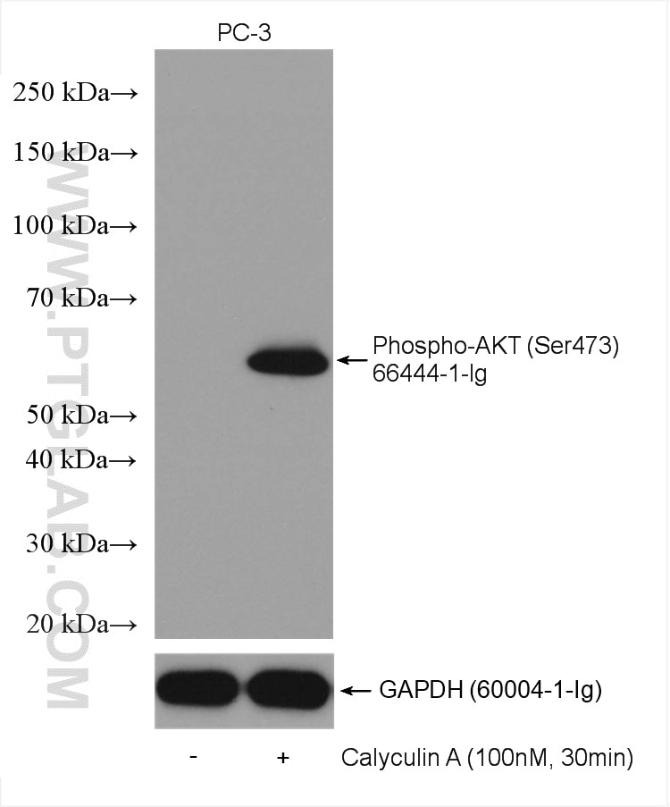
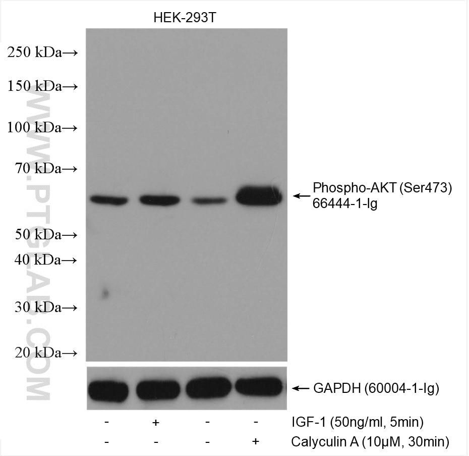
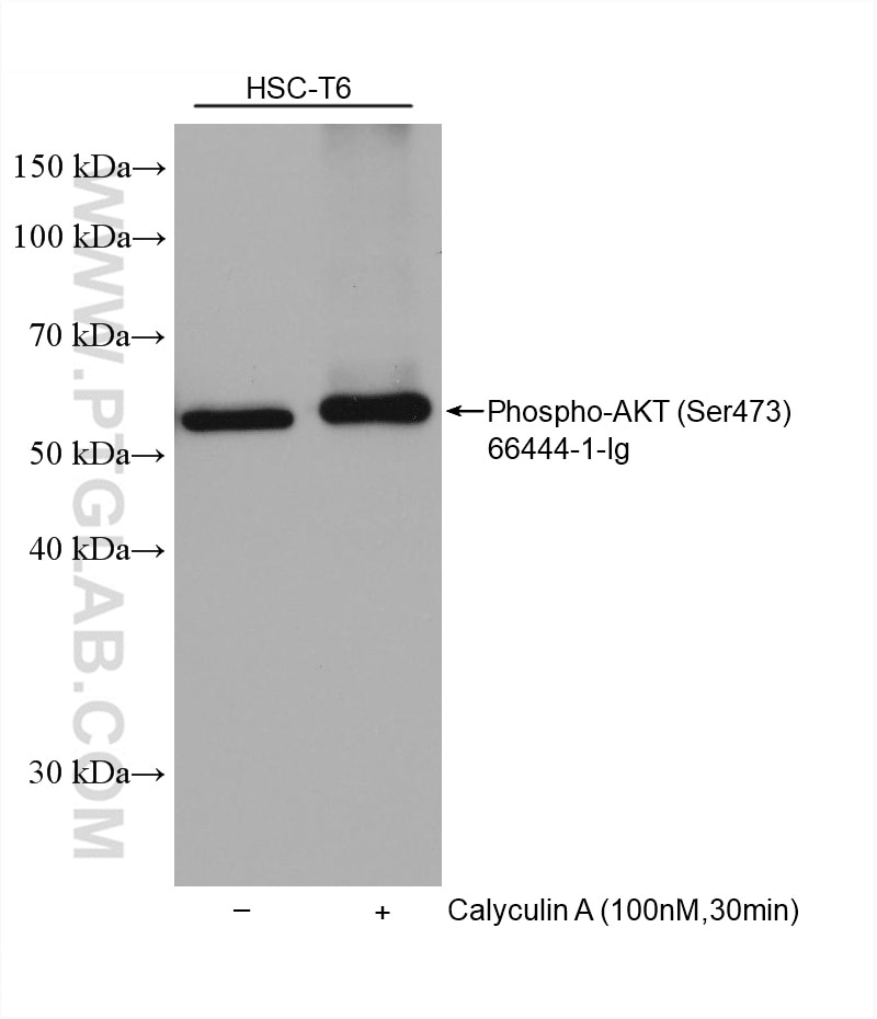
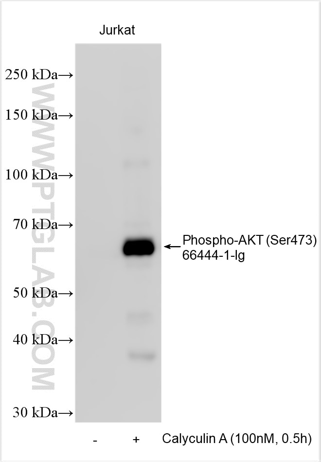
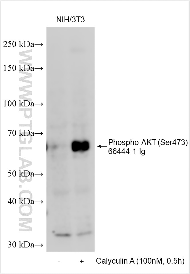
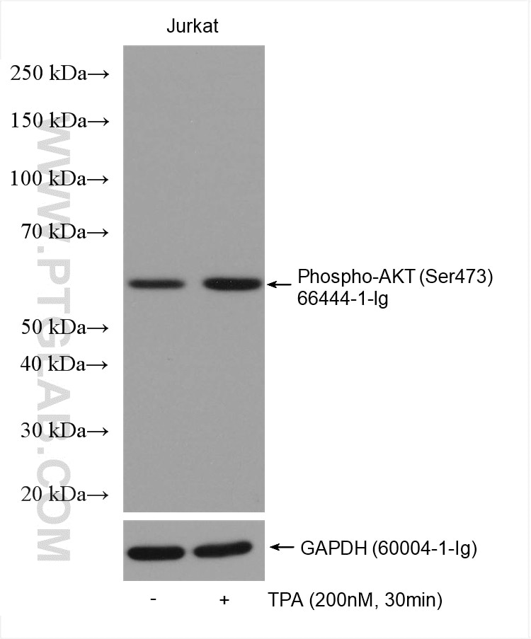
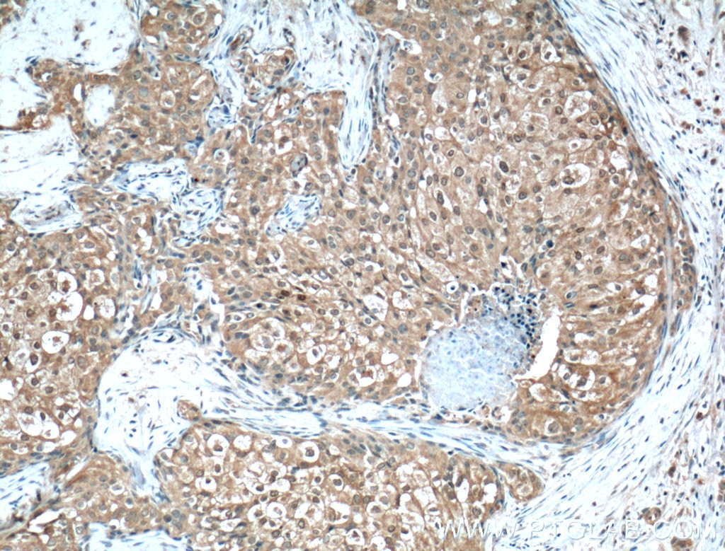
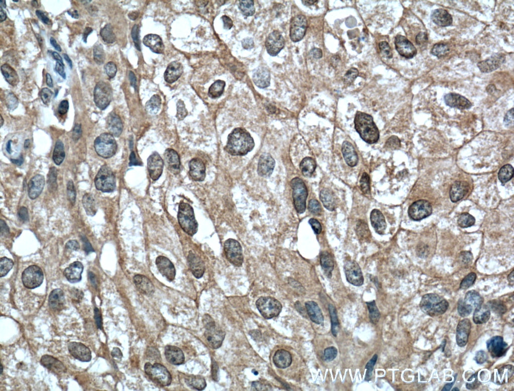
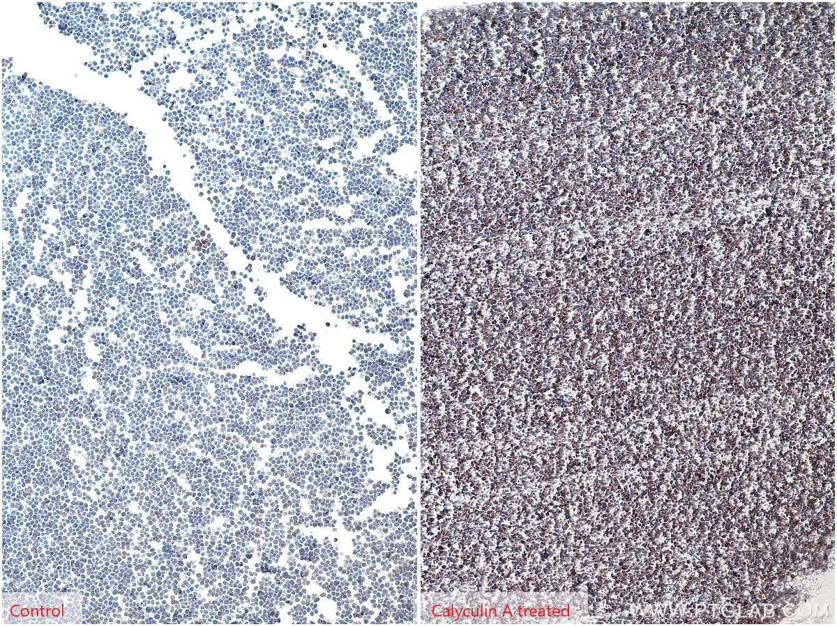
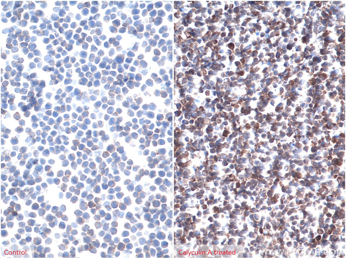
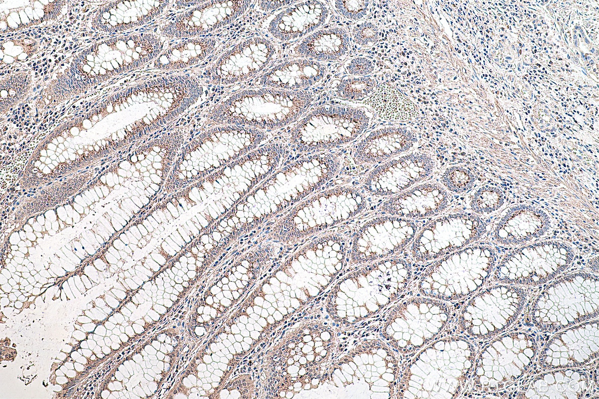
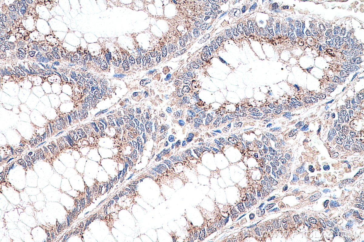
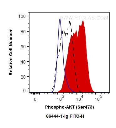
Validation Data Gallery
Tested Applications
Recommended dilution
| Application | Dilution |
|---|---|
| It is recommended that this reagent should be titrated in each testing system to obtain optimal results. | |
Product Information
66444-1-PBS targets Phospho-AKT (Ser473) as part of a matched antibody pair:
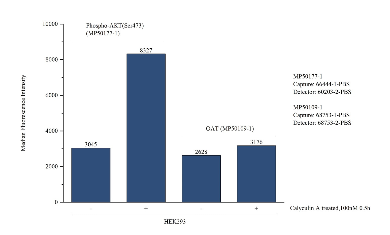
MP50177-1: 66444-1-PBS capture and 60203-2-PBS detection (validated in Cytometric bead array)
Unconjugated mouse monoclonal antibody pair in PBS only (BSA and azide free) storage buffer at a concentration of 1 mg/mL, ready for conjugation.
This conjugation ready format makes antibodies ideal for use in many applications including: ELISAs, multiplex assays requiring matched pairs, mass cytometry, and multiplex imaging applications.Antibody use should be optimized by the end user for each application and assay.
| Tested Reactivity | human, mouse, rat |
| Host / Isotype | Mouse / IgG1 |
| Class | Monoclonal |
| Type | Antibody |
| Immunogen |
Peptide 相同性解析による交差性が予測される生物種 |
| Full Name | v-akt murine thymoma viral oncogene homolog 1 |
| Observed molecular weight | 60-62 kDa |
| GenBank accession number | NM_005163 |
| Gene Symbol | AKT1 |
| Gene ID (NCBI) | 207 |
| RRID | AB_2782958 |
| Conjugate | Unconjugated |
| Form | |
| Form | Liquid |
| Purification Method | Protein G purification |
| UNIPROT ID | P31749 |
| Storage Buffer | PBS only{{ptg:BufferTemp}}7.3 |
| Storage Conditions | Store at -80°C. |
Background Information
1) What is AKT?
The serine/threonine kinase B AKT pathway (also known as the PI3K-Akt pathway) plays a vital role in the regulation of cellular processes, including cell proliferation, survival, and growth - processes that are essential for oncogenesis. Mutation of the regulator proteins PI3K and PTEN causes uncontrolled disruption within the PI3-kinase pathway, leading to the development of human cancers (1,2; see also AKT pathway poster for more details).
2) phospho-AKT and FAQs
A) What is the best way to normalize phosphorylated proteins analyzed by western blot?
Normalize phospho-AKT and total AKT with your loading control (e.g. Actin, tubulin), then calculate the phospho/total ratio using these normalized values.
Put more simply:
1. Calculate the ratio of band intensities of a phospho-AKT band: the loading control.
2. Calculate the ratio of band intensities of total AKT: loading control.
3. Divide ratio obtained #1 by #2 to obtain a normalized value for comparison among different conditions. This procedure allows one to distinguish between a change in AKT expression and a change in the ratio of phospho-AKT.
* If you are looking at the differences in a phospho-AKT expression resulting from an experimental condition (e.g., knockdown), you should also show the expression of total AKT to distinguish between a change in AKT expression (transcription/translation level) and a change in the AKT phosphorylation status.
B) What is the observed molecular weight for AKT and phospho-AKT?
Molecular Weight AKT - 56 kDa
Molecular Weight phospho-AKT - 60 kDa (Figure 1)

Figure 1. WB: HEK-293 cell lysate was subjected to SDS PAGE followed by western blot with 60203-2-Ig (AKT antibody) and 66444-1-Ig (AKT-phospho-S473 antibody) at a dilution of 1:4000 incubated at room temperature for 1.5 hours.
C) Are there any special WB conditions to optimize staining of a phospho-AKT?
Since this is a phosphorylated protein, 5% BSA is recommended over non-fat milk as a blocking agent.
D) What are good positive and negative controls for a phospho-AKT?
- Positive Control: HEK293 cells
- Negative Control: Treatment with PI3K inhibitors (e.g. wortmannin)
E) What species does this antibody react with?
Our internal testing has confirmed that it reacts with the human and mouse forms of phospho-AKT.Reactivity with the human form is also supported by the literature's citations of this antibody.
References:
1. Perturbations of the AKT signaling pathway in human cancer.
2. Targeting the PI3K-Akt pathway in human cancer: rationale and promise.

