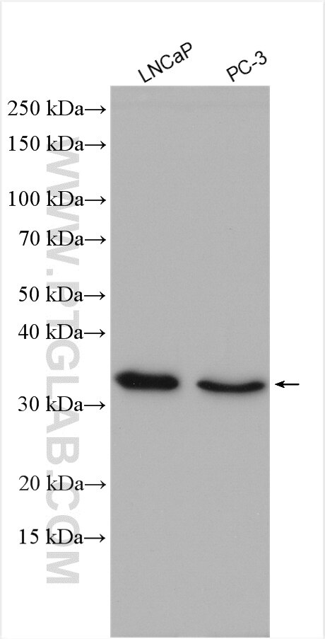
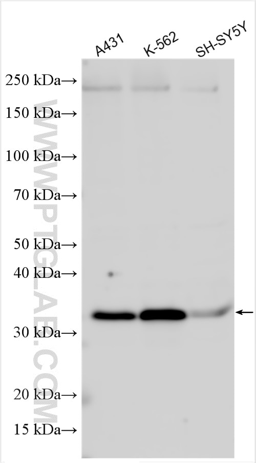
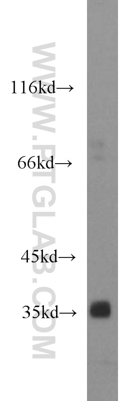
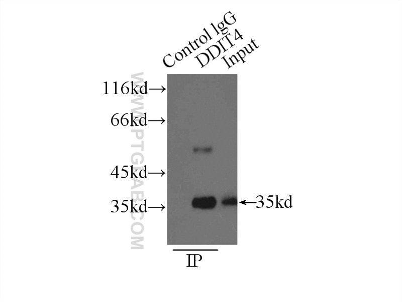
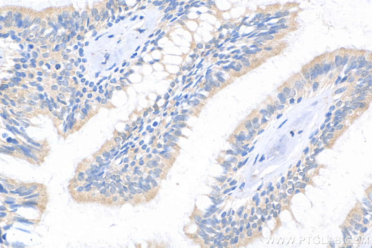
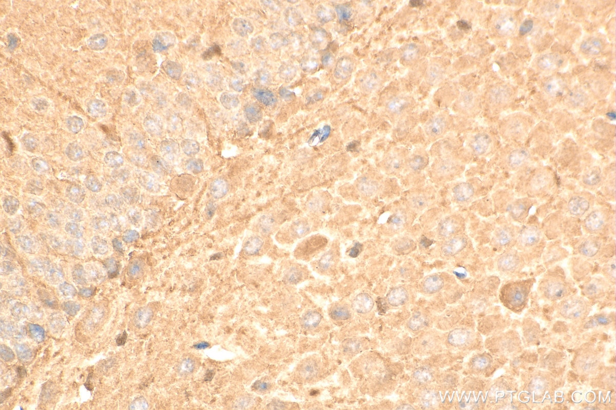
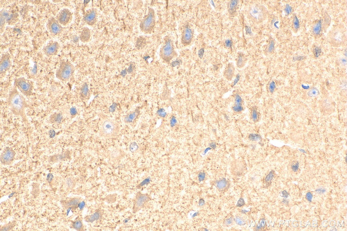
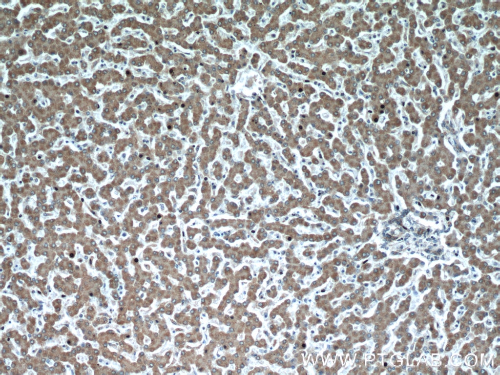
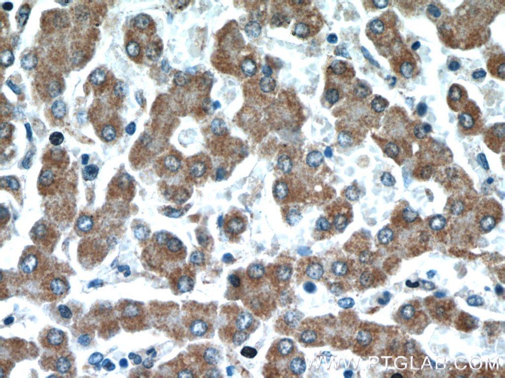
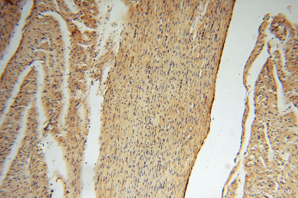
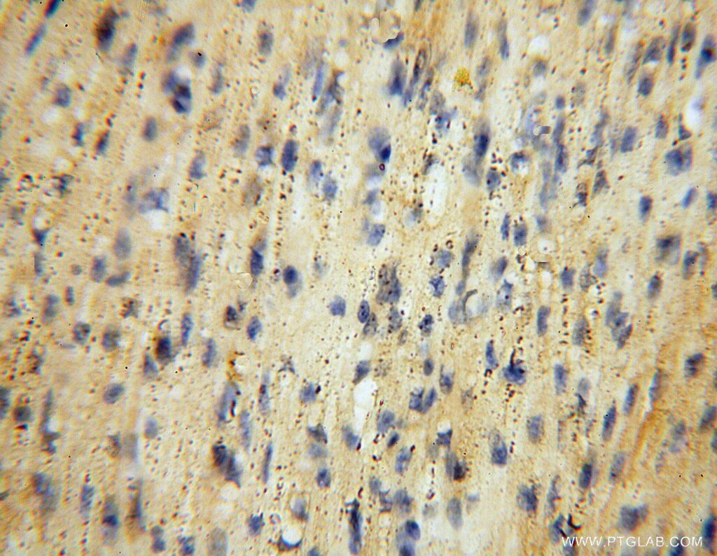
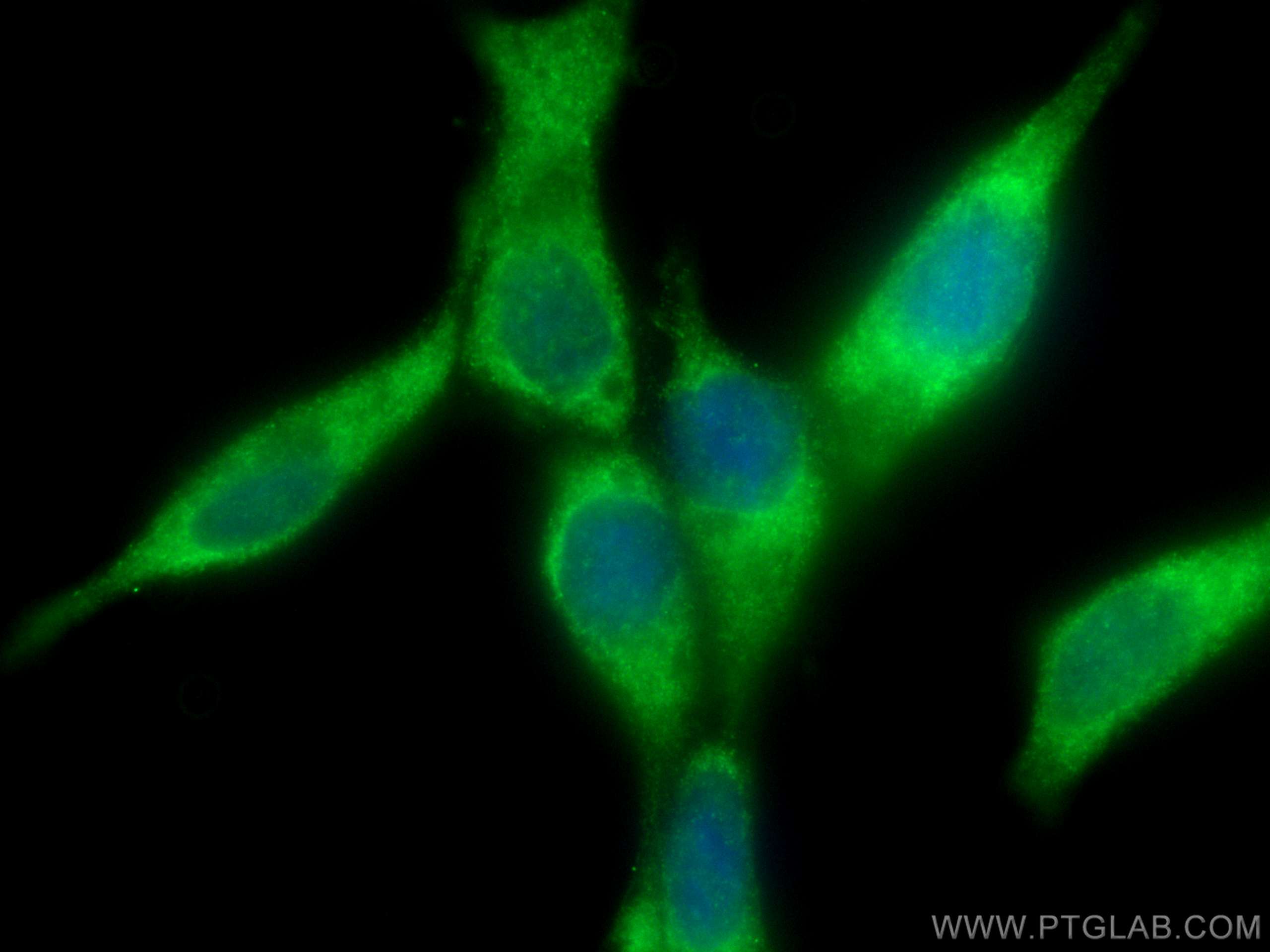
Validation Data Gallery
Tested Applications
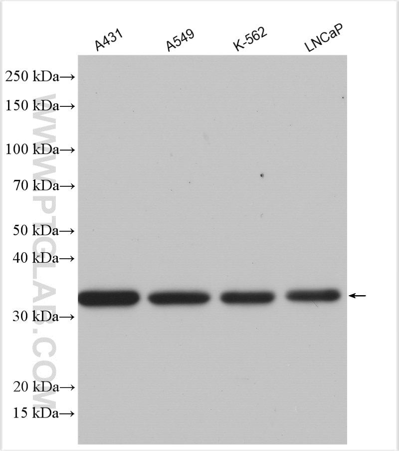
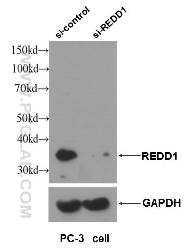
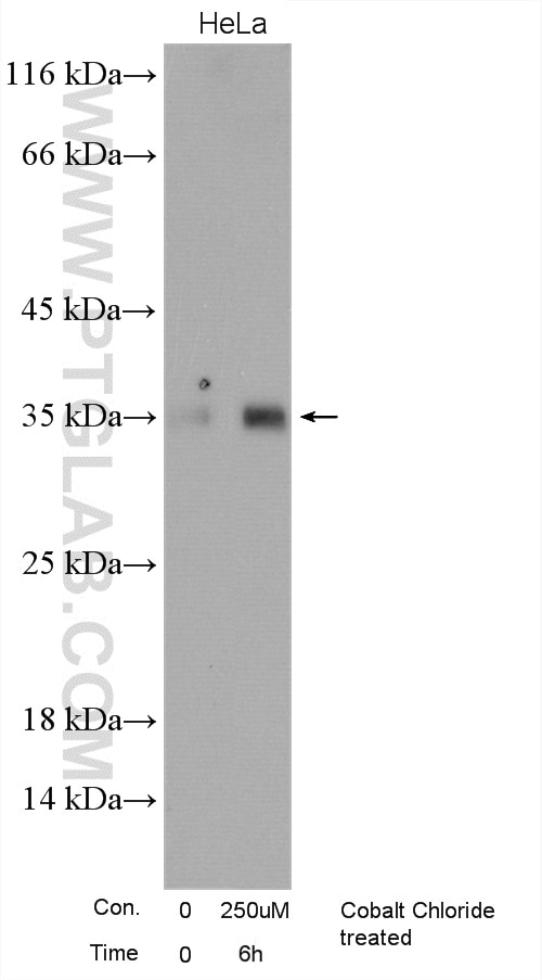
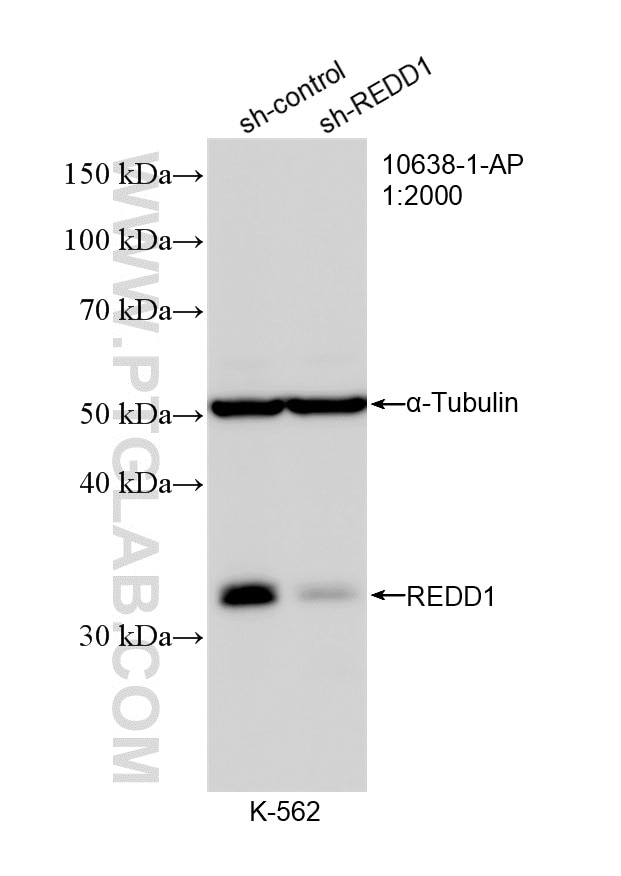
| Positive WB detected in | A431 cells, DU 145 cells, PC-3 cells, Cobalt Chloride treated HeLa cells, LNCaP cells, K-562 cells, A549 cells, SH-SY5Y cells |
| Positive IP detected in | MCF-7 cells |
| Positive IHC detected in | human lung cancer tissue, human heart tissue, human liver tissue Note: suggested antigen retrieval with TE buffer pH 9.0; (*) Alternatively, antigen retrieval may be performed with citrate buffer pH 6.0 |
| Positive IF/ICC detected in | LNCaP cells |
Recommended dilution
| Application | Dilution |
|---|---|
| Western Blot (WB) | WB : 1:2000-1:12000 |
| Immunoprecipitation (IP) | IP : 0.5-4.0 ug for 1.0-3.0 mg of total protein lysate |
| Immunohistochemistry (IHC) | IHC : 1:50-1:500 |
| Immunofluorescence (IF)/ICC | IF/ICC : 1:200-1:800 |
| It is recommended that this reagent should be titrated in each testing system to obtain optimal results. | |
| Sample-dependent, Check data in validation data gallery. | |
Published Applications
| KD/KO | See 38 publications below |
| WB | See 265 publications below |
| IHC | See 25 publications below |
| IF | See 19 publications below |
| IP | See 1 publications below |
| CoIP | See 3 publications below |
| ChIP | See 2 publications below |
Product Information
10638-1-AP targets REDD1 specific in WB, IHC, IF/ICC, IP, CoIP, ChIP, ELISA applications and shows reactivity with human, mouse samples.
| Tested Reactivity | human, mouse |
| Cited Reactivity | human, mouse, rat, pig, rabbit, squirrel, gerbil |
| Host / Isotype | Rabbit / IgG |
| Class | Polyclonal |
| Type | Antibody |
| Immunogen |
CatNo: Ag0965 Product name: Recombinant human REDD1 protein Source: e coli.-derived, PGEX-4T Tag: GST Domain: 1-232 aa of BC007714 Sequence: MPSLWDRFSSSSTSSSPSSLPRTPTPDRPPRSAWGSATREEGFDRSTSLESSDCESLDSSNSGFGPEEDTAYLDGVSLPDFELLSDPEDEHLCANLMQLLQESLAQARLGSRRPARLLMPSQLVSQVGKELLRLAYSEPCGLRGALLDVCVEQGKSCHSVGQLALDPSLVPTFQLTLVLRLDSRLWPKIQGLFSSANSPFLPGFSQSLTLSTGFRVIKKKLYSSEQLLIEEC 相同性解析による交差性が予測される生物種 |
| Full Name | DNA-damage-inducible transcript 4 |
| Calculated molecular weight | 25 kDa |
| Observed molecular weight | 32-35 kDa |
| GenBank accession number | BC007714 |
| Gene Symbol | REDD1/DDIT4 |
| Gene ID (NCBI) | 54541 |
| RRID | AB_2245711 |
| Conjugate | Unconjugated |
| Form | |
| Form | Liquid |
| Purification Method | Antigen affinity purification |
| UNIPROT ID | Q9NX09 |
| Storage Buffer | PBS with 0.02% sodium azide and 50% glycerol{{ptg:BufferTemp}}7.3 |
| Storage Conditions | Store at -20°C. Stable for one year after shipment. Aliquoting is unnecessary for -20oC storage. |
Background Information
REDD1, also named as RTP801 and DDIT4, belongs to the DDIT4 family. REDD1 promotes neuronal cell death. It is a novel transcriptional target of p53 implicated ROS in the p53-dependent DNA damage response. REDD1 controlled cell growth under energy stress, as an essential regulator of TOR activity through the TSC1/2 complex. REDD-1 expression has also been linked to apoptosis, Aβ toxicity and the pathogenesis of ischemic diseases. As an HIF-1-responsive gene, REDD-1 exhibits strong hypoxia-dependent upregulation in ischemic cells of neuronal origin[PMID: 19996311]. In response to stress due to DNA damage and glucocorticoid treatment, REDD-1 is upregulated at the transcriptional level[PMID: 21733849]. REDD-1 negatively regulates the mammalian target of Rapamycin, a serine/threonine kinase often referred to as mTOR[PMID: 22951983]. It is crucial in the coupling of extra- and intracellular cues to mTOR regulation. The absence of REDD-1 is associated with the development of retinopathy, a major cause of blindness[PMID: 22304497]. REDD1 is a new host defense factor, and chemical activation of REDD1 expression represents a potent antiviral intervention strategy[PMID: 21909097]. The calculated molecular weight of REDD1 is 25 kDa. Because of multiple lysines in the proteins, REDD1 offen migrates around 35 kDa on Western blot[PMID: 19221489]. This antibody is a rabbit polyclonal antibody raised against full length human REDD1 antigen. This antibody is specific to the REDD1 from siRNA experiment (PMID:24713927)
Protocols
| Product Specific Protocols | |
|---|---|
| IF protocol for REDD1 specific antibody 10638-1-AP | Download protocol |
| IHC protocol for REDD1 specific antibody 10638-1-AP | Download protocol |
| IP protocol for REDD1 specific antibody 10638-1-AP | Download protocol |
| WB protocol for REDD1 specific antibody 10638-1-AP | Download protocol |
| Standard Protocols | |
|---|---|
| Click here to view our Standard Protocols |
Publications
| Species | Application | Title |
|---|---|---|
Cell Metab Disrupted methionine cycle triggers muscle atrophy in cancer cachexia through epigenetic regulation of REDD1 | ||
Cancer Cell A UBE2O-AMPKα2 Axis that Promotes Tumor Initiation and Progression Offers Opportunities for Therapy. | ||
Nat Immunol The transcription factor Rfx7 limits metabolism of NK cells and promotes their maintenance and immunity. |

