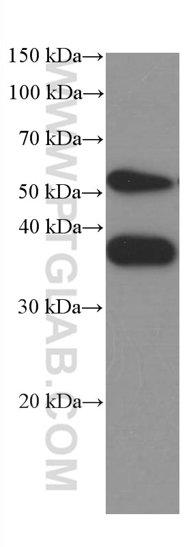
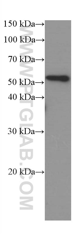
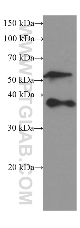
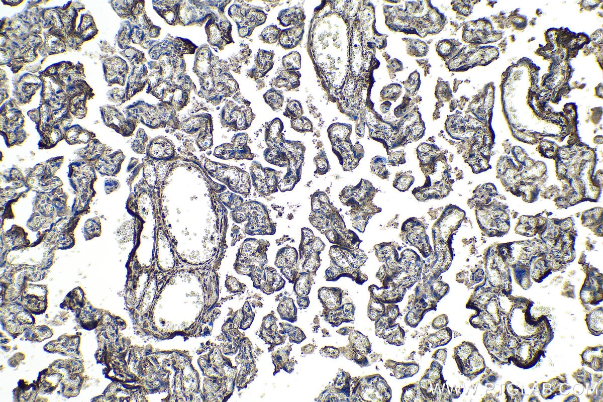
Validation Data Gallery
Tested Applications
| Positive WB detected in | A375 cells, human testis tissue, NCCIT cell |
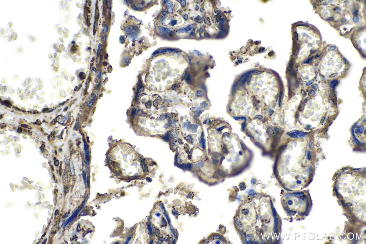
| Positive IHC detected in | human placenta tissue Note: suggested antigen retrieval with TE buffer pH 9.0; (*) Alternatively, antigen retrieval may be performed with citrate buffer pH 6.0 |
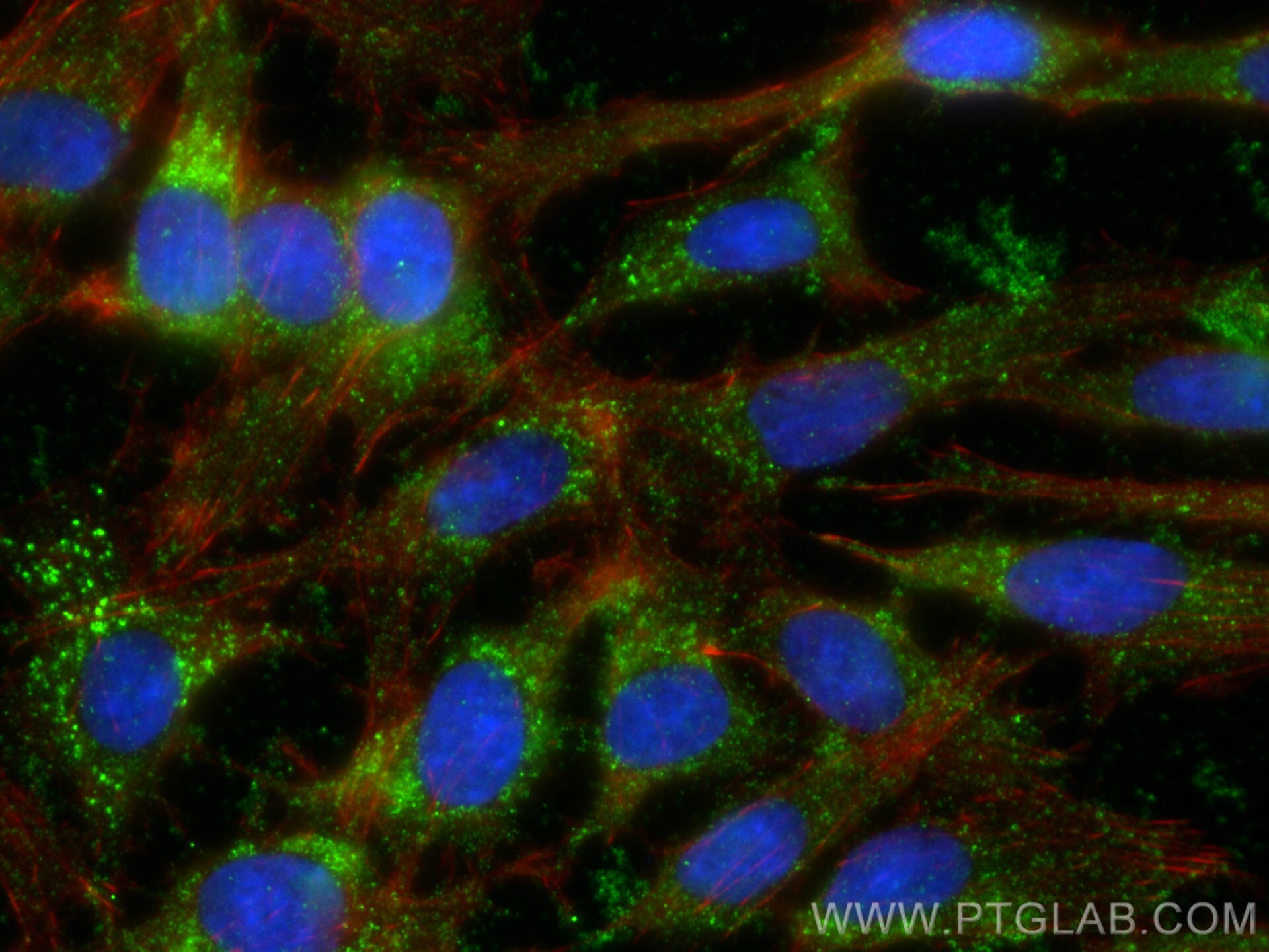
| Positive IF/ICC detected in | C6 cells |
Recommended dilution
| Application | Dilution |
|---|---|
| Western Blot (WB) | WB : 1:2000-1:10000 |
| Immunohistochemistry (IHC) | IHC : 1:1000-1:4000 |
| Immunofluorescence (IF)/ICC | IF/ICC : 1:400-1:1600 |
| It is recommended that this reagent should be titrated in each testing system to obtain optimal results. | |
| Sample-dependent, Check data in validation data gallery. | |
Published Applications
| WB | See 6 publications below |
Product Information
66426-1-Ig targets SPARC in WB, IHC, IF/ICC, ELISA applications and shows reactivity with human, rat samples.
| Tested Reactivity | human, rat |
| Cited Reactivity | human, mouse |
| Host / Isotype | Mouse / IgG2a |
| Class | Monoclonal |
| Type | Antibody |
| Immunogen | SPARC fusion protein Ag7390 相同性解析による交差性が予測される生物種 |
| Full Name | secreted protein, acidic, cysteine-rich (osteonectin) |
| Calculated molecular weight | 35 kDa |
| Observed molecular weight | 38 kDa, 52 kDa |
| GenBank accession number | BC004974 |
| Gene Symbol | SPARC |
| Gene ID (NCBI) | 6678 |
| RRID | AB_2881797 |
| Conjugate | Unconjugated |
| Form | Liquid |
| Purification Method | Protein A purification |
| UNIPROT ID | P09486 |
| Storage Buffer | PBS with 0.02% sodium azide and 50% glycerol , pH 7.3 |
| Storage Conditions | Store at -20°C. Stable for one year after shipment. Aliquoting is unnecessary for -20oC storage. |
Background Information
SPARC, also known as ON (Osteonectin) or BM-40 (Basement-membrane protein 40), is an extracellular glycoprotein with the calculated molecular mass of 35 kDa and the apparent molecular mass of 40-43 kDa and 50 kDa (PMID: 7495300, 12365801). SPARC belongs to a group of matricellular proteins defined as secreted components that do not contribute directly to the formation of structural elements but serve to modulate cell-matrix interactions and cellular functions (PMID: 7542656; 12231357). SPARC is expressed at high levels in bone tissue, is distributed widely in many other tissues and cell types, and is associated generally with tissues undergoing morphogenesis, remodeling and wound repair (PMID: 10567433). It elicits changes in cell shape, inhibits cell-cycle progression, and influences the synthesis of extracellular matrix (PMID: 12721366). Altered expression of SPARC has been reported in a variety of cancers, which include breast, ovarian, colorectal, and pancreatic cancer as well as melanoma and glioblastomas (PMID: 18849185).
Protocols
| Product Specific Protocols | |
|---|---|
| WB protocol for SPARC antibody 66426-1-Ig | Download protocol |
| IHC protocol for SPARC antibody 66426-1-Ig | Download protocol |
| IF protocol for SPARC antibody 66426-1-Ig | Download protocol |
| Standard Protocols | |
|---|---|
| Click here to view our Standard Protocols |
Publications
| Species | Application | Title |
|---|---|---|
Int J Cancer Tissue-resident CXCR4+ macrophage as a poor prognosis signature promotes pancreatic ductal adenocarcinoma progression | ||
Br J Cancer Inactivation of 3-hydroxybutyrate dehydrogenase type 2 promotes proliferation and metastasis of nasopharyngeal carcinoma by iron retention. | ||
Front Endocrinol (Lausanne) Secreted Protein Acidic and Rich in Cysteine Mediates the Development and Progression of Diabetic Retinopathy. | ||
Exp Cell Res Sorafenib derivatives-functionalized gold nanoparticles confer protection against tumor angiogenesis and proliferation via suppression of EGFR and VEGFR-2. | ||
Pathol Res Pract Downregulation of NEBL promotes migration and invasion of clear cell renal cell carcinoma by inducing epithelial-mesenchymal transition | ||
Acta Biomater Targeting Delivery of Mifepristone to Endometrial Dysfunctional Macrophages for Endometriosis Therapy |
