• Featured Product
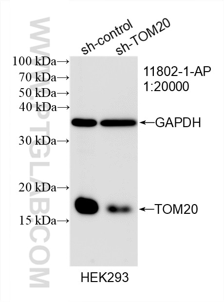
  • KD/KO Validated

TOM20 Polyclonal antibody

TOM20 Polyclonal Antibody for WB, IHC, IF/ICC, IP, ELISA

Host / Isotype

Rabbit / IgG

Reactivity

human, mouse, rat, chicken and More (6)

Applications

WB, IHC, IF/ICC, IP, CoIP, ELISA

Conjugate

Unconjugated

Cat no : 11802-1-AP

Synonyms

TOMM20, TOMM 20, tom 20, MOM19, MAS20


☆1/31(金)までお得に買える! >> IHC・ELISA・IPキット最大30%OFFキャンペーン


Tested Applications

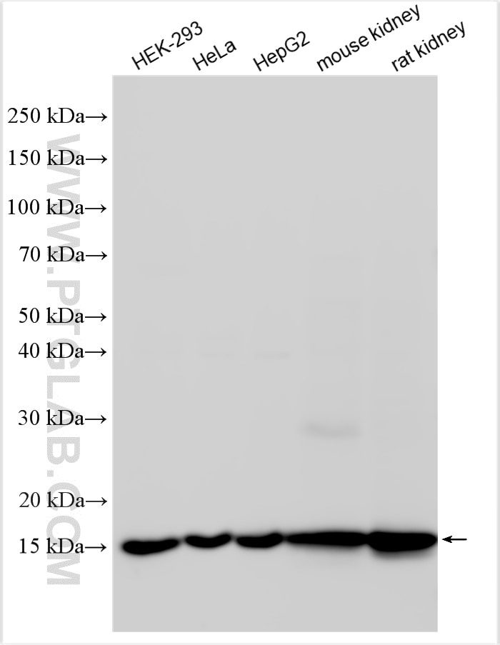
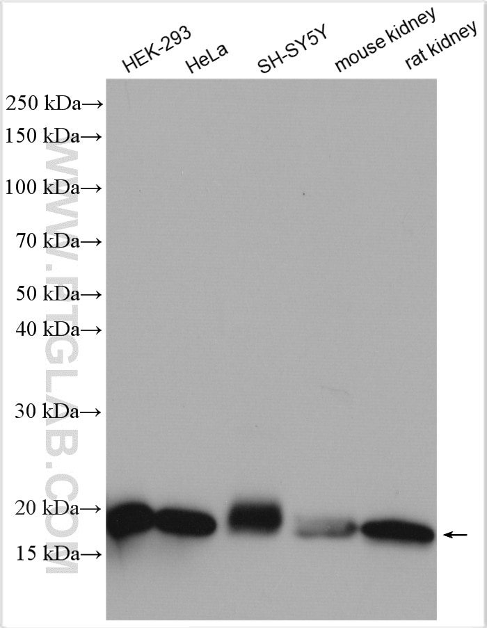
Positive WB detected inHEK-293 cells, HeLa cells, HepG2 cells, mouse kidney tissue, rat kidney tissue, SH-SY5Y cells
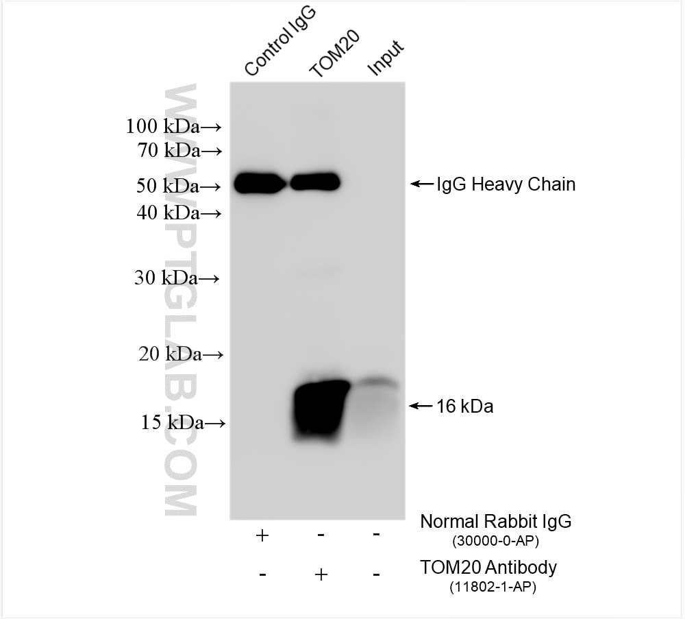
Positive IP detected inHeLa cells
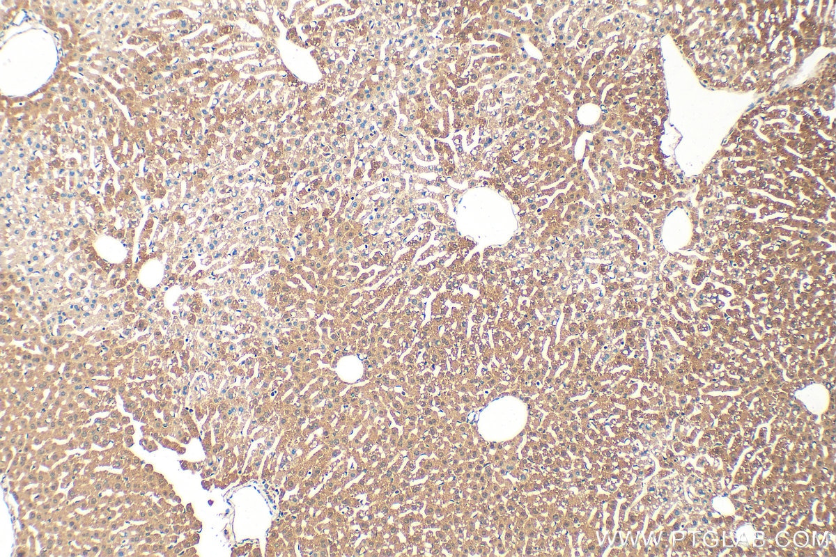
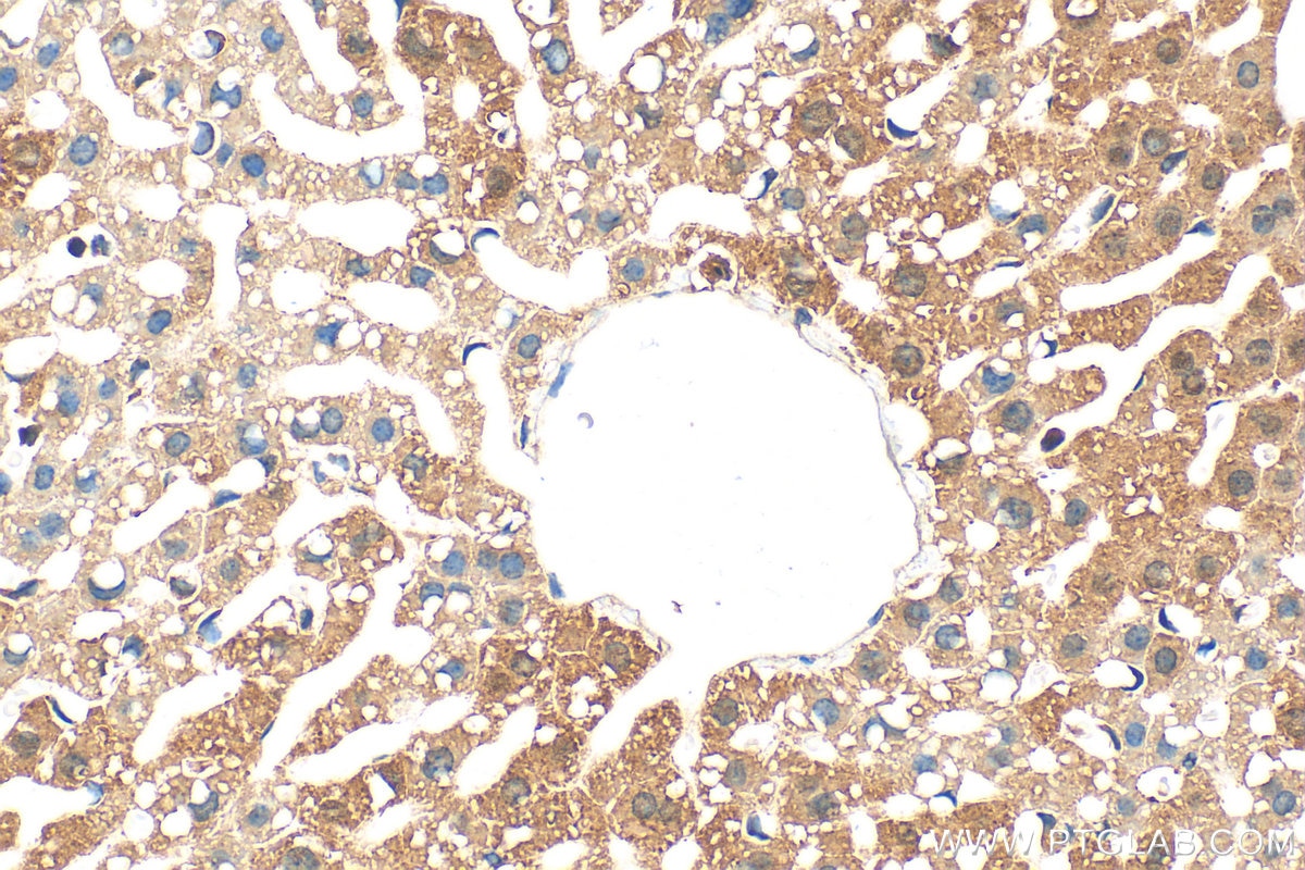
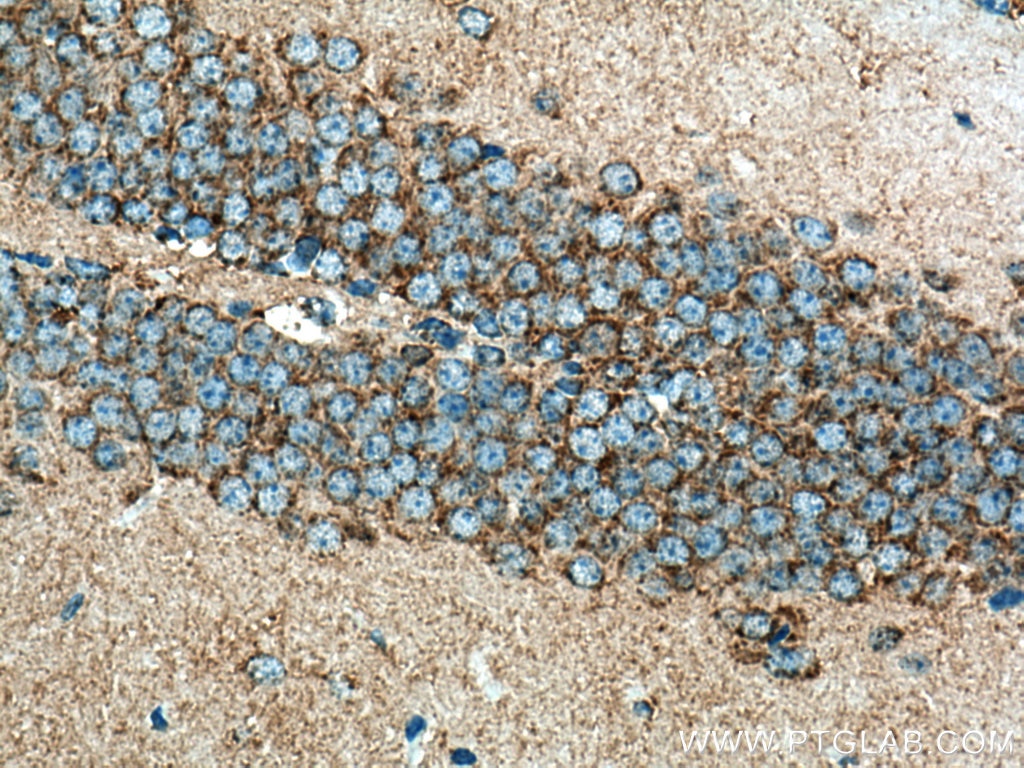
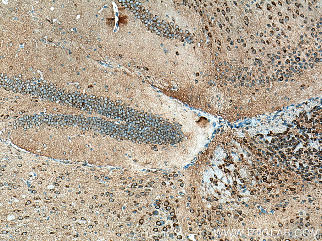
Positive IHC detected inmouse liver tissue, mouse brain tissue
Note: suggested antigen retrieval with TE buffer pH 9.0; (*) Alternatively, antigen retrieval may be performed with citrate buffer pH 6.0
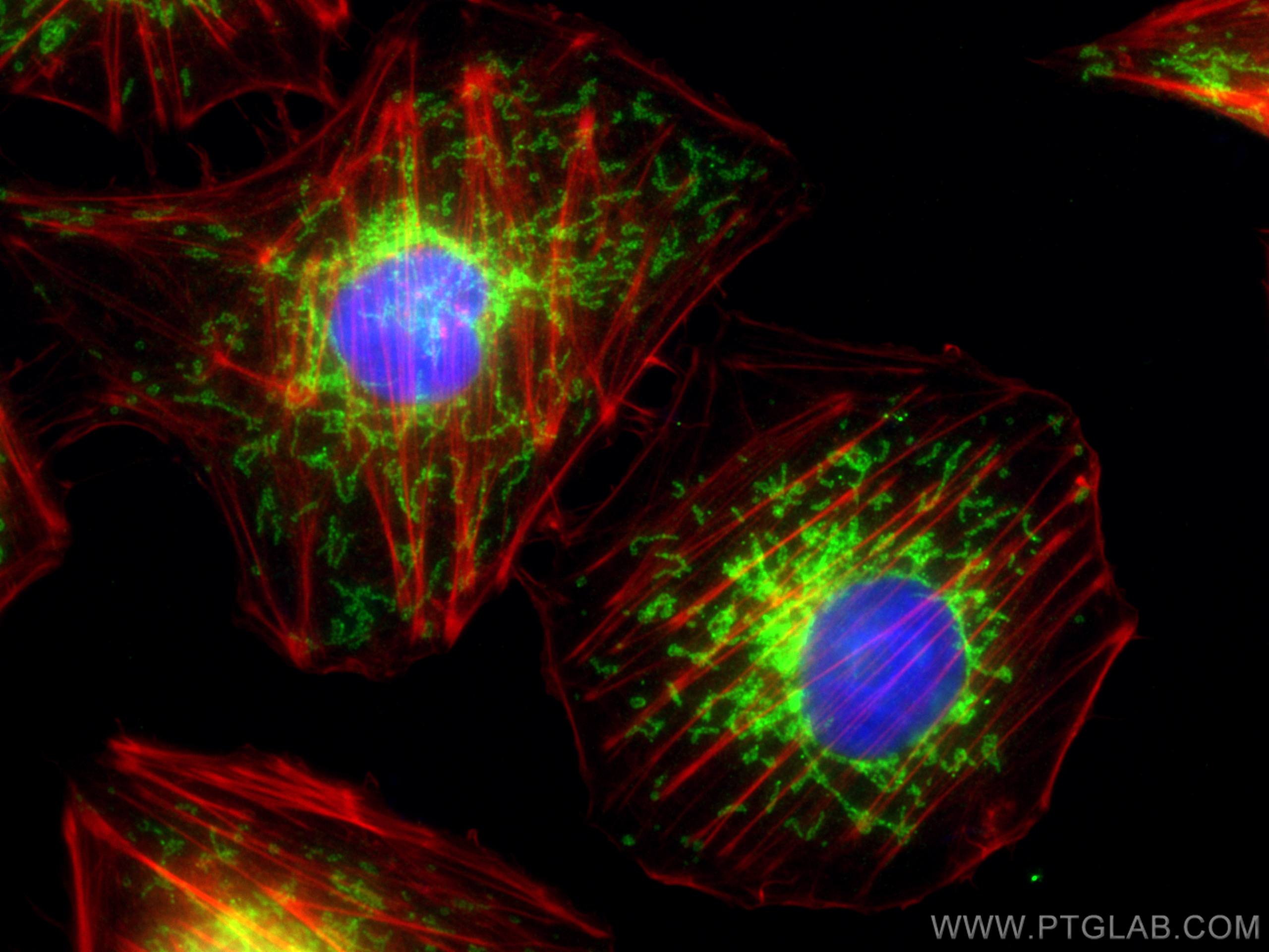
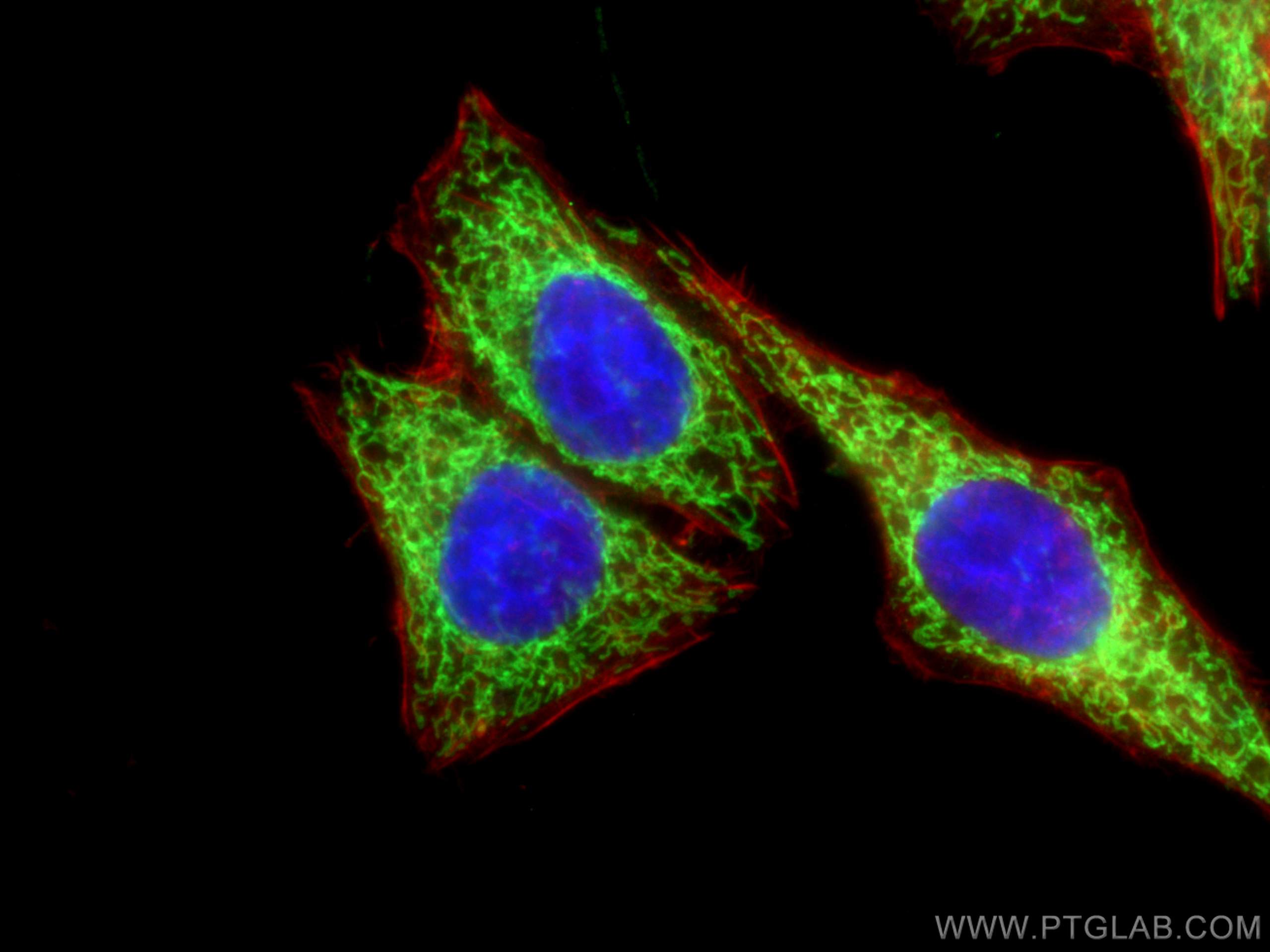
Positive IF/ICC detected inHUVEC cells, HepG2 cells

Recommended dilution

ApplicationDilution
Western Blot (WB)WB : 1:5000-1:50000
Immunoprecipitation (IP)IP : 0.5-4.0 ug for 1.0-3.0 mg of total protein lysate
Immunohistochemistry (IHC)IHC : 1:500-1:2000
Immunofluorescence (IF)/ICCIF/ICC : 1:50-1:500
It is recommended that this reagent should be titrated in each testing system to obtain optimal results.
Sample-dependent, Check data in validation data gallery.

Product Information

11802-1-AP targets TOM20 in WB, IHC, IF/ICC, IP, CoIP, ELISA applications and shows reactivity with human, mouse, rat, chicken samples.

Tested Reactivity human, mouse, rat, chicken
Cited Reactivityhuman, mouse, rat, pig, rabbit, monkey, chicken, zebrafish, hamster, cattle
Host / Isotype Rabbit / IgG
Class Polyclonal
Type Antibody
Immunogen TOM20 fusion protein Ag2378 相同性解析による交差性が予測される生物種
Full Name translocase of outer mitochondrial membrane 20 homolog (yeast)
Calculated molecular weight 145 aa, 16 kDa
Observed molecular weight 16 kDa
GenBank accession numberBC000882
Gene symbol TOM20
Gene ID (NCBI) 9804
RRIDAB_2207530
Conjugate Unconjugated
Form Liquid
Purification MethodAntigen affinity purification
Storage Buffer PBS with 0.02% sodium azide and 50% glycerol pH 7.3.
Storage ConditionsStore at -20°C. Stable for one year after shipment. Aliquoting is unnecessary for -20oC storage.

Background Information

Background

 TOM20 (KIAA0016) belongs to the Tom family. It is a subunit of the mitochondrial import receptor (PMID: 7584026), whose main role is to translocate cytosolically synthesized mitochondrial proteins through the outer mitochondrial membrane and subsequently facilitate the movement of proteins into the TOM40 translocation pore complex (PMID: 21173275).

 What is the molecular weight of TOM20? 

 It is a short 16.3 kDa protein containing several highly conserved regions, including the transmembrane segment, the ligand-binding domain, and flexible segments at the N terminus and the C terminus of the protein crucial for its function (PMID: 15733919).

 What is the subcellular localization of TOM20?

 It specifically localizes in the mitochondrial outer membrane. In malignant cells, strong granular staining in the cytoplasm has been observed.

 What is the tissue specificity of TOM20?

 It is ubiquitously expressed in various tissues, with a particularly high expression in the brain, thyroid, and pancreas.

 What is the function of TOM20 in the mitochondrial membrane?

 Mitochondrial preproteins are generally synthesized in the cytoplasm with signal or targeting sequences, which are recognized by specific receptors on the outer mitochondrial membrane. TOM20, as one of the components of the TOM40 complex, specifically recognizes pre-sequences on target proteins or their unfolded forms. In addition to its translocase activity, TOM20 may act as a chaperone preventing these proteins from aggregation at the surface of mitochondria (PMID: 14699115).

 What is TOM20's involvement in disease?

 Diseases linked to the misregulation of TOM20 include Perry Syndrome and neurodegeneration with brain iron accumulation 2A. Numerous studies also associate deregulation of TOM20 with an array of mitochondrial dysfunctions and mitophagy defects (PMIDs: 30254015, 30160596). 


Protocols

Product Specific Protocols
WB protocol for TOM20 antibody 11802-1-APDownload protocol
IHC protocol for TOM20 antibody 11802-1-APDownload protocol
IF protocol for TOM20 antibody 11802-1-APDownload protocol
IP protocol for TOM20 antibody 11802-1-APDownload protocol
Standard Protocols
Click here to view our Standard Protocols

Publications

SpeciesApplicationTitle
mouseWB

Nature

Gasdermin D-mediated metabolic crosstalk promotes tissue repair

Authors - Zhexu Chi
IF

Nature

Stress response silencing by an E3 ligase mutated in neurodegeneration

Authors - Diane L Haakonsen
mouseWB

Nature

CLSTN3β enforces adipocyte multilocularity to facilitate lipid utilization

Authors - Kevin Qian
mouseWB

Cell Res

AMPK targets PDZD8 to trigger carbon source shift from glucose to glutamine

Authors - Mengqi Li
  • KO Validated
humanWB

Cell Res

Mitochondria-localized cGAS suppresses ferroptosis to promote cancer progression

Authors - Shiqiao Qiu
mouseWB

Nature

BCAA catabolism in brown fat controls energy homeostasis through SLC25A44.

Authors - Takeshi Yoneshiro

Reviews

The reviews below have been submitted by verified Proteintech customers who received an incentive forproviding their feedback.


FH

Durand (Verified Customer) (11-19-2024)

IF on HeLa cells using PFA 4%, 37°C 1h, BSA 10% 30 min, and 1h Ab 1/1000 dilution in BSA 3% Correct mitochondrial network

  • Applications: Immunofluorescence
  • Primary Antibody Dilution: 1/1000
  • Cell Tissue Type: HeLa cells
TOM20 Antibody Immunofluorescence validation (1/1000 dilution) in HeLa cells (Cat no:11802-1-AP)
FH

PK (Verified Customer) (07-05-2024)

Excellent

  • Applications: Western Blot
  • Primary Antibody Dilution: 2000
  • Cell Tissue Type: l2
TOM20 Antibody Western Blot validation (2000 dilution) in l2 (Cat no:11802-1-AP)
FH

SD (Verified Customer) (07-05-2024)

Excellent

  • Applications: Western Blot
  • Primary Antibody Dilution: 1000
  • Cell Tissue Type: HEK
TOM20 Antibody Western Blot validation (1000 dilution) in HEK (Cat no:11802-1-AP)
FH

Joel (Verified Customer) (11-07-2023)

Works well for immunofluorescence (1:200) and Western blotting (1:1000-1:2000)

  • Applications: Western Blot, Immunofluorescence
  • Primary Antibody Dilution: 1:200
  • Cell Tissue Type: Human and mouse cancer cell lines
FH

Lisa (Verified Customer) (03-23-2023)

The antibody worked very well for immunofluorescence, combined with a secondary fluorescent antibody 555.

  • Applications: Immunofluorescence
  • Primary Antibody Dilution: 1:200
  • Cell Tissue Type: COS-7
TOM20 Antibody Immunofluorescence validation (1:200 dilution) in COS-7 (Cat no:11802-1-AP)
FH

S (Verified Customer) (12-12-2022)

excellent

  • Applications: Western Blot
  • Primary Antibody Dilution: 1000
  • Cell Tissue Type: FIBROBLAST
TOM20 Antibody Western Blot validation (1000 dilution) in FIBROBLAST (Cat no:11802-1-AP)
FH

Kazu (Verified Customer) (11-23-2022)

This antibody works and provides robust signals of TOM20 immunoreactivity on feline tissue sections.

  • Applications: Immunofluorescence
  • Primary Antibody Dilution: 1:200
  • Cell Tissue Type: Optic nerve
FH

Wan (Verified Customer) (03-01-2022)

We got very clear and correct bands for tom20., very good antibody.

  • Applications: Western Blot
FH

Süleyman (Verified Customer) (02-07-2022)

It is very specific and nicely labelled mitochondria. I highly suggest it. 4% paraformaldehyde fixation for 10 min at RT and 0.3% Triton-x100 10 minutes for permeabilization at RT. I used 2h at RT or 16h at 4 degree incubation. Alexa fluor 488 1:700 is used for 1 hour at RT for secondary ab.

  • Applications: Immunofluorescence
  • Primary Antibody Dilution: 1:100
  • Cell Tissue Type: HeLa
TOM20 Antibody Immunofluorescence validation (1:100 dilution) in HeLa (Cat no:11802-1-AP)
FH

Sahil (Verified Customer) (10-09-2020)

The tom 20 works great. We wanted to compare and it does stand out

  • Applications: Immunohistochemistry
FH

Thomas (Verified Customer) (09-18-2020)

The antibody allows for exceptionally clear visualisation of the mitochondrial outer membrane. HeLa cells were fixed in 4% PFA for 15 mins and permeabilised in 0.1% Triton-X 100 in PBS. Cells were blocked in 1% BSA. Primary antibody solution was diluted at 1:250 in blocking solution and incubated for 1 hour. Goat anti-rabbit 488 Alexa Fluor secondary antibody (1:250 - green) and DAPI (1:2000 - blue) were incubated with cells for 1 hour.

  • Applications: Immunofluorescence
  • Primary Antibody Dilution: 1:250
  • Cell Tissue Type: HeLa
TOM20 Antibody Immunofluorescence validation (1:250 dilution) in HeLa (Cat no:11802-1-AP)
FH

Maria (Verified Customer) (06-05-2020)

TOM20 1:200 in PBST (0.1% triton) + 10% Donkey Serum O/N 4ºC incubation. Donkey anti-rabbit alexa fluor 555 1:500 1h RT incubation.

  • Applications: Immunofluorescence
  • Primary Antibody Dilution: 1:200
  • Cell Tissue Type: Human fibroblasts
TOM20 Antibody Immunofluorescence validation (1:200 dilution) in Human fibroblasts (Cat no:11802-1-AP)
FH

Mohammad (Verified Customer) (02-25-2020)

For mouse hippocampusWorks well

  • Applications: Immunofluorescence,
FH

Uxoa (Verified Customer) (02-19-2020)

TOM20 ab is great for ICC and WB. Tested in human primary fibroblats. ICC: fixed fibroblats with 4% PFA 15min. TOM20 diluted 1:200 in PBST (0.1% triton) + 10% DS O/N 4ºC incubation. Donkey anti-rabbit alexa fluor 488 1:500 1h RT. DAPI in blue.

  • Applications: Western Blot, Immunofluorescence,
  • Primary Antibody Dilution: 1:200
  • Cell Tissue Type: Human fibroblasts
TOM20 Antibody Western Blot,Immunofluorescence, validation (1:200 dilution) in Human fibroblasts (Cat no:11802-1-AP)
FH

Benjamin (Verified Customer) (01-10-2020)

Reliably used in western blot where it identifies the correct band with high specificity.

  • Applications: Western Blot,
  • Primary Antibody Dilution: 1:500
  • Cell Tissue Type: HEK293T
FH

Aamir (Verified Customer) (01-08-2020)

WB worked fine

  • Applications: Western Blot,
  • Primary Antibody Dilution: 1000
  • Cell Tissue Type: HEKs
FH

Tanusree (Verified Customer) (12-03-2019)

This antibody works good in western blotting analysis using mouse tissues.

  • Applications: Western Blot,
  • Primary Antibody Dilution: 1:500
  • Cell Tissue Type: Mouse brain
FH

Fulong (Verified Customer) (10-24-2019)

It's great for IF.

  • Applications: Immunofluorescence,
  • Cell Tissue Type: Human
FH

Praveen (Verified Customer) (04-15-2019)

Good

  • Applications: Western Blot,
  • Cell Tissue Type: AC16
TOM20 Antibody Western Blot, validation ( dilution) in AC16 (Cat no:11802-1-AP)
FH

Daniela (Verified Customer) (03-19-2019)

A very specific and reliable antibody. For Western Blot, it identifies the correct MW band in cellular and mitochondrial lysate from either murine (N2a) and human (HeLa and U2OS) cell. For immunofluorescence, it is ideal for localization studies: it works perfectly to label mitochondria instead of plasmidic markers.

  • Applications: Western Blot, Immunohistochemistry, Immunofluorescence,
  • Primary Antibody Dilution: Western Blot 1:2000, Immunofluorescence 1:200
  • Cell Tissue Type: N2a, U2OS and HeLa cells
FH

Svitlana (Verified Customer) (03-19-2019)

SDS-PAGE: 15 ug protein/well.Lines on WB1. BioRad Precision Plus Protein standard2. Homogenate of mice brain tissue3. Non-synaptosomal mitochondria from mice brain.Transfer: Immobilon-FL transfer membranes (Millipore) O/N at 30V, 4CBlocking: SEA Block Blocking Buffer 1hSecondary Ab:IRDye 680LT Goat anti-Rabbit, 1h incubation at room temperature.

  • Applications: Western Blot,
  • Primary Antibody Dilution: 1:2000
  • Cell Tissue Type: Mice brain tissue
TOM20 Antibody Western Blot, validation (1:2000 dilution) in Mice brain tissue (Cat no:11802-1-AP)
FH

Ken (Verified Customer) (02-06-2019)

It is an amazing antibody. Very specific and very bright at a 1:1000 dilution.

  • Applications: Immunofluorescence,
  • Primary Antibody Dilution: 1:1000
  • Cell Tissue Type: U2OS cells