Product Information
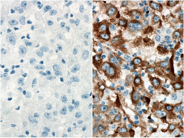
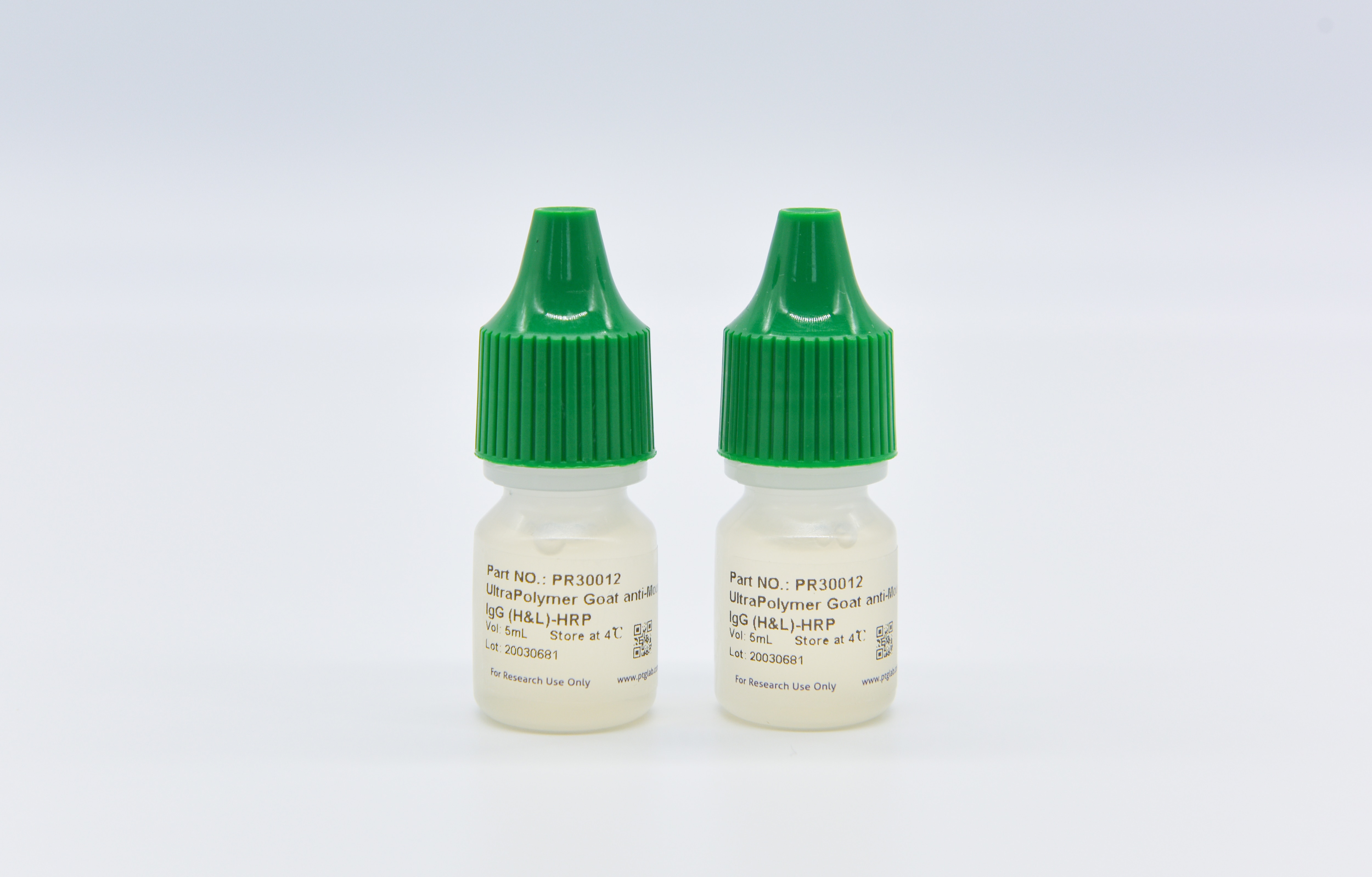
UltraPolymer Goat anti-Mouse IgG (H&L)-HRP is a polymer (secondary antibody) formed by coupling multiple peroxidase (HRP) molecules and anti mouse immunoglobulin (IgG) onto the polymer using the latest polymerization technology. The polymer (secondary antibody) has the characteristics of amplifying signals to increase sensitivity and reducing non-specific background. It can be used together with mouse first antibody, and then react with Chromogen(DAB) to obtain brown color.
Storage
Store the reagent at 2-8°C. It is stable for 6 months from the date of receipt.
Usage
1. Prepare slides from tissues sections following routine methods. Deparaffinize tissues with xylene and re-hydrate using a decreasing ethanol gradient.
2. Perform antigen retrieval following the manufacturer’s recommendations of the primary antibody.
3. Wash the slides with 1x wash buffer. Drain off the liquid on the slides and absorb any residual buffer around the tissue section (making the slides as dry as possible). Use a hydrophobic IHC pen (Cat No. PR30013) to draw a circle on the glass slides around tissue sections (optional).
4. Quenching (optional): Apply the quenching buffer (e.g.,3%H2O2) to the slide, covering the entire tissue. Leave the slides at room temperature for 10 minutes. Briefly wash the slides with 1x wash buffer after quenching.
5. Block the tissues with blocking buffer (e.g., 3% BSA in PBS) for 30 minutes at room temperature. Drain the liquid off the slides and absorb the blocking buffer around the tissue section.
6. Apply the primary antibody working solution to the slide(ONLY primary antibodies raised in mice should be used with this secondary antibody), covering the entire tissue. Incubate at room temperature in a wet box with a lid for 1 hour.
7. Wash the slides three times following primary antibody incubation. Drain the liquid off the slides and absorb any residual buffer around the tissue section. Apply the UltraPolymer Goat anti-Mouse IgG (H&L)-HRP to the slide, covering the entire tissue. Incubate at room temperature in a wet box with a lid for 30 minutes.
8. Wash the slides three times following secondary antibody incubation. Drain the liquid off the slides and absorb any residual buffer around the tissue section. Use pipette to prepare an appropriate volume of Chromogen (DAB or other oxidized by hydrogen peroxide in a reaction typically catalyzed by horseradish peroxidase) in a microcentrifuge tube. Use pipette to add enough Chromogen to cover the tissue. Lay slides flat on the benchtop at room temperature for 5 minutes, or until a brown color develops. If color is visible, stop reaction early by washing with 1x wash buffer.
9. Perform counter staining with hematoxylin.
10. Wash the slides with wash buffer or pure water. Dehydrate tissues using an increasing ethanol gradient followed by xylene. Mount the slides with cover slides and analyze with a light microscope once they are fully dried.
Application Note
This reagent is recommended for FFPE tissues and optimized for manual staining.
Cited in Article as
pr30012, UltraPolymer Goat anti-Mouse IgG (H&L)-HRP, Proteintech, IL, USA
Publications
| Application | Title |
|---|---|
Clin Transl Med Single-cell transcriptome sequencing of B-cell heterogeneity and tertiary lymphoid structure predicts breast cancer prognosis and neoadjuvant therapy efficacy | |
Int J Biol Macromol VDRA downregulate β-catenin/Smad3 and DNA damage and repair associated with improved prognosis in ccRCC patients | |
Free Radic Biol Med Bisphenol A induces placental ferroptosis and fetal growth restriction via the YAP/TAZ-ferritinophagy axis | |
Front Immunol Poly-l-lactic acid microspheres delay aging of epidermal stem cells in rat skin | |
Int Immunopharmacol Fraxetin attenuates DNA damage and inflammation in cisplatin-induced nephrotoxicity via FoxO1 activation | |
Int Immunopharmacol TSLP/TSLPR promotes renal fibrosis by activating STAT3 in renal fibroblasts |


