Validation Data Gallery
Tested Applications
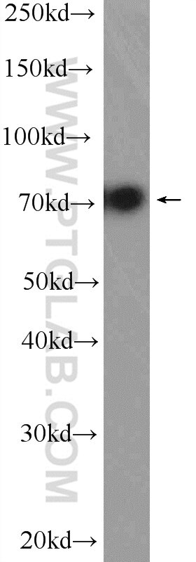
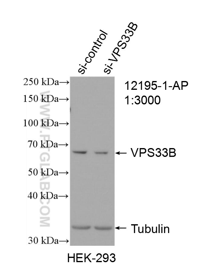
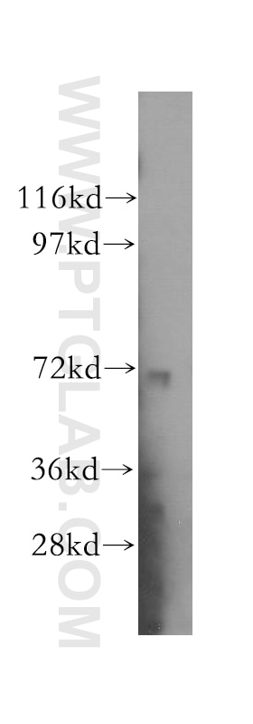
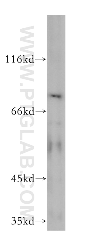
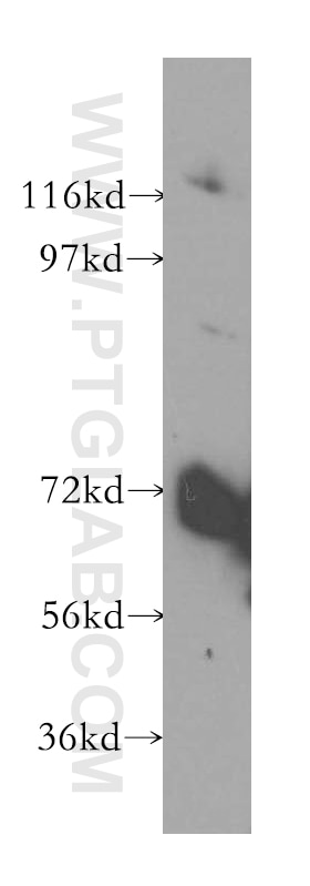
| Positive WB detected in | HEK-293 cells, mouse skeletal muscle tissue, HeLa cells, mouse testis tissue, L02 cells |
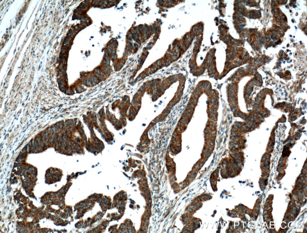
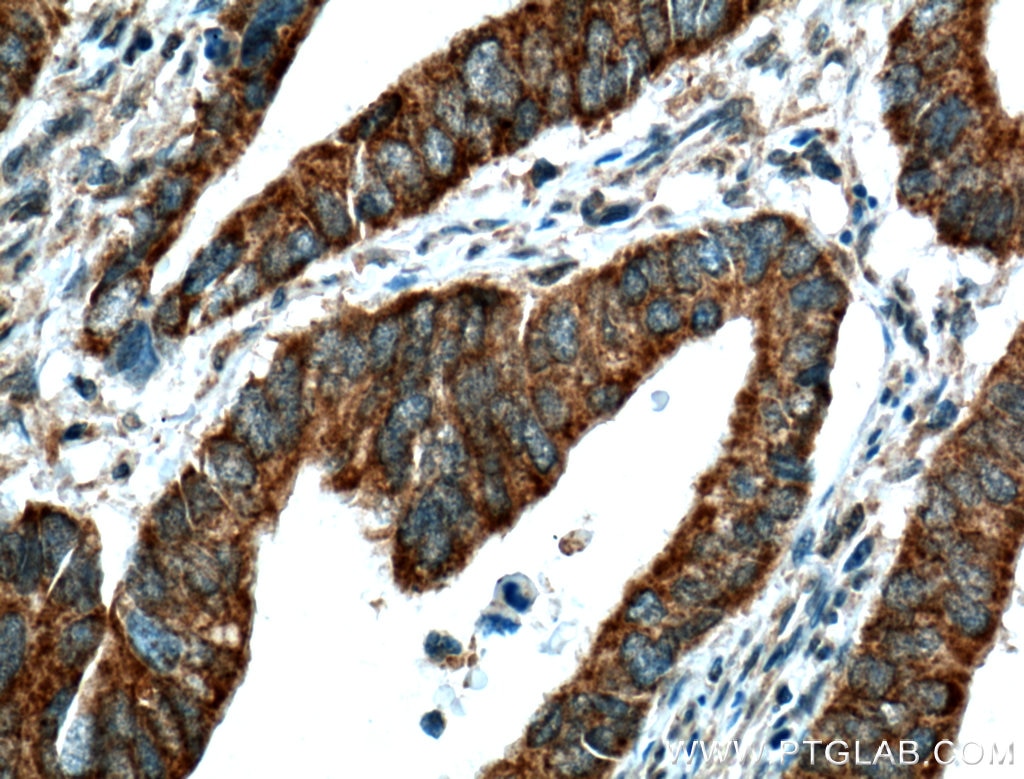
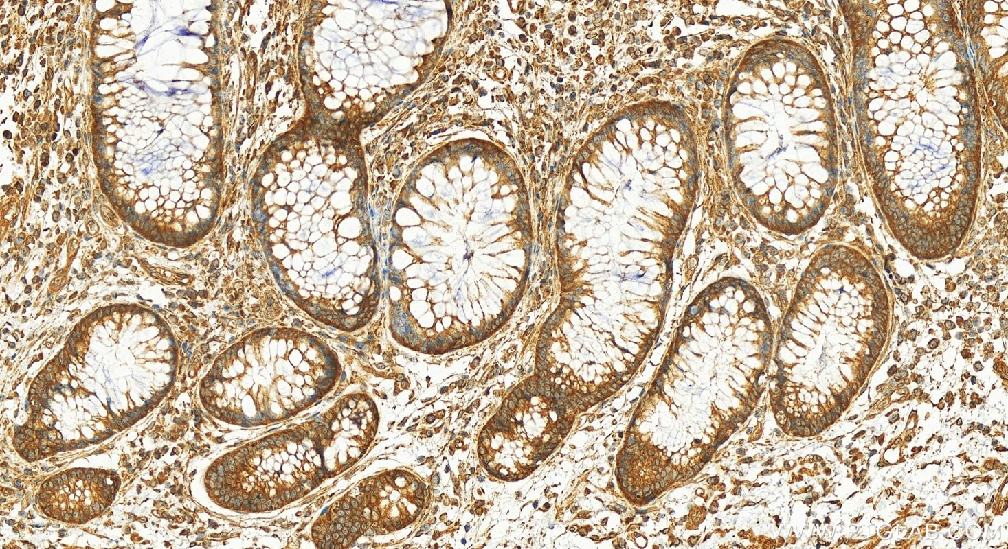
| Positive IHC detected in | human colon cancer tissue Note: suggested antigen retrieval with TE buffer pH 9.0; (*) Alternatively, antigen retrieval may be performed with citrate buffer pH 6.0 |
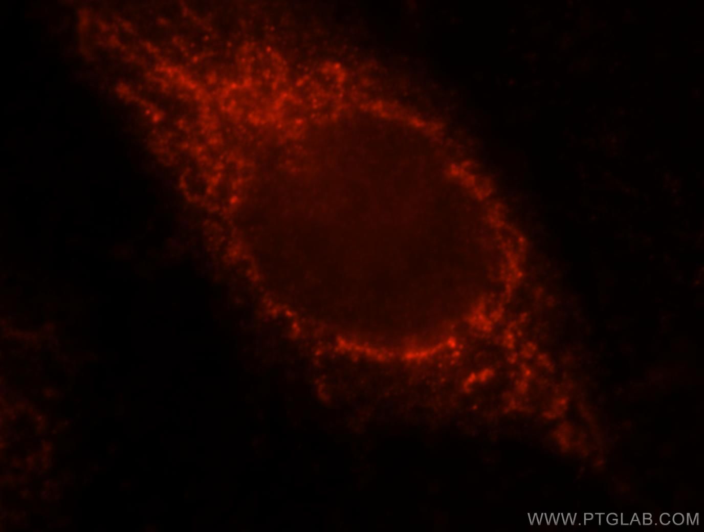
| Positive IF/ICC detected in | MCF-7 cells |
Recommended dilution
| Application | Dilution |
|---|---|
| Western Blot (WB) | WB : 1:500-1:2000 |
| Immunohistochemistry (IHC) | IHC : 1:50-1:500 |
| Immunofluorescence (IF)/ICC | IF/ICC : 1:10-1:100 |
| It is recommended that this reagent should be titrated in each testing system to obtain optimal results. | |
| Sample-dependent, Check data in validation data gallery. | |
Published Applications
| KD/KO | See 1 publications below |
| WB | See 9 publications below |
| IHC | See 4 publications below |
| IF | See 6 publications below |
| IP | See 1 publications below |
| CoIP | See 1 publications below |
| ChIP | See 1 publications below |
Product Information
12195-1-AP targets VPS33B in WB, IHC, IF/ICC, IP, CoIP, ChIP, ELISA applications and shows reactivity with human, mouse, rat samples.
| Tested Reactivity | human, mouse, rat |
| Cited Reactivity | human, mouse, zebrafish |
| Host / Isotype | Rabbit / IgG |
| Class | Polyclonal |
| Type | Antibody |
| Immunogen | VPS33B fusion protein Ag2833 相同性解析による交差性が予測される生物種 |
| Full Name | vacuolar protein sorting 33 homolog B (yeast) |
| Calculated molecular weight | 617 aa, 71 kDa |
| Observed molecular weight | 65-71 kDa |
| GenBank accession number | BC016445 |
| Gene Symbol | VPS33B |
| Gene ID (NCBI) | 26276 |
| RRID | AB_2215198 |
| Conjugate | Unconjugated |
| Form | Liquid |
| Purification Method | Antigen affinity purification |
| UNIPROT ID | Q9H267 |
| Storage Buffer | PBS with 0.02% sodium azide and 50% glycerol , pH 7.3 |
| Storage Conditions | Store at -20°C. Stable for one year after shipment. Aliquoting is unnecessary for -20oC storage. |
Background Information
VPS33B, a homolog of yeast class C vacuolar protein sorting (vps) protein Vps33p, belongs to the STXBP/unc-18/SEC1 family. It may play a role in vesicle-mediated protein trafficking to lysosomal compartments and in membrane docking/fusion reactions of late endosomes/lysosomes. VPS33B mediates phagolysosomal fusion in macrophages (PMID: 18474358). Defects in VPS33B account for most cases of arthrogryposis-renal dysfunction-cholestasis (ARC) syndrome, which is a multisystem disorder associated with abnormalities in polarized liver and kidney cells (PMID: 20190753).
Protocols
| Product Specific Protocols | |
|---|---|
| WB protocol for VPS33B antibody 12195-1-AP | Download protocol |
| IHC protocol for VPS33B antibody 12195-1-AP | Download protocol |
| IF protocol for VPS33B antibody 12195-1-AP | Download protocol |
| Standard Protocols | |
|---|---|
| Click here to view our Standard Protocols |
Publications
| Species | Application | Title |
|---|---|---|
Nat Genet Mutations in VIPAR cause an arthrogryposis, renal dysfunction and cholestasis syndrome phenotype with defects in epithelial polarization.
| ||
Proc Natl Acad Sci U S A Mycobacterium tuberculosis protein tyrosine phosphatase (PtpA) excludes host vacuolar-H+-ATPase to inhibit phagosome acidification. | ||
J Cell Sci Late endosomal transport and tethering are coupled processes controlled by RILP and the cholesterol sensor ORP1L. | ||
Clin Kidney J Glomerular involvement in the arthrogryposis, renal dysfunction and cholestasis syndrome. | ||
Cancer Sci VPS33B interacts with NESG1 to suppress cell growth and cisplatin chemoresistance in ovarian cancer. |
