Validation Data Gallery
Tested Applications
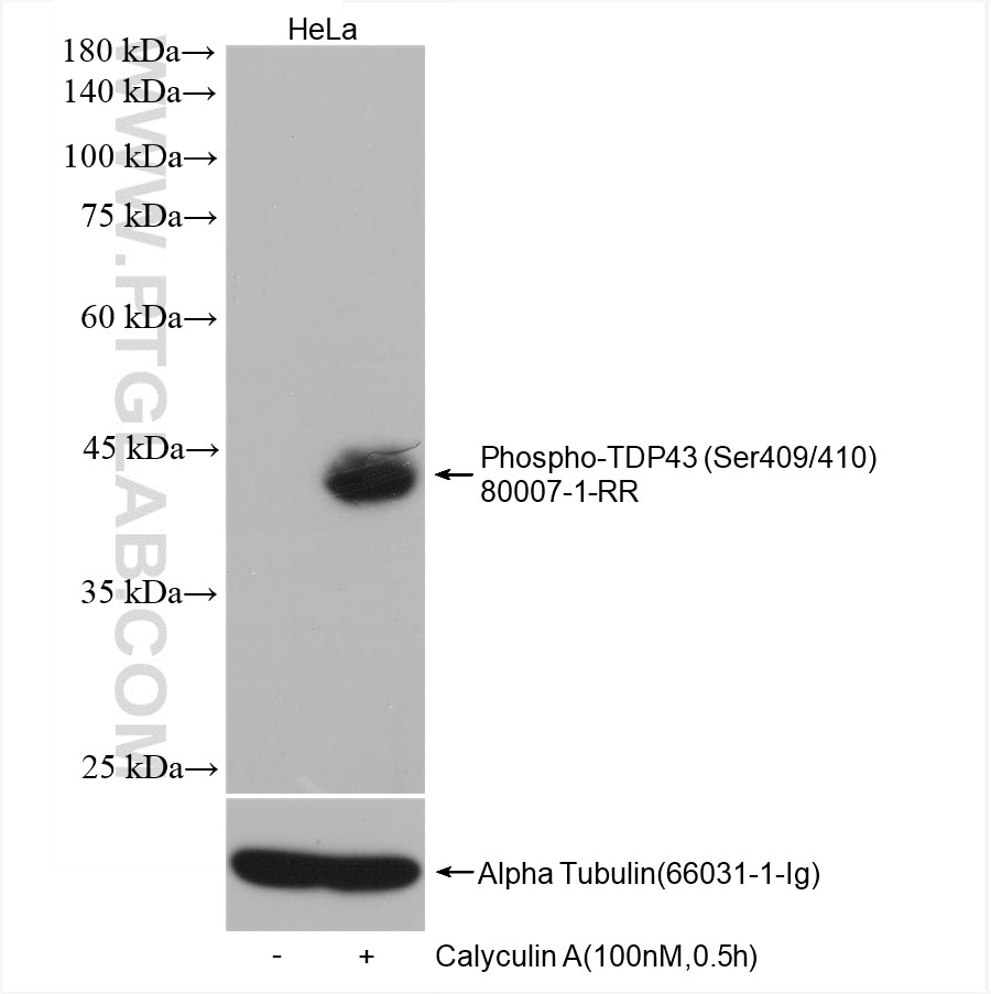
| Positive WB detected in | HeLa cells, Calyculin A treated cells |
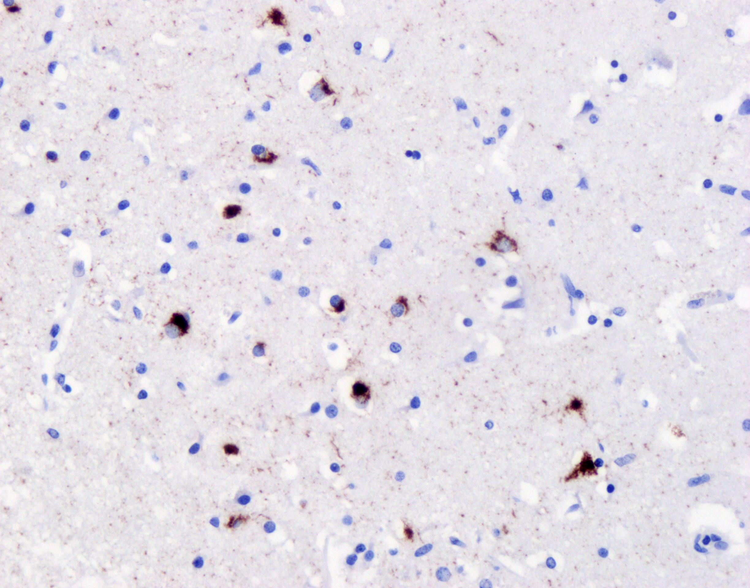
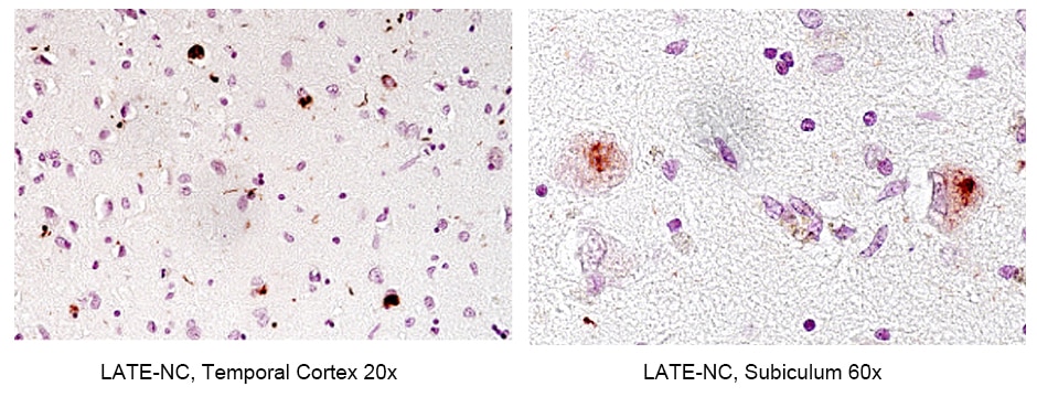
| Positive IHC detected in | frontal cortex of FTLD-TDP type B case, human brain tissue Note: suggested antigen retrieval with TE buffer pH 9.0; (*) Alternatively, antigen retrieval may be performed with citrate buffer pH 6.0 |
Recommended dilution
| Application | Dilution |
|---|---|
| Western Blot (WB) | WB : 1:5000-1:50000 |
| Immunohistochemistry (IHC) | IHC : 1:500-1:2000 |
| It is recommended that this reagent should be titrated in each testing system to obtain optimal results. | |
| Sample-dependent, Check data in validation data gallery. | |
Published Applications
| WB | See 8 publications below |
| IHC | See 8 publications below |
| IF | See 11 publications below |
Product Information
80007-1-RR targets Phospho-TDP43 (Ser409/410) in WB, IHC, IF, ELISA applications and shows reactivity with human, mouse samples.
| Tested Reactivity | human, mouse |
| Cited Reactivity | human, mouse, zebrafish |
| Host / Isotype | Rabbit / IgG |
| Class | Recombinant |
| Type | Antibody |
| Immunogen |
Peptide 相同性解析による交差性が予測される生物種 |
| Full Name | TAR DNA binding protein |
| Calculated molecular weight | 43 kDa |
| Observed molecular weight | 45-50 kDa |
| GenBank accession number | NM_007375 |
| Gene Symbol | TDP-43 |
| Gene ID (NCBI) | 23435 |
| RRID | AB_2882937 |
| Conjugate | Unconjugated |
| Form | |
| Form | Liquid |
| Purification Method | Protein A purification |
| UNIPROT ID | Q13148 |
| Storage Buffer | PBS with 0.02% sodium azide and 50% glycerol{{ptg:BufferTemp}}7.3 |
| Storage Conditions | Store at -20°C. Stable for one year after shipment. Aliquoting is unnecessary for -20oC storage. |
Background Information
Transactivation response (TAR) DNA-binding protein of 43 kDa (also known as TARDBP or TDP-43) was first isolated as a transcriptional inactivator binding to the TAR DNA element of the HIV-1 virus. Neumann et al. (2006) found that a hyperphosphorylated, ubiquitinated, and cleaved form of TARDBP, known as pathologic TDP-43, is the major component of the tau-negative and ubiquitin-positive inclusions that characterize amyotrophic lateral sclerosis (ALS) and the most common pathological subtype of frontotemporal lobar degeneration (FTLD-U). Various forms of TDP-43 exist, including 18-35 kDa of cleaved C-terminal fragments, 45-50 kDa phospho-protein, 55 kDa glycosylated form, 75 kDa hyperphosphorylated form, and 90-300 kDa cross-linked form. (PMID: 17023659,19823856, 21666678, 22193176). 80007-1-RR is a recombinant rabbit monoclonal antibody recognizing TDP-43 only when phosphorylated at 409/410. Immunohistochemical analyses using this antibody only stain the insoluble inclusions in pathologic tissues without normal diffuse nuclear staining.
Protocols
| Product Specific Protocols | |
|---|---|
| WB protocol for Phospho-TDP43 (Ser409/410) antibody 80007-1-RR | Download protocol |
| Standard Protocols | |
|---|---|
| Click here to view our Standard Protocols |
Publications
| Species | Application | Title |
|---|---|---|
J Biol Chem Regulation of TAR DNA binding protein 43 (TDP-43) homeostasis by cytosolic DNA accumulation | ||
Biochim Biophys Acta Mol Basis Dis Phosphorylated TAR DNA-Binding Protein-43: Aggregation and Antibody-Based Inhibition. | ||
bioRxiv Fructose-2,6-bisphosphate restores TDP-43 pathology-driven genome repair deficiency in motor neuron diseases | ||
bioRxiv TDP-43 pathology links innate and adaptive immunity in amyotrophic lateral sclerosis | ||
Acta Neuropathol Commun Endogenous TDP-43 mislocalization in a novel knock-in mouse model reveals DNA repair impairment, inflammation, and neuronal senescence | ||
Ecotoxicol Environ Saf Arsenic exposure activates microglia, inducing neuroinflammation and promoting the occurrence and development of Alzheimer's disease-like neurodegeneration in mice |


