- Featured Product
- KD/KO Validated
Caspase 8/p43/p18 Monoclonal antibody
Caspase 8/p43/p18 Monoclonal Antibody for WB, IHC, IF/ICC, IP, ELISA
Host / Isotype
Mouse / IgG2b
Reactivity
human and More (6)
Applications
WB, IHC, IF/ICC, IP, CoIP, ELISA
Conjugate
Unconjugated
CloneNo.
2B9H8
Cat no : 66093-1-Ig
Synonyms
Validation Data Gallery
Tested Applications
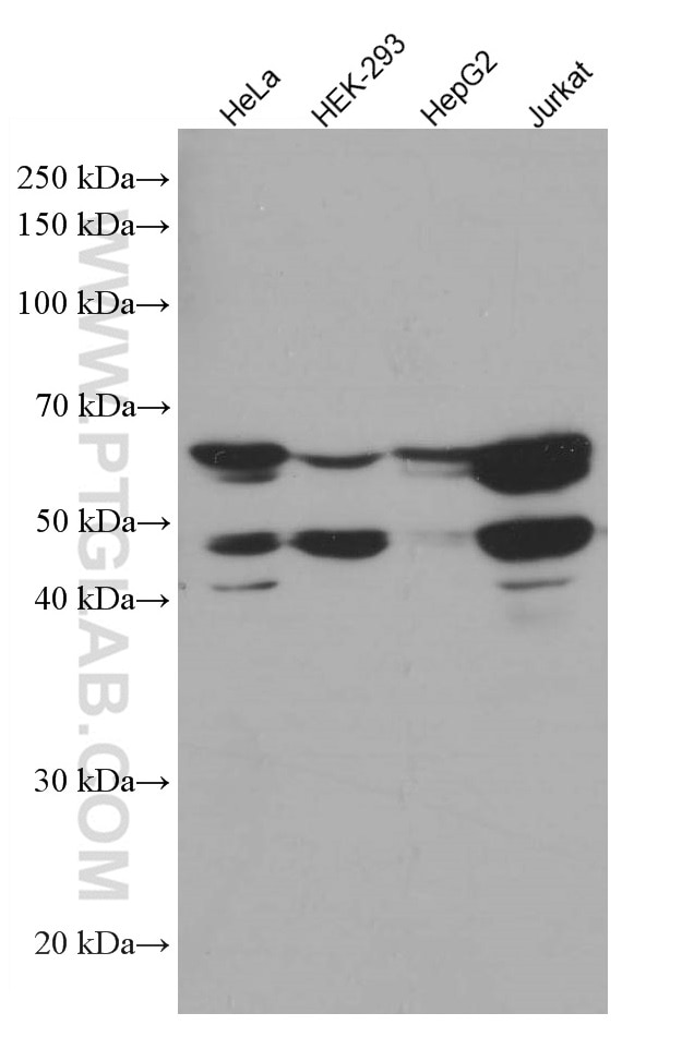
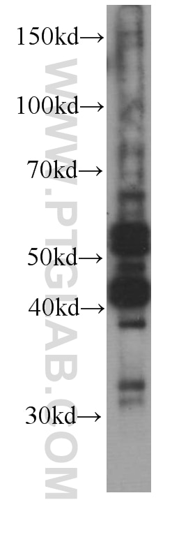
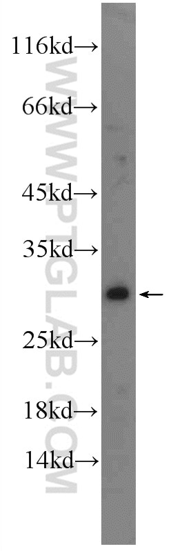
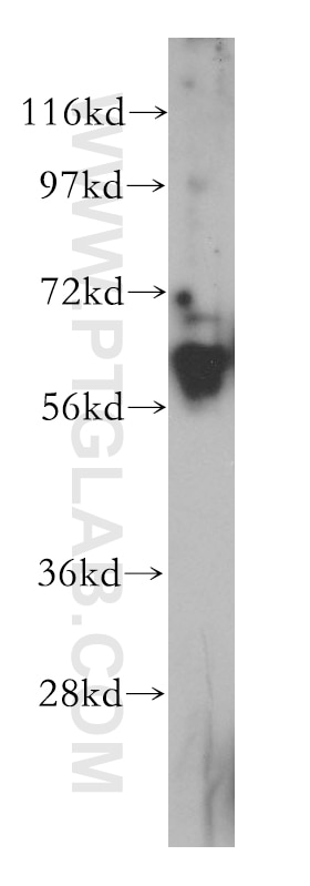
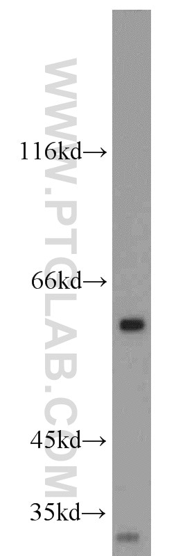
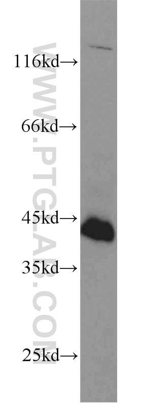
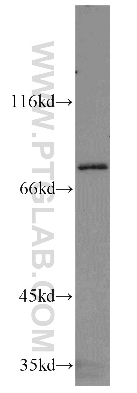
| Positive WB detected in | Jurkat cells, HeLa cells, HepG2 cells, HEK-293 cells |
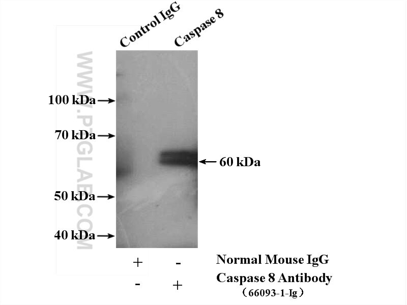
| Positive IP detected in | HepG2 cells |
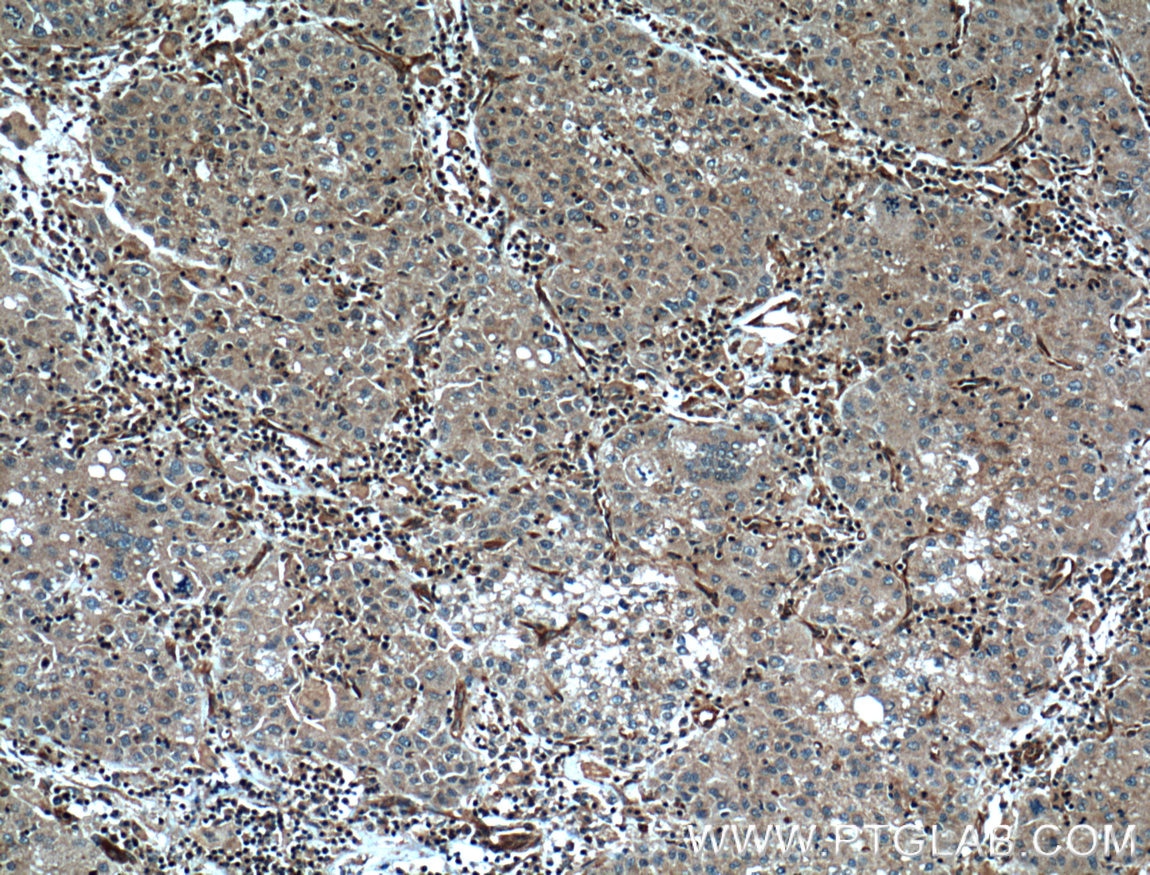
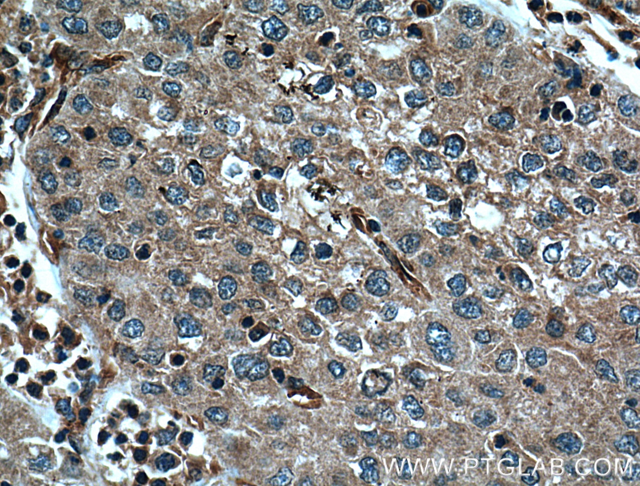
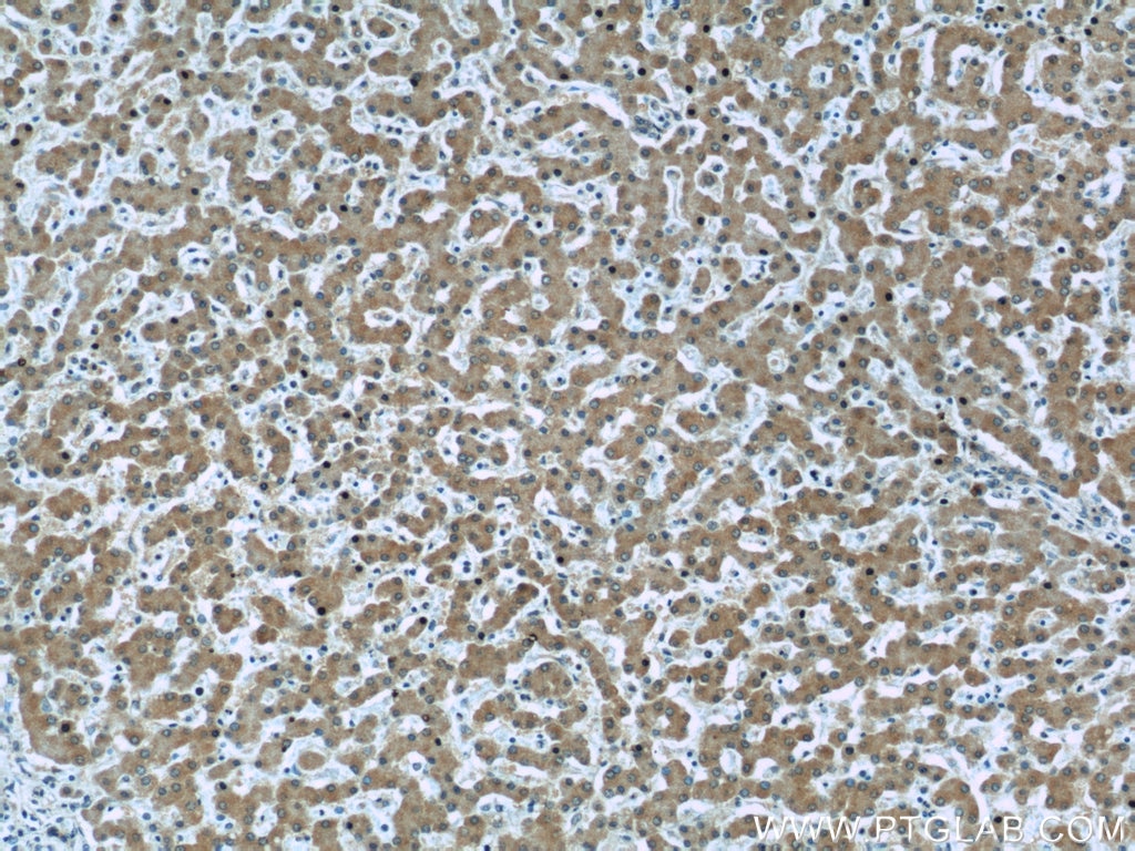
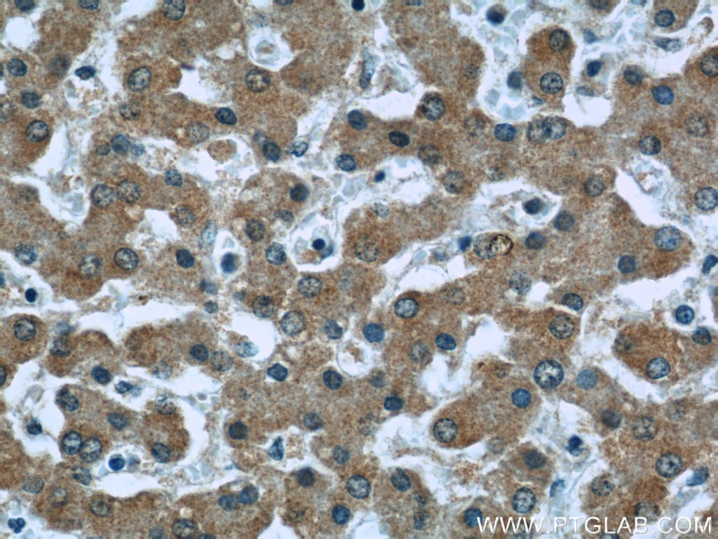
| Positive IHC detected in | human liver cancer tissue, human liver tissue Note: suggested antigen retrieval with TE buffer pH 9.0; (*) Alternatively, antigen retrieval may be performed with citrate buffer pH 6.0 |
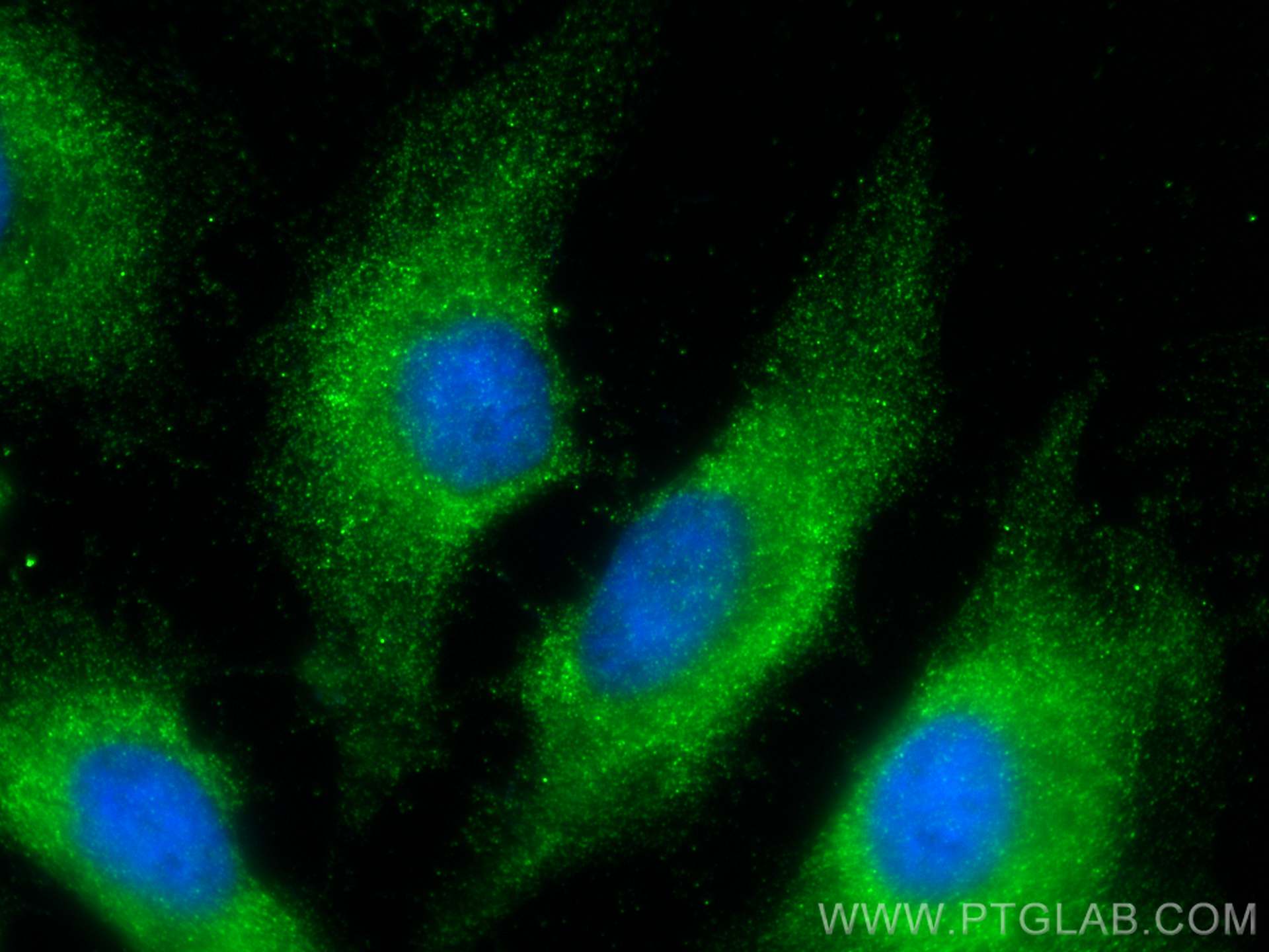
| Positive IF/ICC detected in | HeLa cells |
Recommended dilution
| Application | Dilution |
|---|---|
| Western Blot (WB) | WB : 1:2000-1:10000 |
| Immunoprecipitation (IP) | IP : 0.5-4.0 ug for 1.0-3.0 mg of total protein lysate |
| Immunohistochemistry (IHC) | IHC : 1:100-1:400 |
| Immunofluorescence (IF)/ICC | IF/ICC : 1:400-1:1600 |
| It is recommended that this reagent should be titrated in each testing system to obtain optimal results. | |
| Sample-dependent, Check data in validation data gallery. | |
Published Applications
| KD/KO | See 2 publications below |
| WB | See 71 publications below |
| IHC | See 5 publications below |
| IF | See 6 publications below |
| IP | See 2 publications below |
| ELISA | See 1 publications below |
| CoIP | See 1 publications below |
Product Information
66093-1-Ig targets Caspase 8/p43/p18 in WB, IHC, IF/ICC, IP, CoIP, ELISA applications and shows reactivity with human samples.
| Tested Reactivity | human |
| Cited Reactivity | human, mouse, rat, pig, monkey, chicken, sheep |
| Host / Isotype | Mouse / IgG2b |
| Class | Monoclonal |
| Type | Antibody |
| Immunogen | Caspase 8/p43/p18 fusion protein Ag20524 相同性解析による交差性が予測される生物種 |
| Full Name | caspase 8, apoptosis-related cysteine peptidase |
| Calculated molecular weight | 538 aa, 62 kDa |
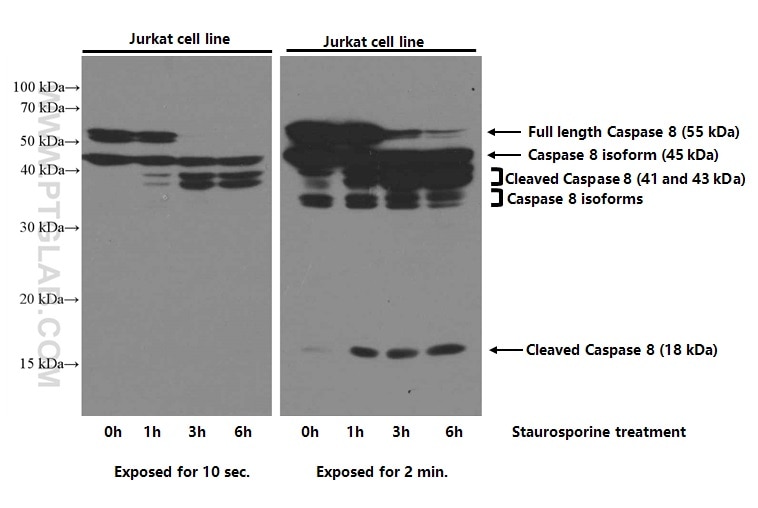
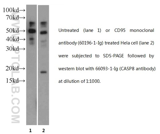
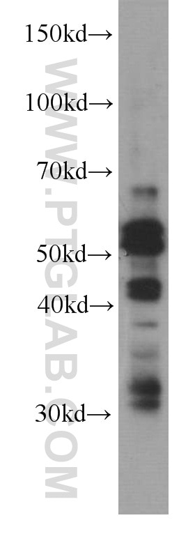
| Observed molecular weight | 53-57 kDa, 32-45 kDa, 18 kDa |
| GenBank accession number | BC028223 |
| Gene symbol | Caspase 8 |
| Gene ID (NCBI) | 841 |
| RRID | AB_11232214 |
| Conjugate | Unconjugated |
| Form | Liquid |
| Purification Method | Protein A purification |
| Storage Buffer | PBS with 0.02% sodium azide and 50% glycerol pH 7.3. |
| Storage Conditions | Store at -20°C. Stable for one year after shipment. Aliquoting is unnecessary for -20oC storage. |
Background Information
Caspase 8, also named as MCH5, MACH, FLICE, and CAP4, belongs to the peptidase C14A family. It may participate in the GZMB apoptotic pathways and functions as an upstream regulator in a-bisabolol-induced apoptosis. Caspase 8 catalyzes an essential intermediate step in the ubiquitination and proteasome-mediated degradation of IRF3 (PMID:21816816). It may control diabetic embryopathy-associated apoptosis via regulation of the Bid-stimulated mitochondrion/caspase-9 pathway (PMID:19194987). Caspase 8 is expressed as nine isoforms by alternative splicing with the molecular mass from 26 kDa to 62 kDa. This antibody can recognize pro- and cleaved-caspase 8.
Protocols
| Product Specific Protocols | |
|---|---|
| WB protocol for Caspase 8/p43/p18 antibody 66093-1-Ig | Download protocol |
| IHC protocol for Caspase 8/p43/p18 antibody 66093-1-Ig | Download protocol |
| IF protocol for Caspase 8/p43/p18 antibody 66093-1-Ig | Download protocol |
| IP protocol for Caspase 8/p43/p18 antibody 66093-1-Ig | Download protocol |
| Standard Protocols | |
|---|---|
| Click here to view our Standard Protocols |
Publications
| Species | Application | Title |
|---|---|---|
Cancer Commun (Lond) Targeting autophagy overcomes cancer-intrinsic resistance to CAR-T immunotherapy in B-cell malignancies | ||
Mol Cell Estrogen-Related Hormones Induce Apoptosis by Stabilizing Schlafen-12 Protein Turnover.
| ||
Acta Biomater TRAIL-modified, doxorubicin-embedded periodic mesoporous organosilica nanoparticles for targeted drug delivery and efficient antitumor immunotherapy. | ||
J Pineal Res Melatonin alleviates weanling stress in mice: involvement of intestinal microbiota. | ||
Chem Sci Integration of exonuclease III-powered three-dimensional DNA walker with single-molecule detection for multiple initiator caspases assay. | ||
