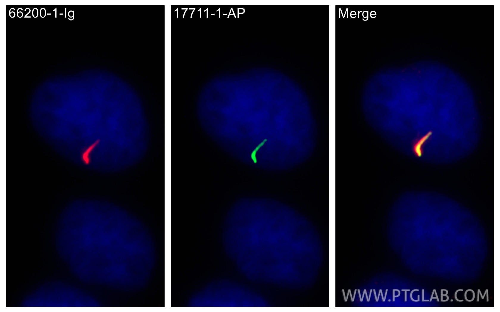
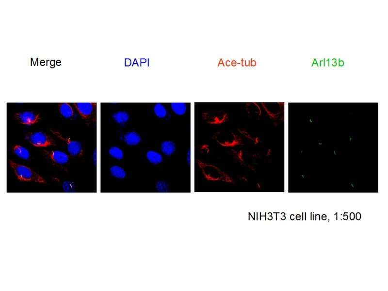
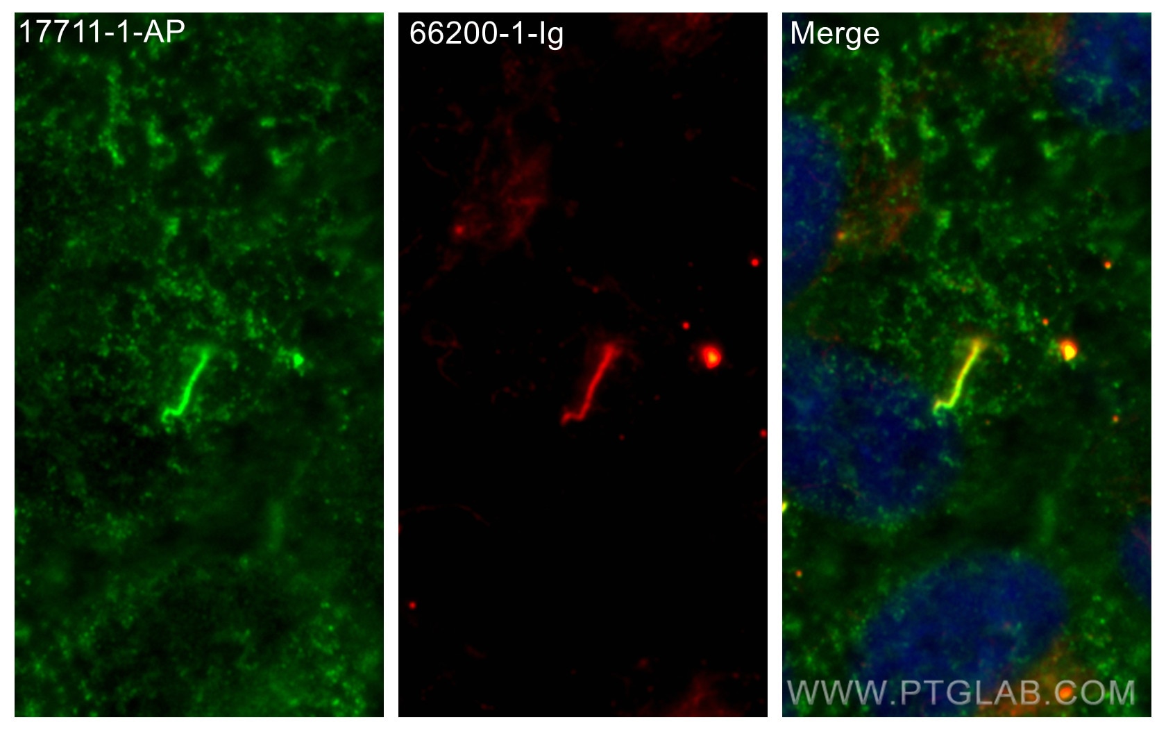
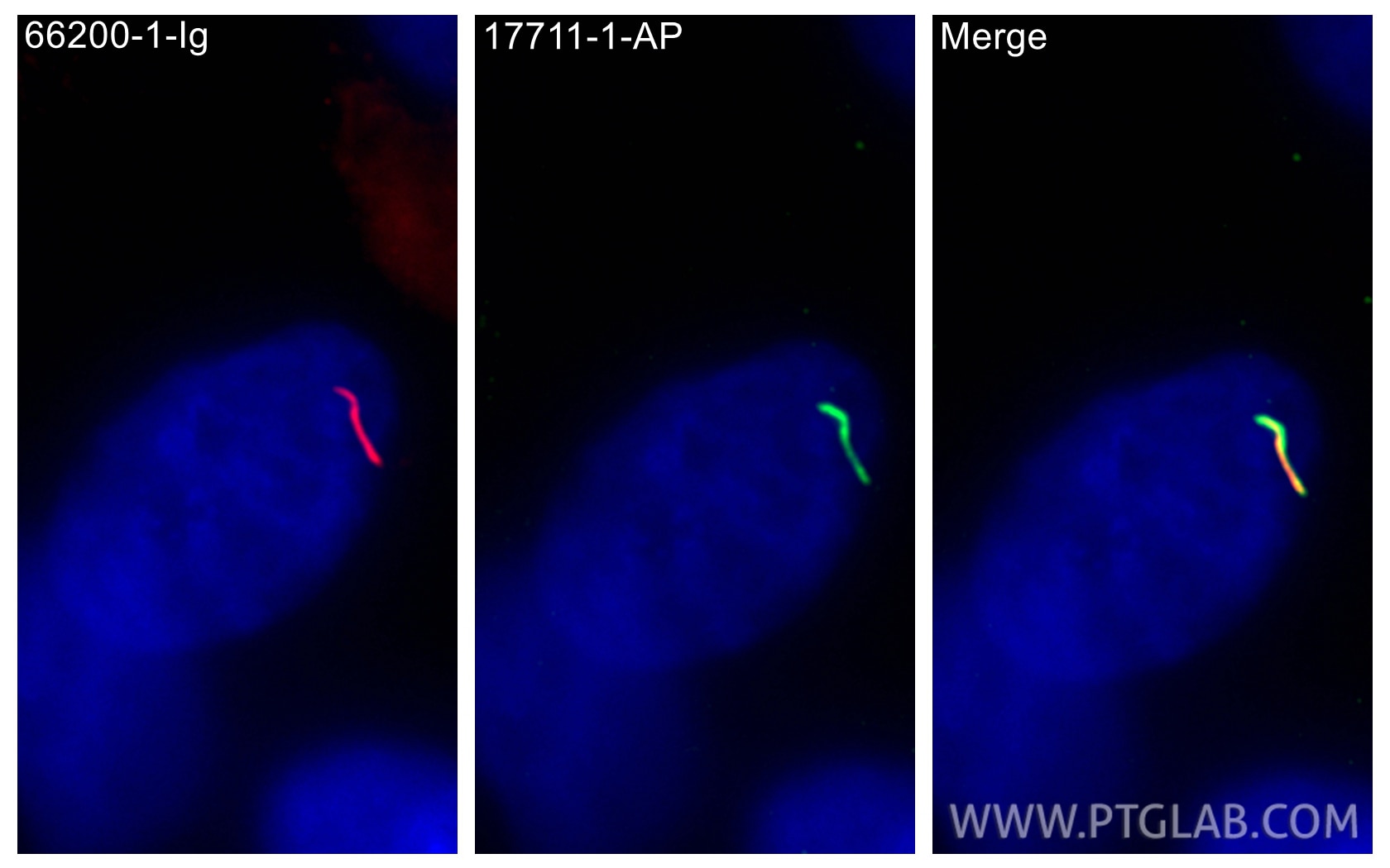
Validation Data Gallery
Tested Applications
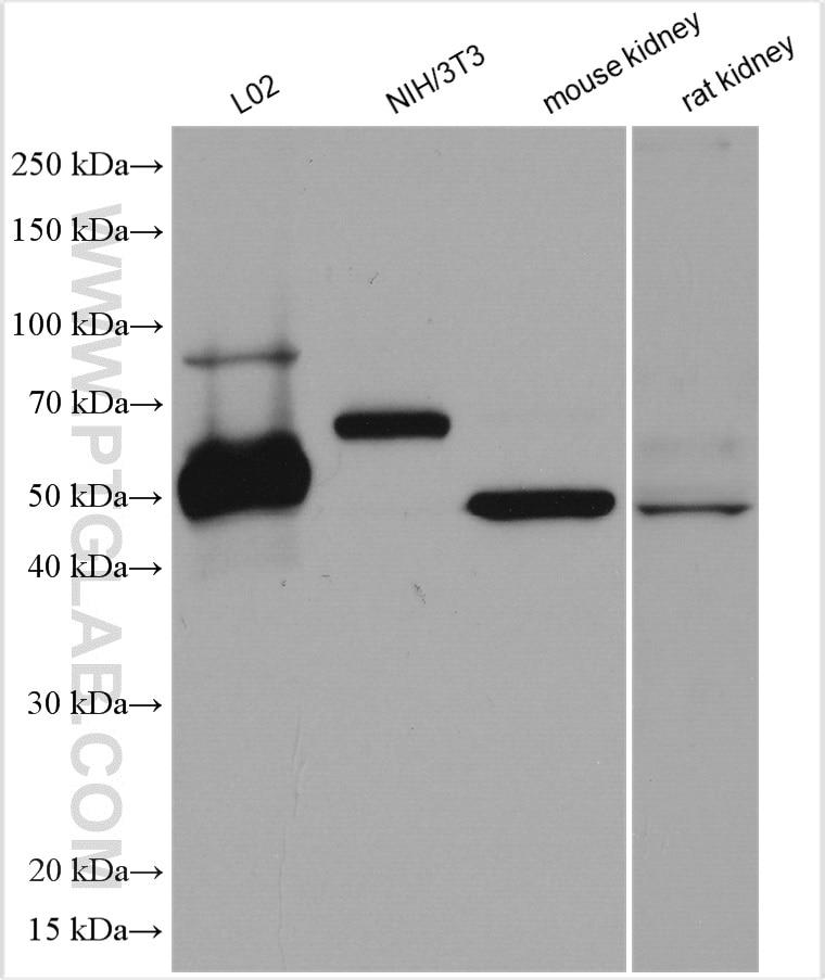
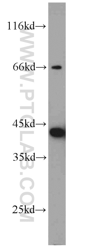
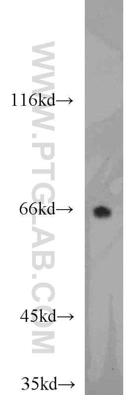
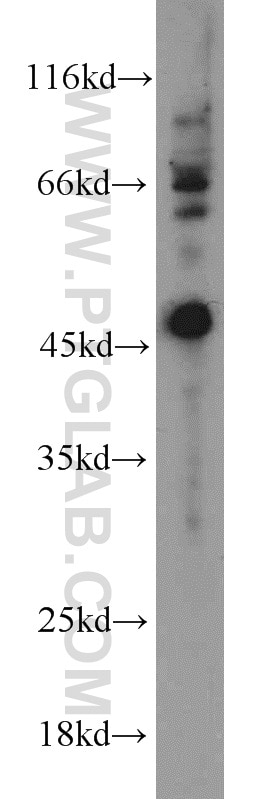
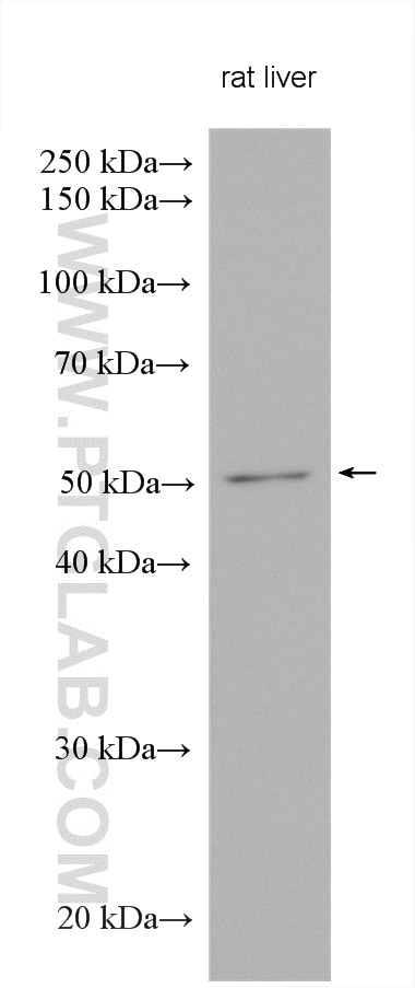
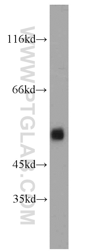
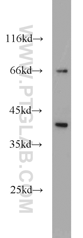
| Positive WB detected in | L02 cells, mouse brain tissue, mouse kidney tissue, mouse liver tissue, NIH/3T3 cells, rat liver tissue, rat kidney tissue |
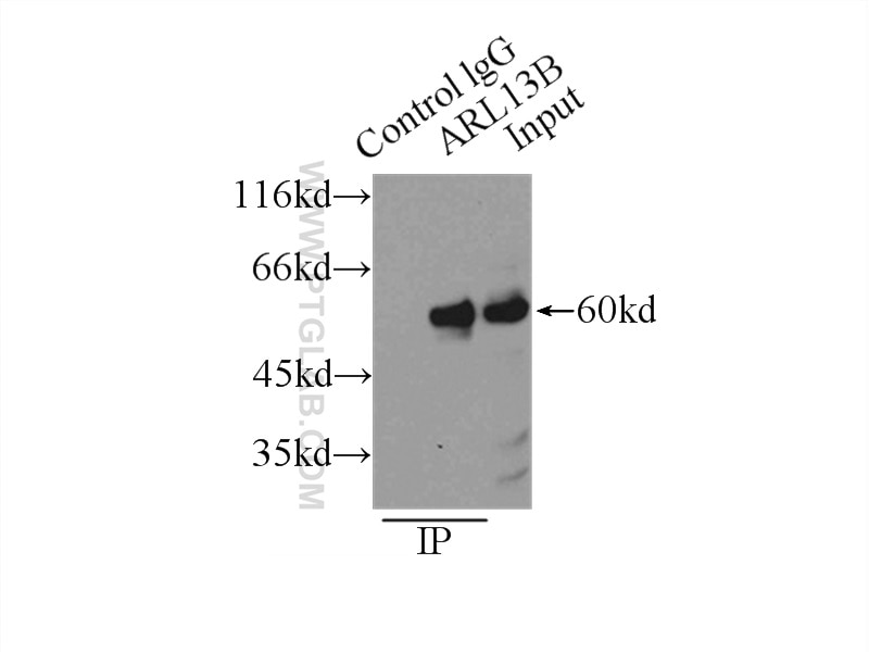
| Positive IP detected in | L02 cells |
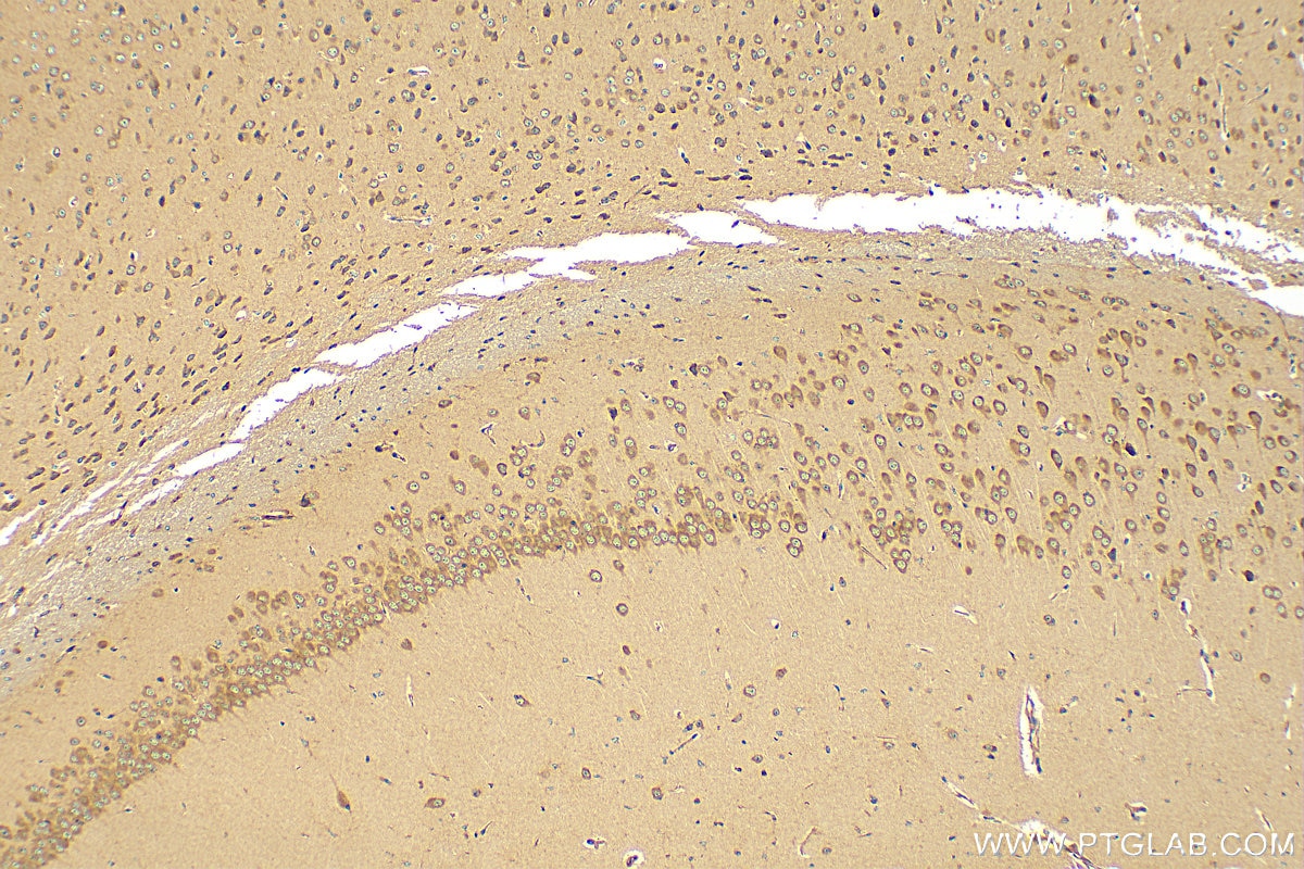
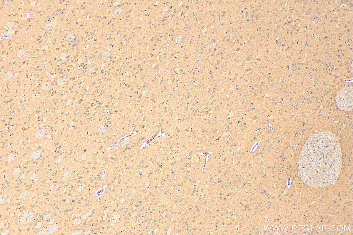
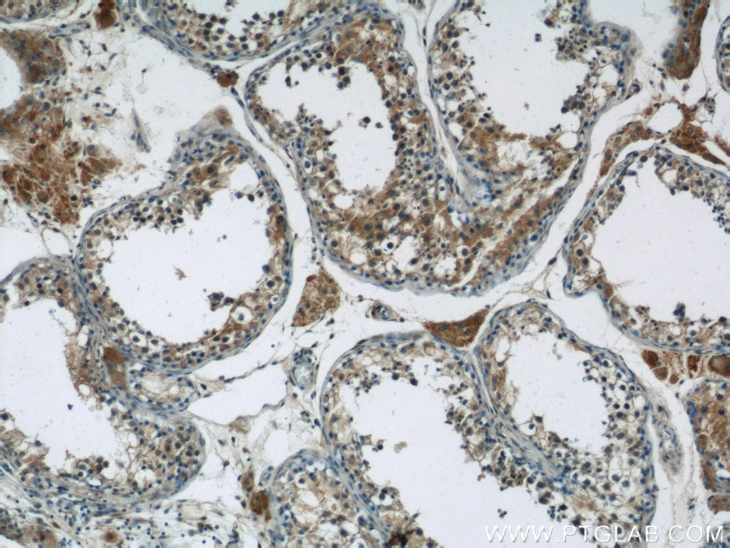
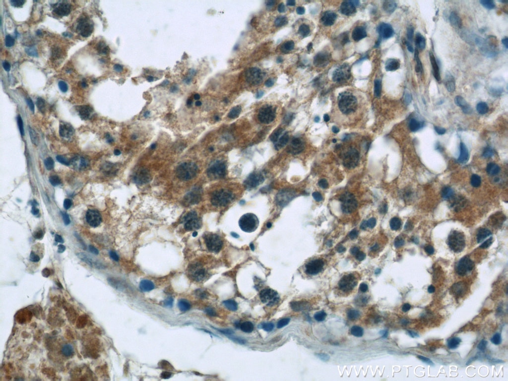
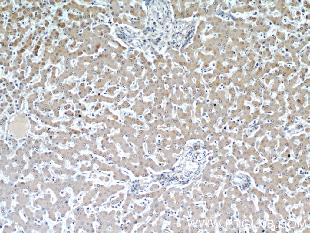
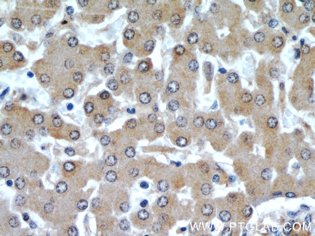
| Positive IHC detected in | mouse brain tissue, human liver tissue, human testis tissue Note: suggested antigen retrieval with TE buffer pH 9.0; (*) Alternatively, antigen retrieval may be performed with citrate buffer pH 6.0 |
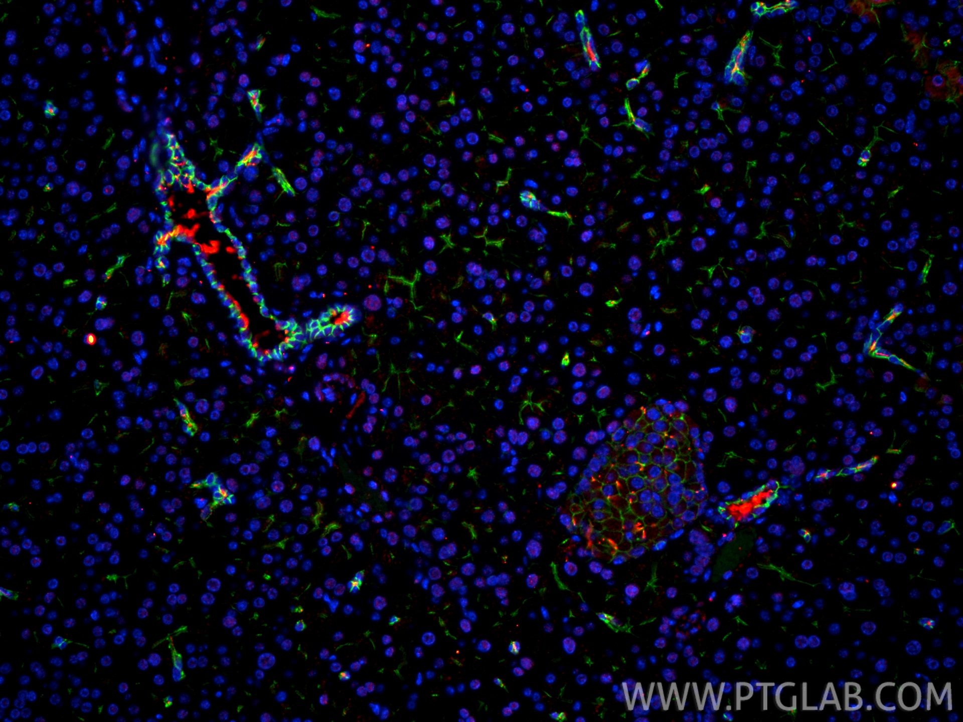
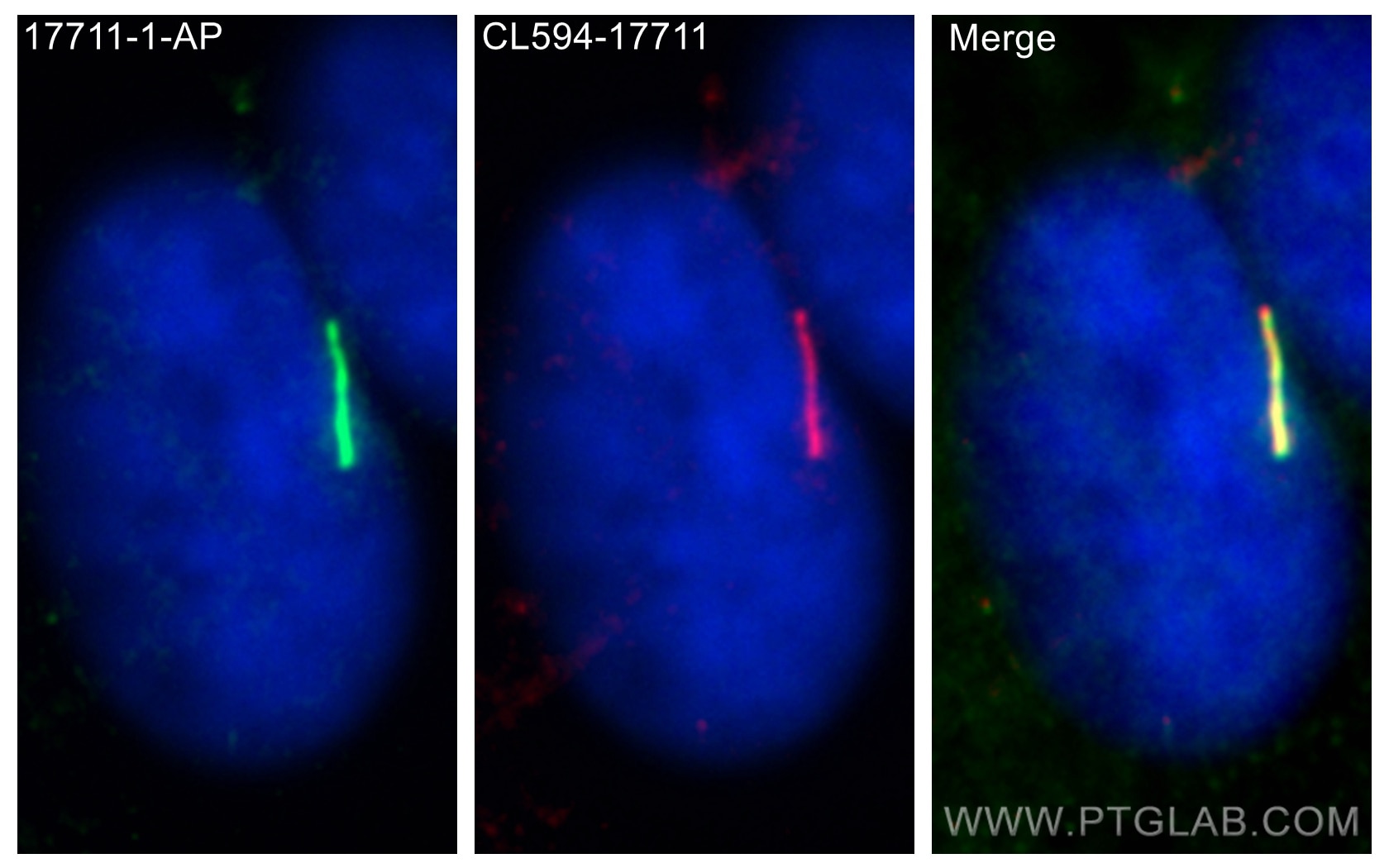
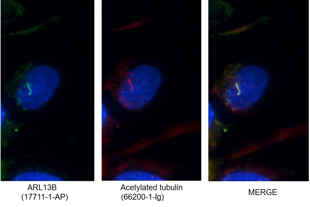
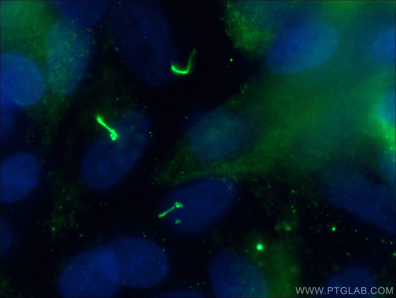
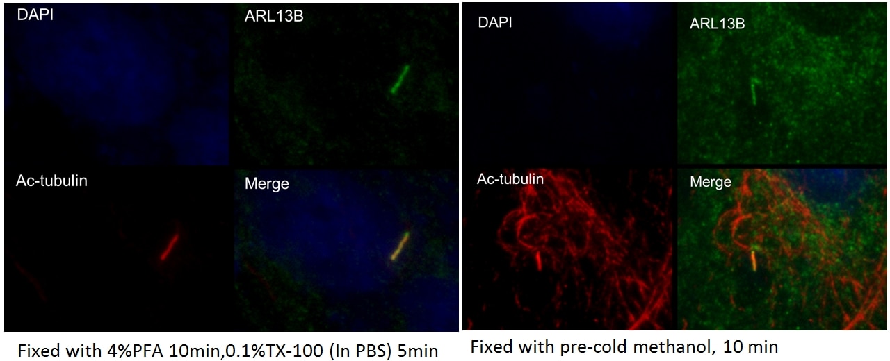
| Positive IF/ICC detected in | hTERT-RPE1 cells, NIH3T3 cells, MDCK cells |
Recommended dilution
| Application | Dilution |
|---|---|
| Western Blot (WB) | WB : 1:1000-1:6000 |
| Immunoprecipitation (IP) | IP : 0.5-4.0 ug for 1.0-3.0 mg of total protein lysate |
| Immunohistochemistry (IHC) | IHC : 1:50-1:500 |
| Immunofluorescence (IF)/ICC | IF/ICC : 1:200-1:800 |
| It is recommended that this reagent should be titrated in each testing system to obtain optimal results. | |
| Sample-dependent, Check data in validation data gallery. | |
Published Applications
| KD/KO | See 13 publications below |
| WB | See 99 publications below |
| IHC | See 39 publications below |
| IF | See 771 publications below |
| IP | See 3 publications below |
Product Information
17711-1-AP targets ARL13B in WB, IHC, IF/ICC, IP, ELISA applications and shows reactivity with human, mouse, rat, canine samples.
| Tested Reactivity | human, mouse, rat, canine |
| Cited Reactivity | human, mouse, rat, pig, canine, monkey, chicken, zebrafish, sheep, xenopus |
| Host / Isotype | Rabbit / IgG |
| Class | Polyclonal |
| Type | Antibody |
| Immunogen |
CatNo: Ag12015 Product name: Recombinant human ARL13B protein Source: e coli.-derived, PGEX-4T Tag: GST Domain: 1-321 aa of BC094725 Sequence: MFSLMASCCGWFKRWREPVRLANKQDKEGALGEADVIECLSLEKLVNEHKCLCQIEPCSAISGYGKKIDKSIKKGLYWLLHVIARDFDALNERIQKETTEQRALEEQEKQERAERVRKLREERKQNEQEQAELDGTSGLAELDPEPTNPFQPIASVIIENEGKLEREKKNQKMEKDSDGCHLKHKMEHEQIETQGQVNHNGQKNNEFGLVENYKEALTQQLKNEDETDRPSLESANGKKKTKKLRMKRNHRVEPLNIDDCAPESPTPPPPPPPVGWGTPKVTRLPKLEPLGETHHNDFYRKPLPPLAVPQRPNSDAHDVIS 相同性解析による交差性が予測される生物種 |
| Full Name | ADP-ribosylation factor-like 13B |
| Calculated molecular weight | 48 kDa |
| Observed molecular weight | 40-48 kDa, 60 kDa |
| GenBank accession number | BC094725 |
| Gene Symbol | ARL13B |
| Gene ID (NCBI) | 200894 |
| RRID | AB_2060867 |
| Conjugate | Unconjugated |
| Form | |
| Form | Liquid |
| Purification Method | Antigen affinity purification |
| UNIPROT ID | Q3SXY8 |
| Storage Buffer | PBS with 0.02% sodium azide and 50% glycerol{{ptg:BufferTemp}}7.3 |
| Storage Conditions | Store at -20°C. Stable for one year after shipment. Aliquoting is unnecessary for -20oC storage. |
Background Information
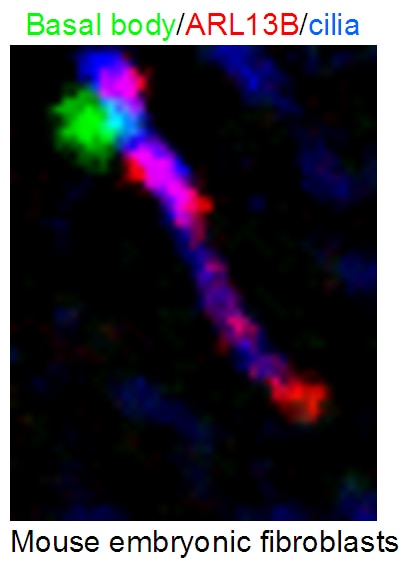
ARL13B, also named ARL2L1, is the Ras superfamily's small ciliary G protein. Localized in the cilia, it is required for cilium biogenesis and sonic hedgehog signaling. Defects in ARL13B cause Joubert syndrome (JS), an autosomal recessive disorder characterized by a distinctive cerebellar malformation (PMID: 19906870). This antibody detects two specific bands at 60 kDa and 48 kDa. Arl13b is predicted to be a 48 kDa protein, and the 60 kDa band will likely represent a modified form of Arl13b. ARL13B can be used to mark the cilia (PMID:22072986).
Protocols
| Product Specific Protocols | |
|---|---|
| IF protocol for ARL13B antibody 17711-1-AP | Download protocol |
| IHC protocol for ARL13B antibody 17711-1-AP | Download protocol |
| IP protocol for ARL13B antibody 17711-1-AP | Download protocol |
| WB protocol for ARL13B antibody 17711-1-AP | Download protocol |
| Standard Protocols | |
|---|---|
| Click here to view our Standard Protocols |
Publications
| Species | Application | Title |
|---|---|---|
Nat Biotechnol Guided self-organization and cortical plate formation in human brain organoids. | ||
Nature Quantitative lineage analysis identifies a hepato-pancreato-biliary progenitor niche. | ||
Science Control of meiotic chromosomal bouquet and germ cell morphogenesis by the zygotene cilium. | ||
Nature A slow-cycling LGR5 tumour population mediates basal cell carcinoma relapse after therapy. | ||
Nature Aspm knockout ferret reveals an evolutionary mechanism governing cerebral cortical size. |

