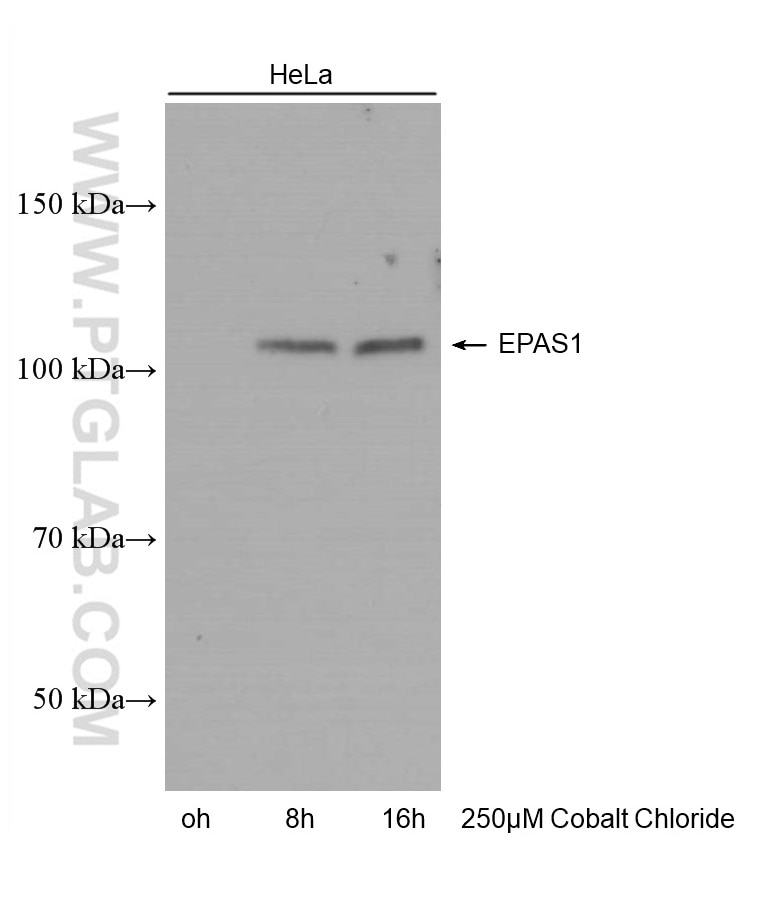
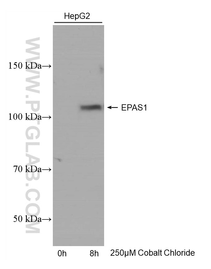
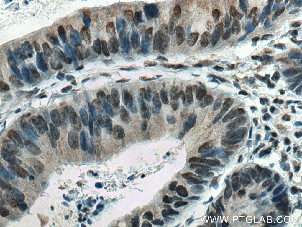
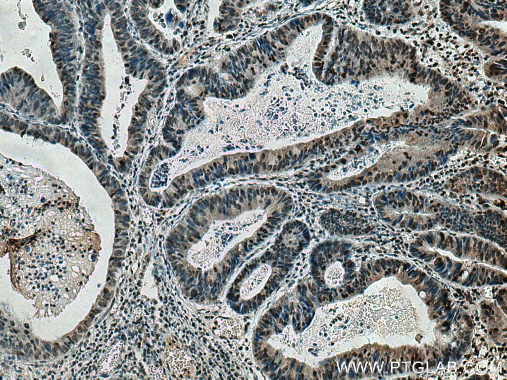
Validation Data Gallery
Tested Applications
| Positive WB detected in | HeLa cells, HepG2 cells |
| Positive IHC detected in | human colon cancer tissue Note: suggested antigen retrieval with TE buffer pH 9.0; (*) Alternatively, antigen retrieval may be performed with citrate buffer pH 6.0 |
Recommended dilution
| Application | Dilution |
|---|---|
| Western Blot (WB) | WB : 1:1000-1:5000 |
| Immunohistochemistry (IHC) | IHC : 1:250-1:1000 |
| It is recommended that this reagent should be titrated in each testing system to obtain optimal results. | |
| Sample-dependent, Check data in validation data gallery. | |
Published Applications
| KD/KO | See 2 publications below |
| WB | See 7 publications below |
| IHC | See 1 publications below |
| IF | See 2 publications below |
Product Information
66731-1-Ig targets HIF2α/EPAS1 in WB, IHC, IF, ELISA applications and shows reactivity with Human samples.
| Tested Reactivity | Human |
| Cited Reactivity | human, bovine |
| Host / Isotype | Mouse / IgG2a |
| Class | Monoclonal |
| Type | Antibody |
| Immunogen | HIF2α/EPAS1 fusion protein Ag24886 相同性解析による交差性が予測される生物種 |
| Full Name | endothelial PAS domain protein 1 |
| Calculated molecular weight | 96 kDa |
| Observed molecular weight | 120 kDa |
| GenBank accession number | BC051338 |
| Gene Symbol | HIF2α |
| Gene ID (NCBI) | 2034 |
| RRID | AB_2882081 |
| Conjugate | Unconjugated |
| Form | Liquid |
| Purification Method | Protein A purification |
| UNIPROT ID | Q99814 |
| Storage Buffer | PBS with 0.02% sodium azide and 50% glycerol{{ptg:BufferTemp}}7.3 |
| Storage Conditions | Store at -20°C. Stable for one year after shipment. Aliquoting is unnecessary for -20oC storage. |
Background Information
Endothelial PAS domain-containing protein 1 (EPAS1), also known as Hypoxia-inducible factor 2-alpha (HIF2-alpha,HIF2A), is a transcription factor involved in the induction of oxygen regulated genes. Binds to core DNA sequence 5'-[AG]CGTG-3' within the hypoxia response element (HRE) of target gene promoters. Regulates the vascular endothelial growth factor (VEGF) expression and seems to be implicated in the development of blood vessels and the tubular system of lung. May also play a role in the formation of the endothelium that gives rise to the blood brain barrier. Potent activator of the Tie-2 tyrosine kinase expression. Activation seems to require recruitment of transcriptional coactivators such as CREBPB and probably EP300. Interaction with redox regulatory protein APEX seems to activate CTAD. EPAS1 is expressed in most tissues, with highest levels in placenta, lung and heart. Selectively expressed in endothelial cells.
Protocols
| Product Specific Protocols | |
|---|---|
| WB protocol for HIF2α/EPAS1 antibody 66731-1-Ig | Download protocol |
| IHC protocol for HIF2α/EPAS1 antibody 66731-1-Ig | Download protocol |
| Standard Protocols | |
|---|---|
| Click here to view our Standard Protocols |
Publications
| Species | Application | Title |
|---|---|---|
Nat Commun Extravillous trophoblast cell lineage development is associated with active remodeling of the chromatin landscape
| ||
Cell Mol Gastroenterol Hepatol A Mitochondrial DNA Variant Elevates the Risk of Gallstone Disease by Altering Mitochondrial Function. | ||
Front Vet Sci miRNA-150_R-1 mediates the HIF-1/ErbB signaling pathway to regulate the adhesion of endometrial epithelial cells in cows experiencing retained placenta | ||
bioRxiv NOTUM-MEDIATED WNT SILENCING DRIVES EXTRAVILLOUS TROPHOBLAST CELL LINEAGE DEVELOPMENT | ||
Oncoimmunology HIF2A mediates lineage transition to aggressive phenotype of cancer-associated fibroblasts in lung cancer brain metastasis | ||
Front Biosci (Landmark Ed) The PLCG2 Inhibits Tumor Progression and Mediates Angiogenesis by VEGF Signaling Pathway in Clear Cell Renal Cell Carcinoma |


