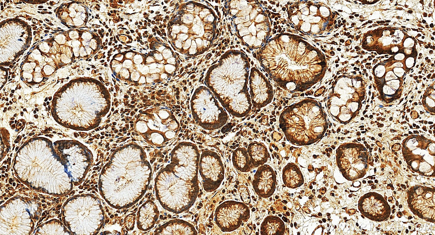
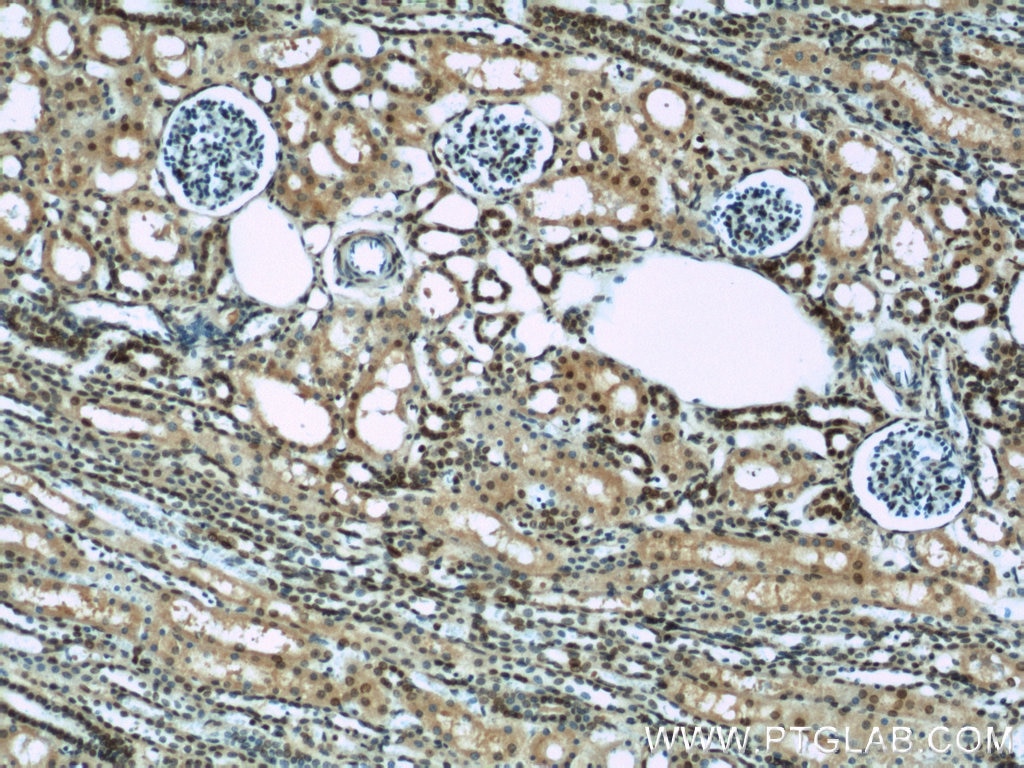
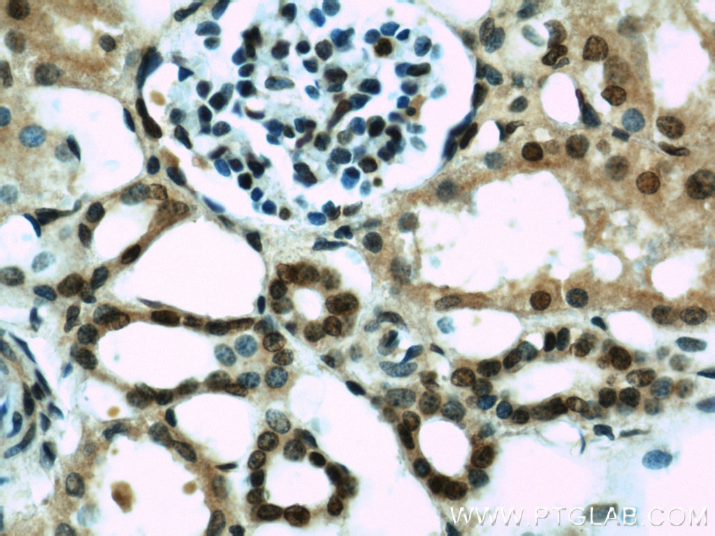
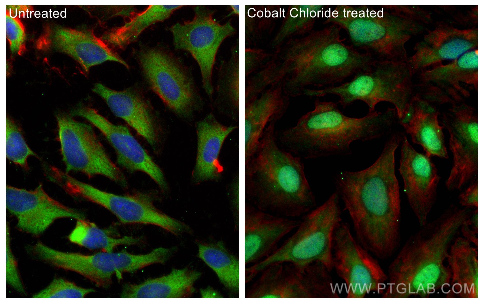
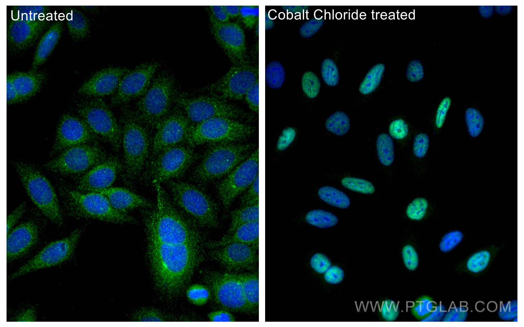
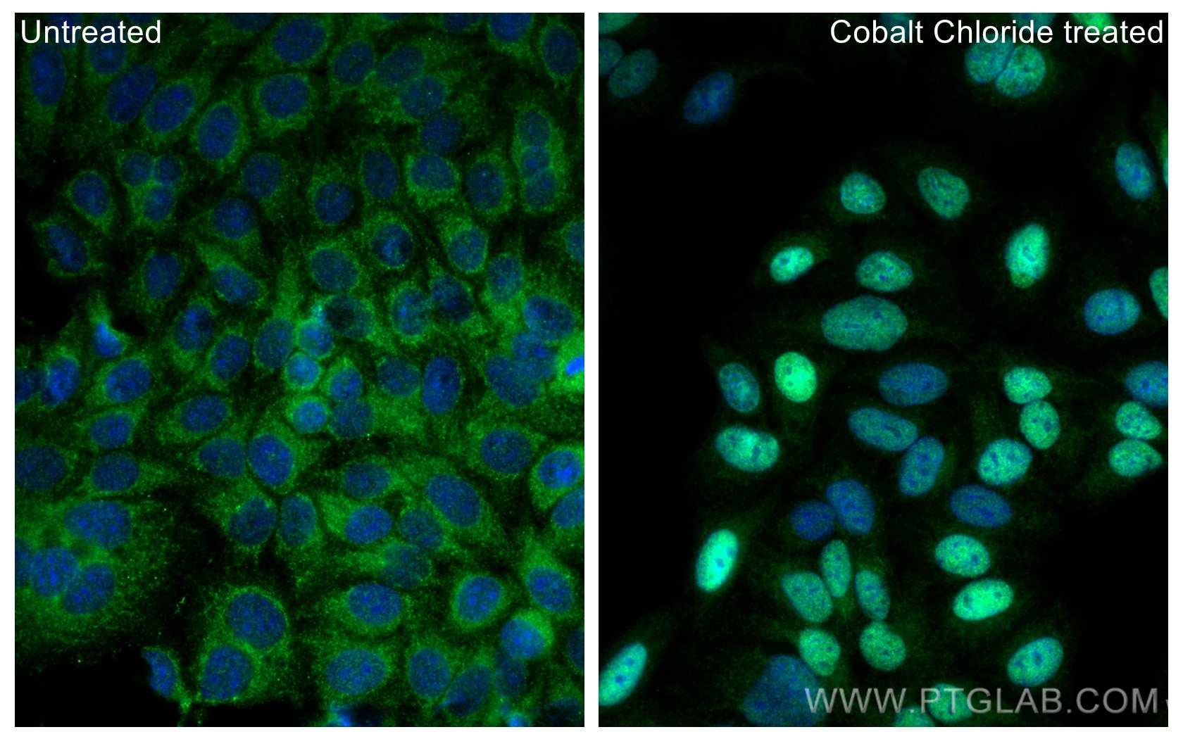
Validation Data Gallery
Tested Applications
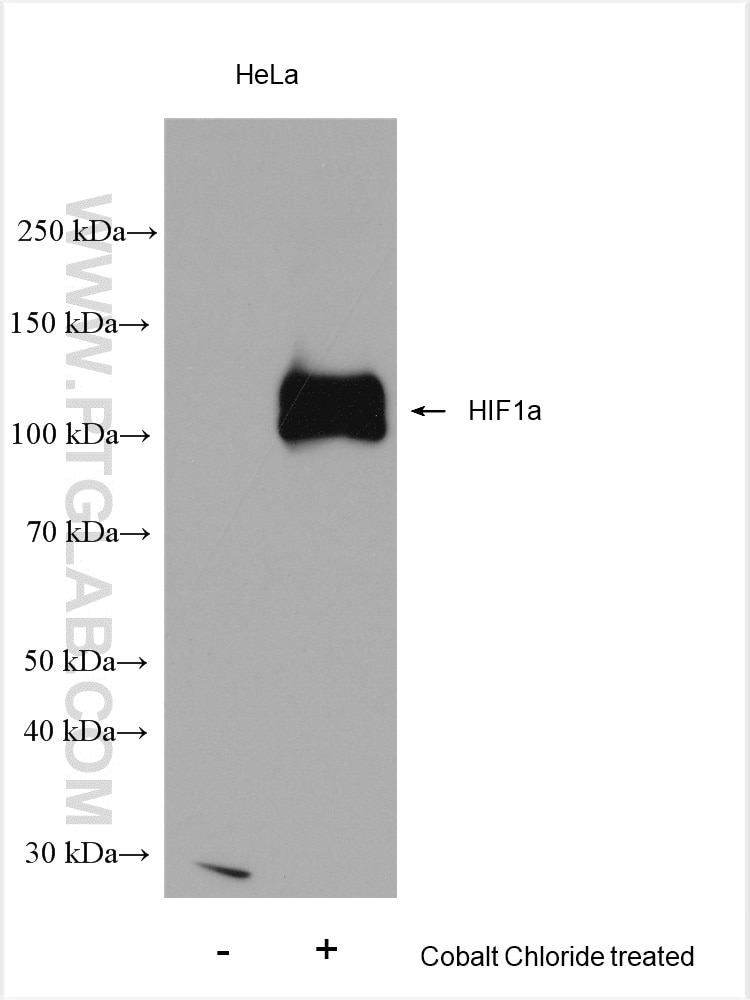
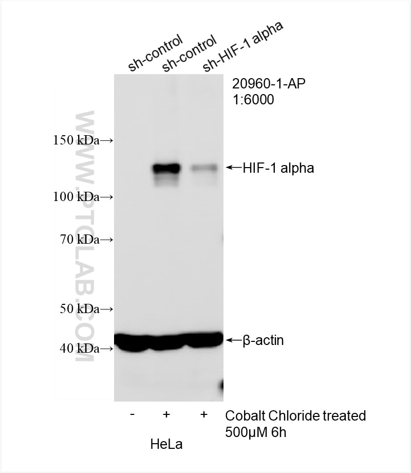
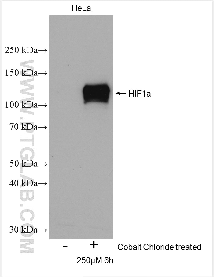
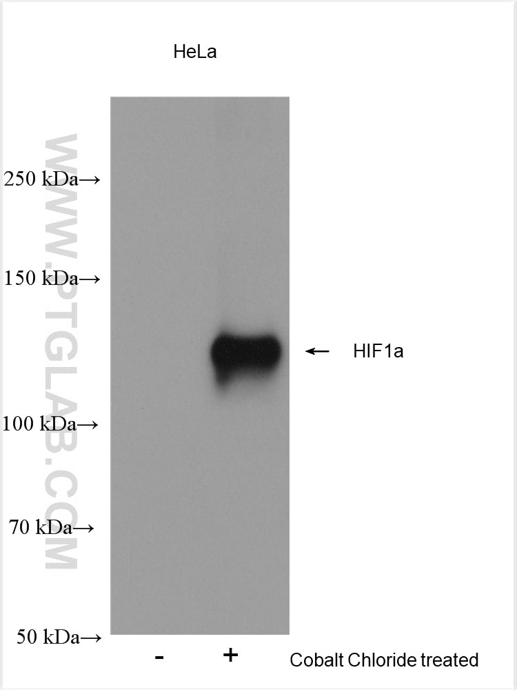
| Positive WB detected in | Cobalt Chloride treated HeLa cells |
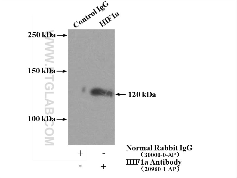
| Positive IP detected in | HeLa cells |
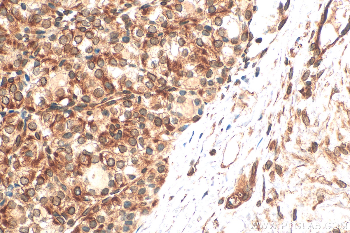
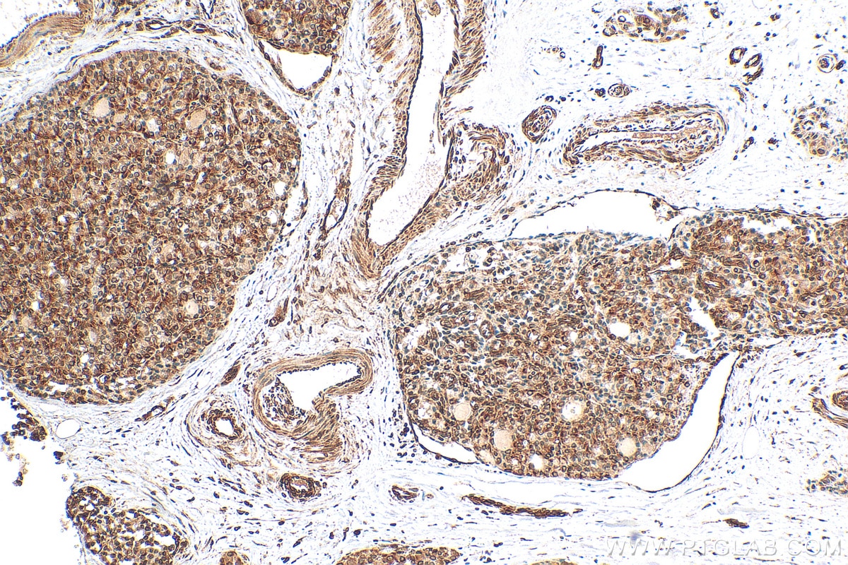
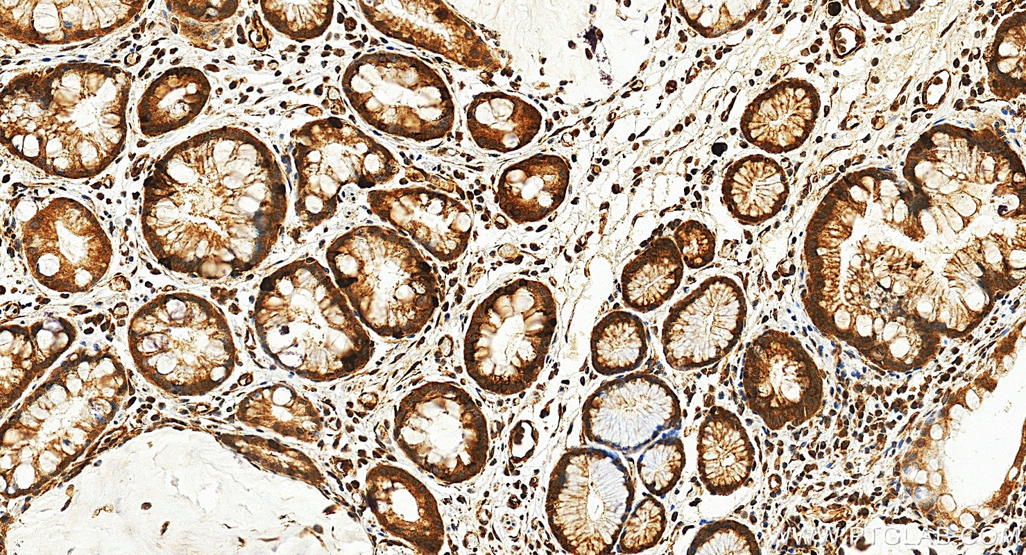
| Positive IHC detected in | human thyroid cancer tissue, human kidney tissue, human stomach cancer tissue Note: suggested antigen retrieval with TE buffer pH 9.0; (*) Alternatively, antigen retrieval may be performed with citrate buffer pH 6.0 |
| Positive IF/ICC detected in | Cobalt Chloride treated HeLa cells, Cobalt Chloride treated HepG2 cells |
Samples need to be treated with hypoxia stimulation.
Recommended dilution
| Application | Dilution |
|---|---|
| Western Blot (WB) | WB : 1:2000-1:12000 |
| Immunoprecipitation (IP) | IP : 0.5-4.0 ug for 1.0-3.0 mg of total protein lysate |
| Immunohistochemistry (IHC) | IHC : 1:50-1:500 |
| Immunofluorescence (IF)/ICC | IF/ICC : 1:200-1:800 |
| It is recommended that this reagent should be titrated in each testing system to obtain optimal results. | |
| Sample-dependent, Check data in validation data gallery. | |
Product Information
20960-1-AP targets HIF-1 alpha in WB, IHC, IF/ICC, IP, CoIP, ChIP, RIP, ELISA applications and shows reactivity with human samples.
| Tested Reactivity | human |
| Cited Reactivity | human, pig, chicken, goat |
| Host / Isotype | Rabbit / IgG |
| Class | Polyclonal |
| Type | Antibody |
| Immunogen |
CatNo: Ag15198 Product name: Recombinant human HIF1a protein Source: e coli.-derived, PGEX-4T Tag: GST Domain: 574-799 aa of BC012527 Sequence: LRSFDQLSPLESSSASPESASPQSTVTVFQQTQIQEPTANATTTTATTDELKTVTKDRMEDIKILIASPSPTHIHKETTSATSSPYRDTQSRTASPNRAGKGVIEQTEKSHPRSPNVLSVALSQRTTVPEEELNPKILALQNAQRKRKMEHDGSLFQAVGIGTLLQQPDDHAATTSLSWKRVKGCKSSEQNGMEQKTIILIPSDLACRLLGQSMDESGLPQLTSYD 相同性解析による交差性が予測される生物種 |
| Full Name | hypoxia inducible factor 1, alpha subunit (basic helix-loop-helix transcription factor) |
| Calculated molecular weight | 826 aa, 93 kDa |
| Observed molecular weight | 120 kDa |
| GenBank accession number | BC012527 |
| Gene Symbol | HIF1A |
| Gene ID (NCBI) | 3091 |
| RRID | AB_10732601 |
| Conjugate | Unconjugated |
| Form | |
| Form | Liquid |
| Purification Method | Antigen affinity purification |
| UNIPROT ID | Q16665 |
| Storage Buffer | PBS with 0.02% sodium azide and 50% glycerol{{ptg:BufferTemp}}7.3 |
| Storage Conditions | Store at -20°C. Stable for one year after shipment. Aliquoting is unnecessary for -20oC storage. |
Background Information
HIF1a, the major regulator of the cellular responses to hypoxia, consists of an oxygen-sensitive subunit, HIF1 alpha (HIF1A), and an oxygen-insensitive subunit, HIF1 beta (arylhydrocarbon receptor nuclear transporter [ARNT]). Under normal oxygen conditions, HIF1a is continuously produced and destroyed, in a process involving hydroxylation, interaction with von Hippel-Lindau (VHL) protein, polyubiquitylation and subsequent proteasomal degradation. Under hypoxic conditions, hydroxylation is impaired and HIF1a is stabilized. HIF1a localizes in cytoplasm in normoxia, but it can translocate into nuclear in response to hypoxia. The calculated molecular weight of HIF1a is 93 kDa, but the modified protein HIF1a is about 110-120kDa (PMID: 11698256, .PMID: 7539918). .
Protocols
| Product Specific Protocols | |
|---|---|
| IF protocol for HIF-1 alpha antibody 20960-1-AP | Download protocol |
| IHC protocol for HIF-1 alpha antibody 20960-1-AP | Download protocol |
| IP protocol for HIF-1 alpha antibody 20960-1-AP | Download protocol |
| WB protocol for HIF-1 alpha antibody 20960-1-AP | Download protocol |
| Standard Protocols | |
|---|---|
| Click here to view our Standard Protocols |
Publications
| Species | Application | Title |
|---|---|---|
Gastroenterology Pancreatic acinar cells-derived sphingosine-1-phosphate contributes to fibrosis of chronic pancreatitis via inducing autophagy and activation of pancreatic stellate cells | ||
Cancer Cell Cancer cell autophagy, reprogrammed macrophages, and remodeled vasculature in glioblastoma triggers tumor immunity | ||
Nat Cell Biol A single-cell transcriptomic landscape of the lungs of patients with COVID-19. | ||
ACS Nano Augmenting Intracellular Cargo Delivery of Extracellular Vesicles in Hypoxic Tissues through Inhibiting Hypoxia-Induced Endocytic Recycling | ||
Mol Cell The mitochondrial DNAJC co-chaperone TCAIM reduces α-ketoglutarate dehydrogenase protein levels to regulate metabolism |

