Validation Data Gallery
Tested Applications
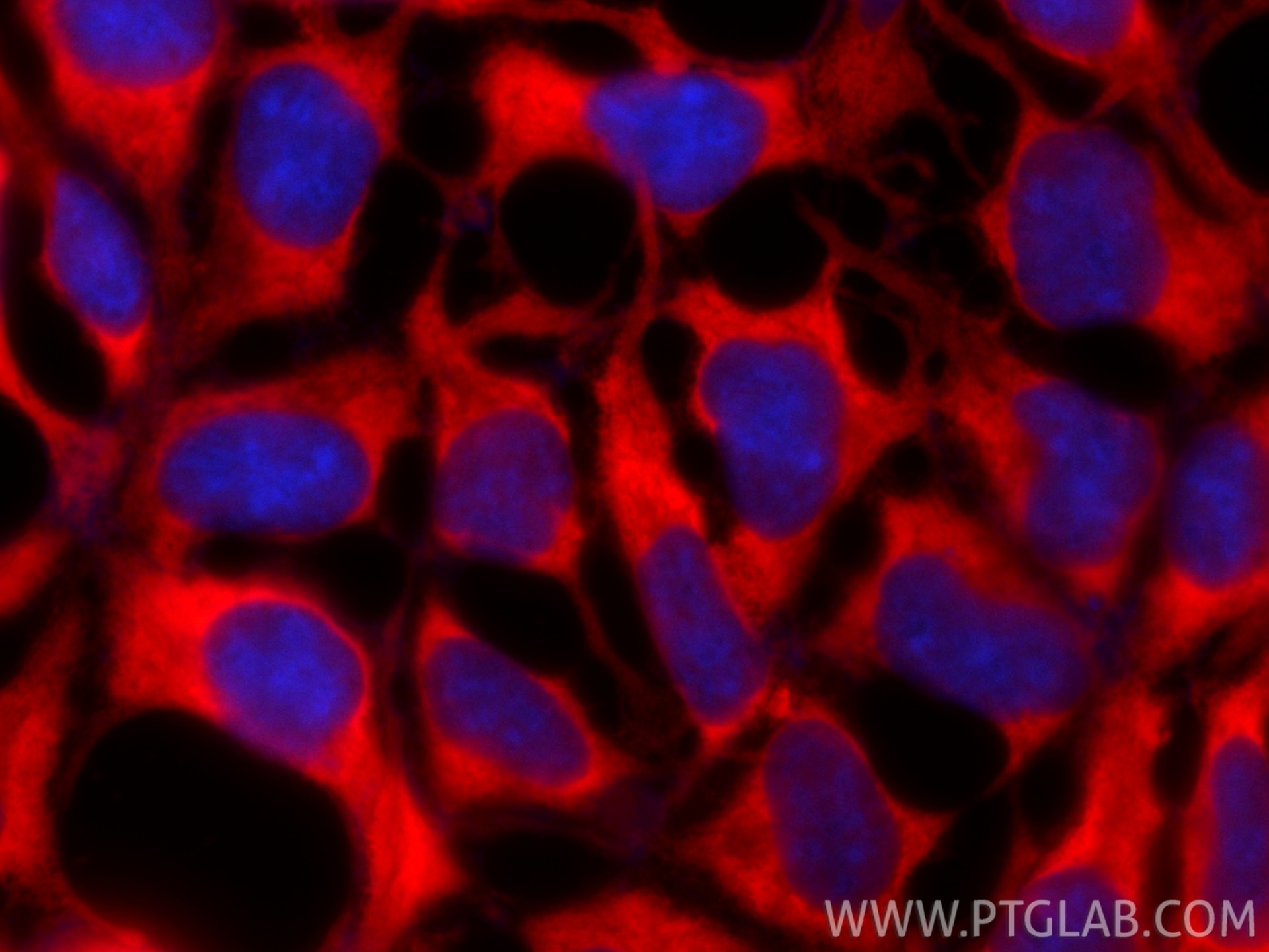
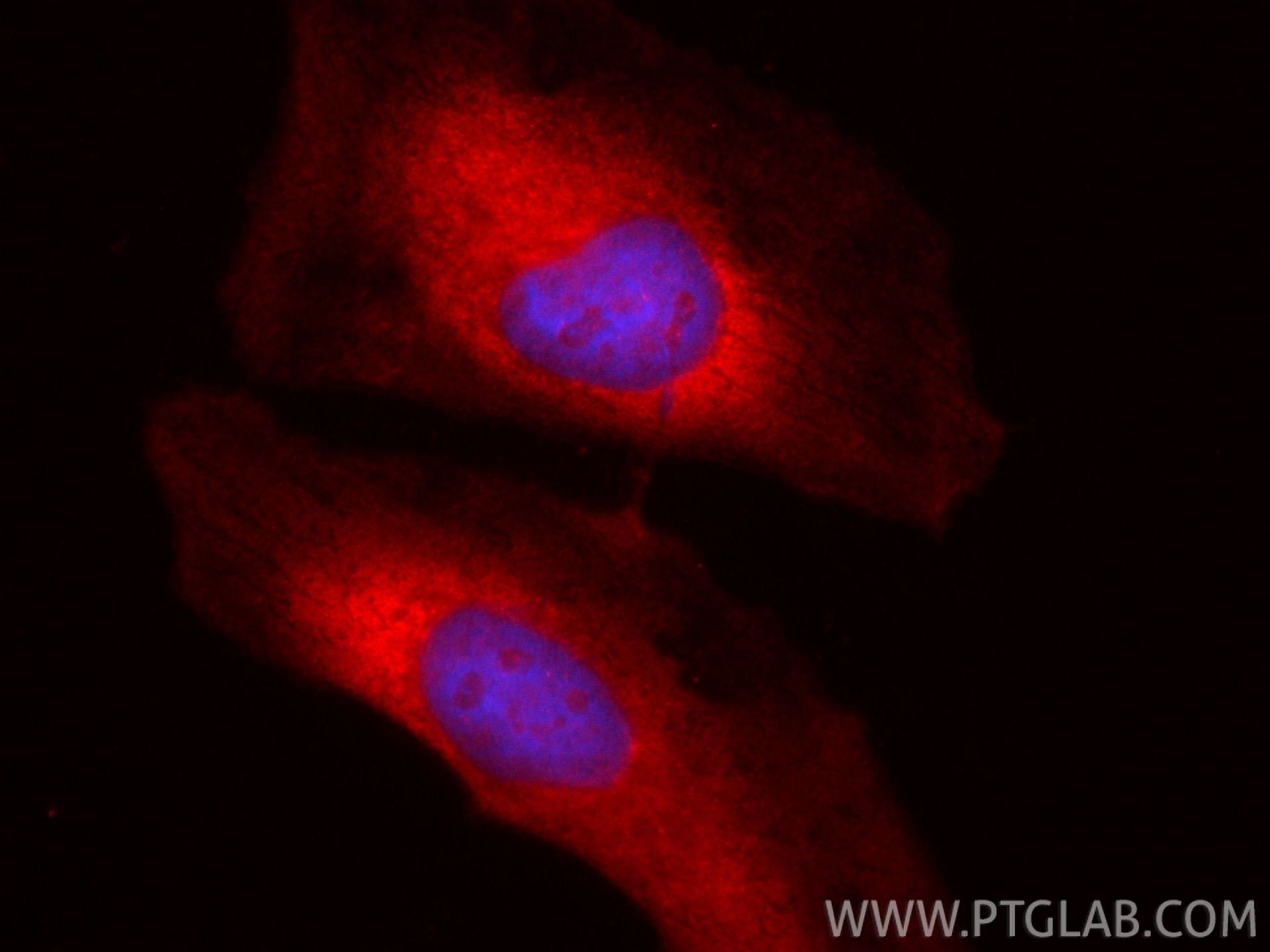
| Positive IF/ICC detected in | HEK-293 cells, HeLa cells |
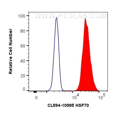
| Positive FC (Intra) detected in | HeLa cells |
Recommended dilution
| Application | Dilution |
|---|---|
| Immunofluorescence (IF)/ICC | IF/ICC : 1:50-1:500 |
| Flow Cytometry (FC) (INTRA) | FC (INTRA) : 0.40 ug per 10^6 cells in a 100 µl suspension |
| It is recommended that this reagent should be titrated in each testing system to obtain optimal results. | |
| Sample-dependent, Check data in validation data gallery. | |
Product Information
CL594-10995 targets HSP70 in IF/ICC, FC (Intra) applications and shows reactivity with human, mouse, rat samples.
| Tested Reactivity | human, mouse, rat |
| Host / Isotype | Rabbit / IgG |
| Class | Polyclonal |
| Type | Antibody |
| Immunogen |
CatNo: Ag1446 Product name: Recombinant human HSP70 protein Source: e coli.-derived, PGEX-4T Tag: GST Domain: 291-641 aa of BC009322 Sequence: IDFYTSITRARFEELCSDLFRSTLEPVEKALRDAKLDKAQIHDLVLVGGSTRIPKVQKLLQDFFNGRDLNKSINPDEAVAYGAAVQAAILMGDKSENVQDLLLLDVAPLSLGLETAGGVMTALIKRNSTIPTKQTQIFTTYSDNQPGVLIQVYEGERAMTKDNNLLGRFELSGIPPAPRGVPQIEVTFDIDANGILNVTATDKSTGKANKITITNDKGRLSKEEIERMVQEAEKYKAEDEVQRERVSAKNALESYAFNMKSAVEDEGLKGKISEADKKKVLDKCQEVISWLDANTLAEKDEFEHKRKELEQVCNPIISGLYQGAGGPGPGGFGAQGPKGGSGSGPTIEEVD 相同性解析による交差性が予測される生物種 |
| Full Name | heat shock 70kDa protein 1A |
| Calculated molecular weight | 70 kDa |
| Observed molecular weight | 66-70 kDa |
| GenBank accession number | BC009322 |
| Gene Symbol | HSP70 |
| Gene ID (NCBI) | 3303 |
| RRID | AB_3084639 |
| Conjugate | CoraLite®594 Fluorescent Dye |
| Excitation/Emission maxima wavelengths | 588 nm / 604 nm |
| Form | |
| Form | Liquid |
| Purification Method | Antigen affinity purification |
| UNIPROT ID | P0DMV8 |
| Storage Buffer | PBS with 50% glycerol, 0.05% Proclin300, 0.5% BSA{{ptg:BufferTemp}}7.3 |
| Storage Conditions | Store at -20°C. Avoid exposure to light. Stable for one year after shipment. Aliquoting is unnecessary for -20oC storage. |
Background Information
-
What is Hsp70/HSP1A?
HSP1A is a member of the Hsp70 (heat shock protein 70) proteins that act as molecular chaperones ensuring correct protein folding and preventing protein aggregation. Hsp70 protein production is greatly induced by various stress stimuli, including high temperature and toxins. Its expression is often elevated in various cancers.
-
FAQs for Hsp70
a. I cannot detect Hsp70 by western blotting
HSP70s are typically expressed at low levels under normal physiological conditions but are dramatically up-regulated in response to cellular stress. Try to always include cell lysate from cells subjected to stress conditions as a positive control.
b. What loading control can I use for cellular stress experiments with Hsp70?
Choosing a loading control antibody is an important step in western blotting experimental setup. We highly recommend using more than one loading control while developing new cellular stress assays to ensure that a given treatment does not alter expression of house-keeping genes. Hsp70 has a molecular size of 70 kDa, so we recommend using GAPDH (36 kDa), actin (42 kDa), or tubulin (50-55 kDa). More information on our control antibodies can be found here: https://www.ptglab.com/news/blog/loading-control-antibodies-for-western-blotting/.
Protocols
| Product Specific Protocols | |
|---|---|
| IF protocol for CL594 HSP70 antibody CL594-10995 | Download protocol |
| Standard Protocols | |
|---|---|
| Click here to view our Standard Protocols |


