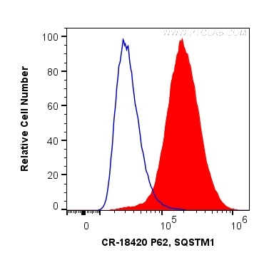
Validation Data Gallery
Tested Applications
| Positive FC (Intra) detected in | HeLa cells |
Recommended dilution
| Application | Dilution |
|---|---|
| Flow Cytometry (FC) (INTRA) | FC (INTRA) : 0.20 ug per 10^6 cells in a 100 µl suspension |
| It is recommended that this reagent should be titrated in each testing system to obtain optimal results. | |
| Sample-dependent, Check data in validation data gallery. | |
Product Information
CR-18420 targets P62/SQSTM1 in FC (Intra) applications and shows reactivity with human samples.
| Tested Reactivity | human |
| Host / Isotype | Rabbit / IgG |
| Class | Polyclonal |
| Type | Antibody |
| Immunogen |
CatNo: Ag13131 Product name: Recombinant human P62;SQSTM1 protein Source: e coli.-derived, PGEX-4T Tag: GST Domain: 1-440 aa of BC017222 Sequence: MASLTVKAYLLGKEDAAREIRRFSFCCSPEPEAEAEAAAGPGPCERLLSRVAALFPALRPGGFQAHYRDEDGDLVAFSSDEELTMAMSYVKDDIFRIYIKEKKECRRDHRPPCAQEAPRNMVHPNVICDGCNGPVVGTRYKCSVCPDYDLCSVCEGKGLHRGHTKLAFPSPFGHLSEGFSHSRWLRKVKHGHFGWPGWEMGPPGNWSPRPPRAGEARPGPTAESASGPSEDPSVNFLKNVGESVAAALSPLGIEVDIDVEHGGKRSRLTPVSPESSSTEEKSSSQPSSCCSDPSKPGGNVEGATQSLAEQMRKIALESEGRPEEQMESDNCSGGDDDWTHLSSKEVDPSTGELQSLQMPESEGPSSLDPSQEGPTGLKEAALYPHLPPEADPRLIESLSQMLSMGFSDEGGWLTRLLQTKNYDIGAALDTIQYSKHPPPL 相同性解析による交差性が予測される生物種 |
| Full Name | sequestosome 1 |
| Calculated molecular weight | 48 kDa |
| Observed molecular weight | 62 kDa |
| GenBank accession number | BC017222 |
| Gene Symbol | P62/SQSTM1 |
| Gene ID (NCBI) | 8878 |
| RRID | AB_2934338 |
| Conjugate | Cardinal Red™ Fluorescent Dye |
| Excitation/Emission maxima wavelengths | 592 nm / 611 nm |
| Form | |
| Form | Liquid |
| Purification Method | Antigen affinity purification |
| UNIPROT ID | Q13501 |
| Storage Buffer | PBS with 50% glycerol, 0.05% Proclin300, 0.5% BSA{{ptg:BufferTemp}}7.3 |
| Storage Conditions | Store at -20°C. Avoid exposure to light. Stable for one year after shipment. Aliquoting is unnecessary for -20oC storage. |
Background Information
Background
P62 (ubiquitin-binding protein P62), also known as Sequestosome-1, is a multifunctional adaptor protein most widely known for its role as an autophagosome cargo protein (PMID: 8551575). P62 via specific interactions with polyubiquitylated target proteins induces their selective autophagy (PMID: 17580304). It also plays an important role in the regulation of the NFkB signaling pathway, senescence, cell differentiation, apoptosis, and immune responses (PMID: 26404812).
What is the molecular weight of P62?
The observed molecular weight of the protein can vary from as low as 8 kDa (for the smallest isoforms) to 48 kDa.
What is the subcellular localization of P62?
P62 is mainly localized in the cytoplasm; however, upon autophagy induction, e.g., via starvation or selective inhibitor treatment, it localizes in vesicular structures - autophagosomes.
What is the tissue specificity of P62?
It is ubiquitously expressed in various tissues.
What is the function of P62 in the regulation of cell death and autophagy?
It is a selective autophagy receptor that forms a bridge between polyubiquitylated cargo (via its UBA domains) and an autophagy modifier such as LC3 (via LIR domains) (PMIDs: 16286508, 20168092, 24128730, 28404643, 22622177). The process of selective autophagy is tightly regulated at many levels, including the posttranslational modifications (PTMs) of various proteins in the cascade, P62 among others (PMID: 29233872). P62 is involved in the regulation of cell death induction in response to various stimuli, e.g., via activation of caspase-8 at the autophagosome membrane (PMID: 29480462). In addition, P62 is degraded during the autophagic process, which makes its intracellular level a marker for autophagy progression.
What is P62's involvement in disease?
Mutations in P62 have been associated with the following diseases: sporadic and familial Paget's disease of bone, neurodegenerative diseases, diabetes, and obesity (PMID: 29480462). A growing number of reports suggest the implication of P62 in the induction of multiple cellular oncogenic transformations. Indeed, increased levels of P62 have been linked to tumor formation, cancer promotion, and resistance to therapy (PMID: 29738493). Moreover, P62 is an unfavorable prognostic marker in liver cancer.
Protocols
| Product Specific Protocols | |
|---|---|
| FC protocol for Cardinal Red™ P62/SQSTM1 antibody CR-18420 | Download protocol |
| Standard Protocols | |
|---|---|
| Click here to view our Standard Protocols |

