Cell types & culture characteristics
| Cell culture cell types | |
| Primary culture cells | Working with adherent cells |
| Cell line culture | Working with cells in suspension |
Working with the primary cell culture
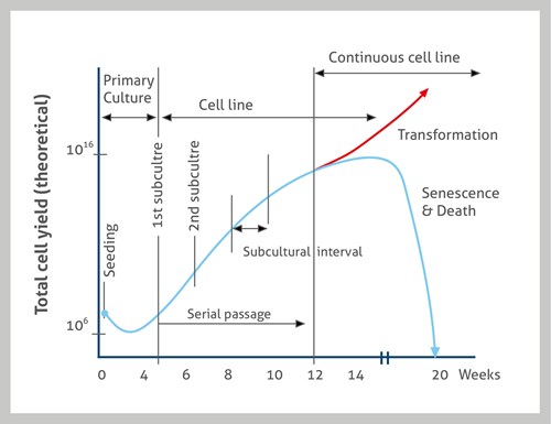
Primary culture cells are isolated from the tissue and proliferated under the appropriate conditions until they occupy all of the available substrate (i.e., reach confluence). To allow growth of the culture to continue past this point, cells need to be subcultured by transferring them to a new culture dish with fresh growth medium. Primary cell culture: cell lines directly expanded from tissues. Unless they undergo an immortalization procedure, primary cells have a limited lifespan and usually reach senescence after 10–20 passages.
Working with the cell line culture
After the first subculture, the primary cells start to become a cell line or subclone. Cell lines derived from primary cultures have a limited lifespan as a consequence of being outside of their tissue niche. While cell culturing, those cells with the highest growth capacity predominate, resulting in a degree of genotypic and phenotypic uniformity in the population over time.
Continuous cell culture: Cells are immortalized and can be grown for many passages with no significant loss of viability. Many are well-known cell lines with very defined characteristics.
Primary cells culture vs. cell line culture.
Working with adherent cells
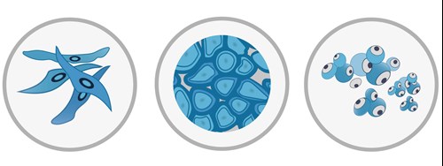
Adherent cells: types of cell lines that grow in the monolayer attached to the surface. While passaged (see standard passaging), a detaching agent (e.g., trypsin) needs to be used to detach them from the surface. They re-attach to the surface within a few hours upon plating. Based on their morphology, adherent cells can be divided into two main categories:
-
Fibroblast-like: elongated shape, usually migrate on the dish
-
Epithelial-like: polygonal shape, stationary, grow in patches
Suspension cells (lymphoblast-like): type of cell lines that grow in suspension and do not form monolayers on the surface. Cells form clumps, especially at high density (summary Figure 5).
Working with cells in suspension
Figure 5. Mammalian cell lines differ in shape. Left: adherent fibroblast-like cells with an elongated shape; Middle: adherent epithelial-like with a polygonal shape; Right: suspension lymphoblast-like cells with a rounded shape.