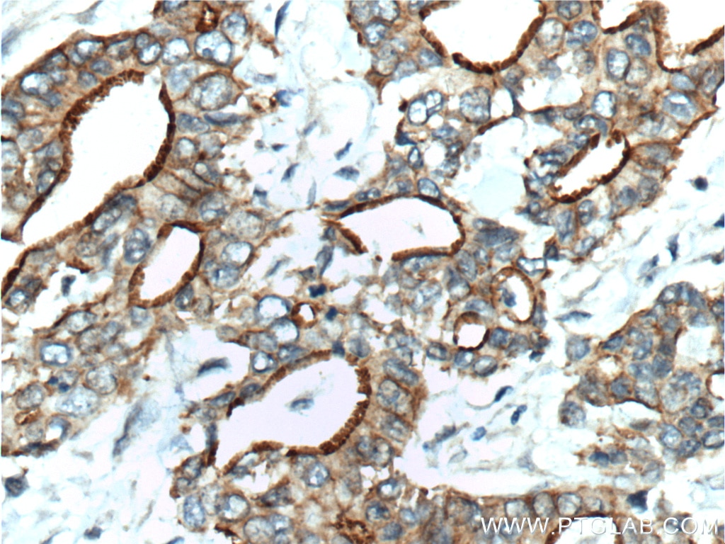
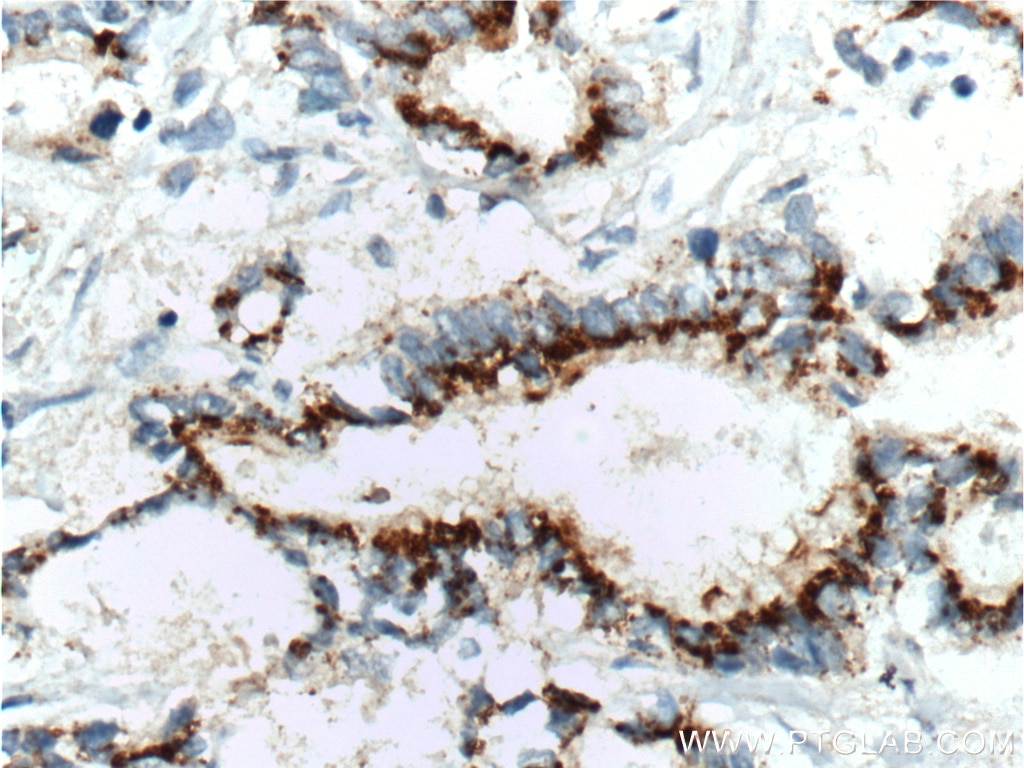
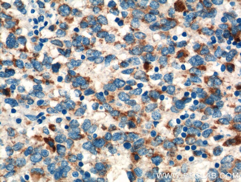
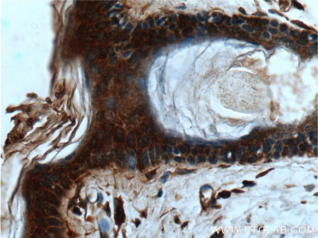
TNF-alpha Polyclonal antibody
TNF-alpha Polyclonal Antibody for WB, ELISA
Host / Isotype
Rabbit / IgG
Reactivity
human, mouse and More (5)
Applications
WB, ELISA, Cell treatment, Transwell Assay
Conjugate
Unconjugated
700
Cat no : 17590-1-AP
Synonyms
Validation Data Gallery
Tested Applications
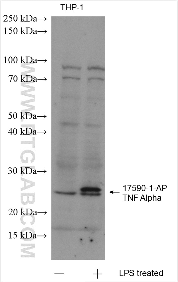
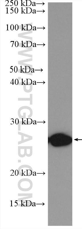
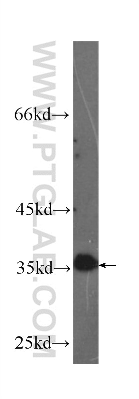
| Positive WB detected in | THP-1 cells, LPS treated RAW 264.7 cells |
Recommended dilution
| Application | Dilution |
|---|---|
| Western Blot (WB) | WB : 1:500-1:2000 |
| It is recommended that this reagent should be titrated in each testing system to obtain optimal results. | |
| Sample-dependent, Check data in validation data gallery. | |
Published Applications
| WB | See 561 publications below |
| ELISA | See 3 publications below |
Product Information
17590-1-AP targets TNF-alpha in WB, ELISA, Cell treatment, Transwell Assay applications and shows reactivity with human, mouse samples.
| Tested Reactivity | human, mouse |
| Cited Reactivity | human, mouse, rat, rabbit, bovine, sheep, fish |
| Host / Isotype | Rabbit / IgG |
| Class | Polyclonal |
| Type | Antibody |
| Immunogen | TNF-alpha fusion protein Ag11433 相同性解析による交差性が予測される生物種 |
| Full Name | tumor necrosis factor (TNF superfamily, member 2) |
| Calculated molecular weight | 233 aa, 26 kDa |
| Observed molecular weight | 26 kDa |
| GenBank accession number | BC028148 |
| Gene symbol | TNF-alpha |
| Gene ID (NCBI) | 7124 |
| RRID | AB_2271853 |
| Conjugate | Unconjugated |
| Form | Liquid |
| Purification Method | Antigen affinity purification |
| Storage Buffer | PBS with 0.02% sodium azide and 50% glycerol pH 7.3. |
| Storage Conditions | Store at -20°C. Stable for one year after shipment. Aliquoting is unnecessary for -20oC storage. |
Background Information
TNF, as also known as TNF-alpha, or cachectin, is a multifunctional proinflammatory cytokine that belongs to the tumor necrosis factor (TNF) superfamily. It is expressed as a 26 kDa membrane bound protein and is then cleaved by TNF-alpha converting enzyme (TACE) to release the soluble 17 kDa monomer, which forms homotrimers in circulation. It is produced chiefly by activated macrophages, although it can be produced by many other cell types such as CD4+ lymphocytes, NK cells, neutrophils, mast cells, eosinophils, and neurons. It can bind to, and thus functions through its receptors TNFRSF1A/TNFR1 and TNFRSF1B/TNFBR. This cytokine is involved in the regulation of a wide spectrum of biological processes including cell proliferation, differentiation, apoptosis, lipid metabolism, and coagulation. This cytokine has been implicated in a variety of diseases, including autoimmune diseases, ins resistance, and cancer.
Protocols
| Product Specific Protocols | |
|---|---|
| WB protocol for TNF-alpha antibody 17590-1-AP | Download protocol |
| Standard Protocols | |
|---|---|
| Click here to view our Standard Protocols |
Publications
| Species | Application | Title |
|---|---|---|
Brain Behav Immun HMGB1 mediates depressive behavior induced by chronic stress through activating the kynurenine pathway. | ||
Bioact Mater A bioactive composite hydrogel dressing that promotes healing of both acute and chronic diabetic skin wounds | ||
ACS Cent Sci Macrophage Inactivation by Small Molecule Wedelolactone via Targeting sEH for the Treatment of LPS-Induced Acute Lung Injury | ||
Adv Sci (Weinh) 3D Printing of a Vascularized Mini-Liver Based on the Size-Dependent Functional Enhancements of Cell Spheroids for Rescue of Liver Failure | ||
Small M2 Macrophage Membrane-Mediated Biomimetic-Nanoparticle Carrying COX-siRNA Targeted Delivery for Prevention of Tendon Adhesions by Inhibiting Inflammation | ||
Gut Microbes S-amlodipine induces liver inflammation and dysfunction through the alteration of intestinal microbiome in a rat model |
Reviews
The reviews below have been submitted by verified Proteintech customers who received an incentive forproviding their feedback.
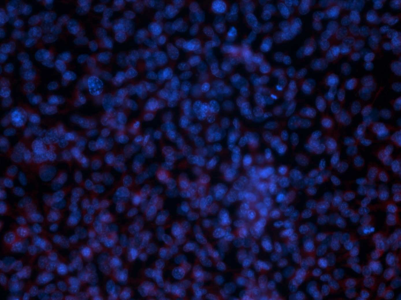
FH Margarita (Verified Customer) (11-22-2023) | Prostate Cancer cell line, overnight incubation, Mouse
 |
FH Isha (Verified Customer) (09-20-2021) | signal was very good. My sample was renal tissue lysate after ischemia reperfusion
|
