Validation Data Gallery
Tested Applications
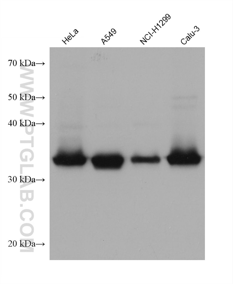
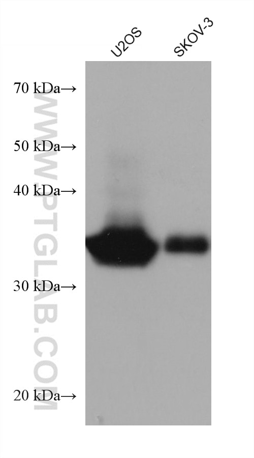
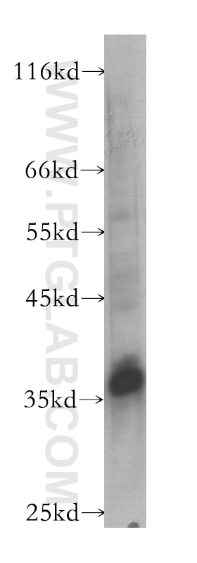
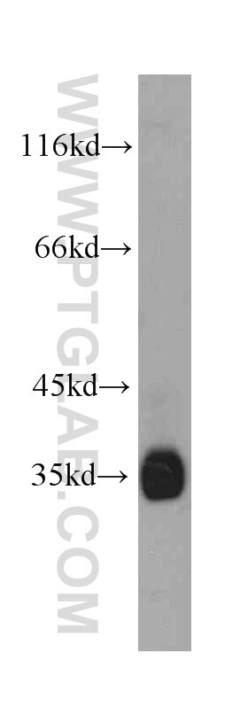
| Positive WB detected in | HeLa cells, HepG2 cells, Raji cells, U2OS cells, SKOV-3 cells, A549 cells, NCI-H1299 cells, Calu-3 cells |
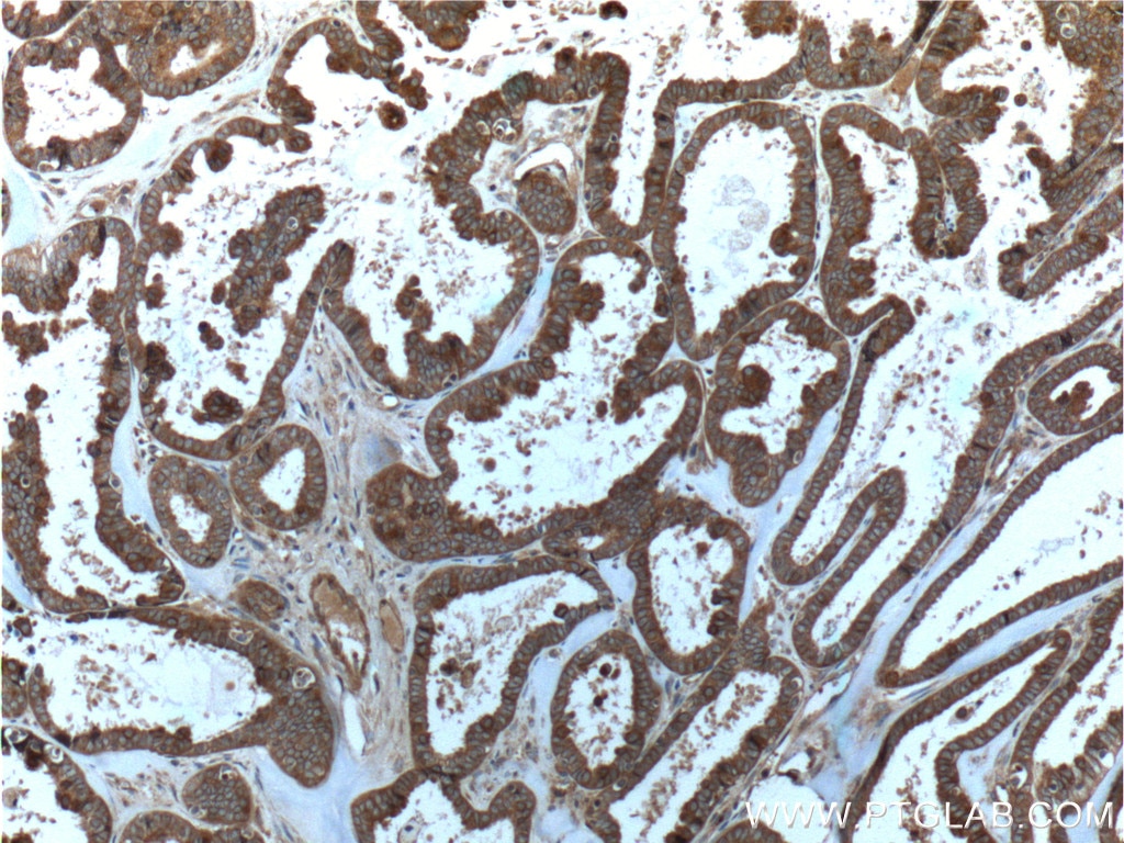
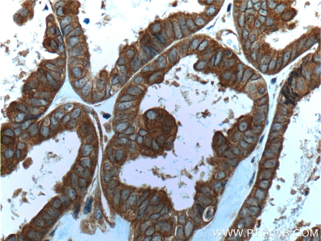
| Positive IHC detected in | human ovary tumor tissue Note: suggested antigen retrieval with TE buffer pH 9.0; (*) Alternatively, antigen retrieval may be performed with citrate buffer pH 6.0 |
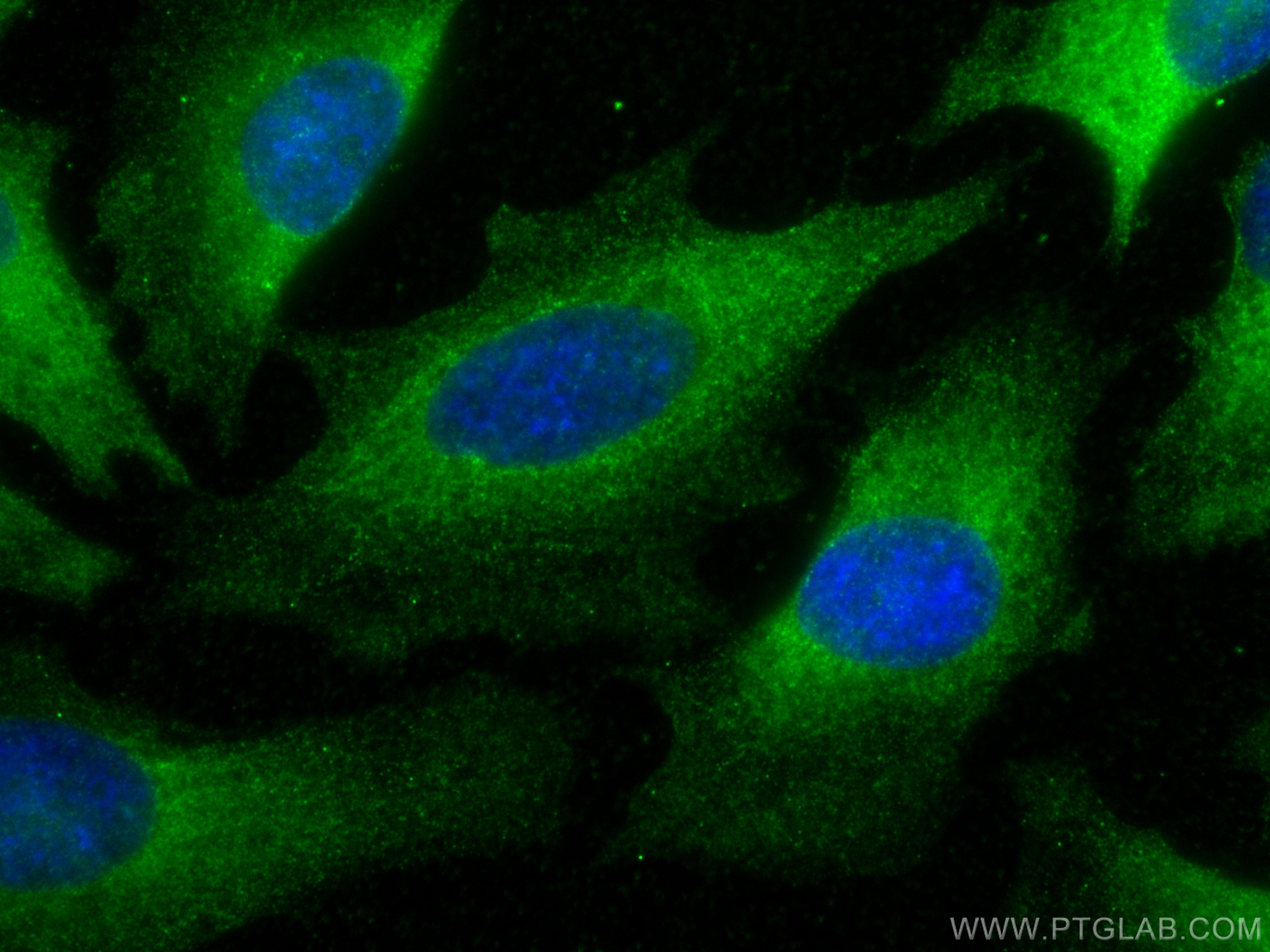
| Positive IF/ICC detected in | HeLa cells |
Recommended dilution
| Application | Dilution |
|---|---|
| Western Blot (WB) | WB : 1:1000-1:6000 |
| Immunohistochemistry (IHC) | IHC : 1:50-1:500 |
| Immunofluorescence (IF)/ICC | IF/ICC : 1:400-1:1600 |
| It is recommended that this reagent should be titrated in each testing system to obtain optimal results. | |
| Sample-dependent, Check data in validation data gallery. | |
Published Applications
| KD/KO | See 1 publications below |
| WB | See 4 publications below |
| IHC | See 3 publications below |
| IF | See 4 publications below |
Product Information
60060-1-Ig targets Follistatin in WB, IHC, IF/ICC, ELISA applications and shows reactivity with human samples.
| Tested Reactivity | human |
| Cited Reactivity | human, mouse, rat, camel |
| Host / Isotype | Mouse / IgG2b |
| Class | Monoclonal |
| Type | Antibody |
| Immunogen | Follistatin fusion protein Ag0775 相同性解析による交差性が予測される生物種 |
| Full Name | follistatin |
| Calculated molecular weight | 33 aa, 7 kDa |
| Observed molecular weight | 35 kDa |
| GenBank accession number | BC004107 |
| Gene Symbol | Follistatin |
| Gene ID (NCBI) | 10468 |
| RRID | AB_2106706 |
| Conjugate | Unconjugated |
| Form | Liquid |
| Purification Method | Protein A purification |
| UNIPROT ID | P19883 |
| Storage Buffer | PBS with 0.02% sodium azide and 50% glycerol , pH 7.3 |
| Storage Conditions | Store at -20°C. Stable for one year after shipment. Aliquoting is unnecessary for -20oC storage. |
Background Information
Follistatin (FST) is a member of the tissue growth factor β family and is a secreted glycoprotein that antagonizes many members of the family, including activin A, growth differentiation factor11 , and myostatin. It binds activin A with high affinity and whose expression can be induced by activin A and several other pro-inflammatory cytokines. Activin A-follistatin complexes are biologically inactive and bind to cell surface heparan sulphate-containing proteoglycans for internalisation and degradation. It has also been found to play a significant role in the management of skeletal muscle size and mass. It also has important roles in early embryonic development, differentiation of ovarian granulosa cells, liver fibrosis and polycystic ovarian syndrome. FS 288 glycosylation results in a major species of 35 kDa and a less abundant 40 kDa and minor products of 40 and 42 kDa (PMID: 9785474). Three typical follistatin bands are at 32, 35, and 39 kDa (PMID: 2036994).
Protocols
| Product Specific Protocols | |
|---|---|
| WB protocol for Follistatin antibody 60060-1-Ig | Download protocol |
| IHC protocol for Follistatin antibody 60060-1-Ig | Download protocol |
| IF protocol for Follistatin antibody 60060-1-Ig | Download protocol |
| Standard Protocols | |
|---|---|
| Click here to view our Standard Protocols |
Publications
| Species | Application | Title |
|---|---|---|
Cells Tubule-Derived Follistatin Is Increased in the Urine of Rats with Renal Ischemia and Reflects the Severity of Acute Tubular Damage | ||
Antioxid Redox Signal Follistatin Protects against Glomerular Mesangial Cell Apoptosis and Oxidative Stress to Ameliorate Chronic Kidney Disease. | ||
Front Pharmacol Follistatin Attenuates Myocardial Fibrosis in Diabetic Cardiomyopathy via the TGF-β-Smad3 Pathway. | ||
Fertil Steril Myostatin, follistatin and activin type II receptors are highly expressed in adenomyosis. | ||
PeerJ Development of a gene doping detection method to detect overexpressed human follistatin using an adenovirus vector in mice. | ||
Animals (Basel) Screening and Identification of Differential Ovarian Proteins before and after Induced Ovulation via Seminal Plasma in Bactrian Camels. |
