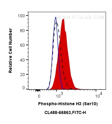
Validation Data Gallery
Tested Applications
| Positive FC (Intra) detected in | nocodazole treated HeLa cells |
Recommended dilution
| Application | Dilution |
|---|---|
| Flow Cytometry (FC) (INTRA) | FC (INTRA) : 0.50 ug per 10^6 cells in a 100 µl suspension |
| It is recommended that this reagent should be titrated in each testing system to obtain optimal results. | |
| Sample-dependent, Check data in validation data gallery. | |
Product Information
CL488-66863 targets Phospho-Histone H3 (Ser10) in FC (Intra) applications and shows reactivity with human, mouse, rat samples.
| Tested Reactivity | human, mouse, rat |
| Host / Isotype | Mouse / IgG1 |
| Class | Monoclonal |
| Type | Antibody |
| Immunogen | Peptide 相同性解析による交差性が予測される生物種 |
| Full Name | histone cluster 1, H3a |
| Calculated molecular weight | 15 kDa |
| Observed molecular weight | 15-17 kDa |
| GenBank accession number | NM_003529 |
| Gene Symbol | HIST1H3A |
| Gene ID (NCBI) | 8350 |
| RRID | AB_2923760 |
| Conjugate | CoraLite® Plus 488 Fluorescent Dye |
| Excitation/Emission maxima wavelengths | 493 nm / 522 nm |
| Form | Liquid |
| Purification Method | Protein G purification |
| Storage Buffer | PBS with 50% glycerol, 0.05% Proclin300, 0.5% BSA , pH 7.3 |
| Storage Conditions | Store at -20°C. Avoid exposure to light. Stable for one year after shipment. Aliquoting is unnecessary for -20oC storage. |
Background Information
Phospho-histone-H3 (PHH3) is a core histone protein, which in its phosphorylated state forms the principal constituents of eukaryotic chromatin, with histone H3 being phosphorylated at serine (Ser) 10 or Ser28 as well as its phosphorylation of Ser10 being strongly correlated with the late G2 to M-phase transition in mammalian mitotic cells. On the basis of previous research, a few cell line- and animal model-based researches have displayed an increase in phosphorylation of histone H3 at Ser10 (H3S10ph), the only histone marker that is involved in carcinogenesis and cellular transformation. Histone H3 phosphorylation on serine-10 is specific to mitosis and phosphorylated histone H3 (PHH3) proliferation markers (as counts defined per area or as indices defined per cell numbers) are increasingly being used to evaluate proliferation in various tumors.
