• Featured Product
  • KD/KO Validated

YY1 Monoclonal antibody

YY1 Monoclonal Antibody for WB, IHC, IF/ICC, IP, ELISA

Host / Isotype

Mouse / IgG2a

Reactivity

human, mouse, rat, monkey and More (1)

Applications

WB, IHC, IF/ICC, IP, CoIP, ChIP, RIP, ELISA

Conjugate

Unconjugated

CloneNo.

2E11C5

Cat no : 66281-1-Ig

Synonyms

DELTA, Delta transcription factor, INO80 complex subunit S, INO80S, NF E1, UCRBP, Yin and yang 1, YIN YANG 1, YY 1, YY1, YY1 transcription factor


"YY1 Antibodies" Comparison

View side-by-side comparison of YY1 antibodies from other vendors to find the one that best suits your research needs.
Filter:
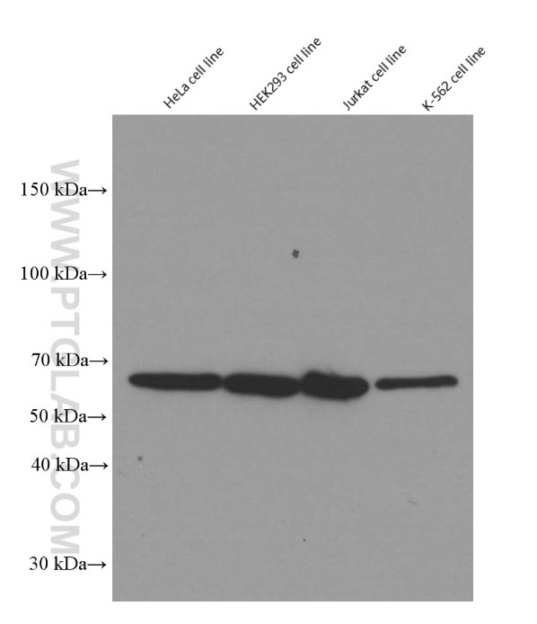
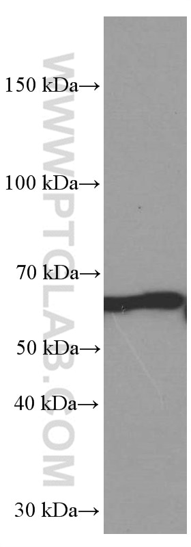
  • Western Blot (WB) analysis of HeLa cells using YY1 Monoclonal antibody (66281-1-Ig)

    WB analysis of HeLa using 66281-1-Ig

    Western blot of YY1 in various human cell lines with 66281-1-Ig at dilution of 1:50000 incubated at room temperature for 1.5 hours.

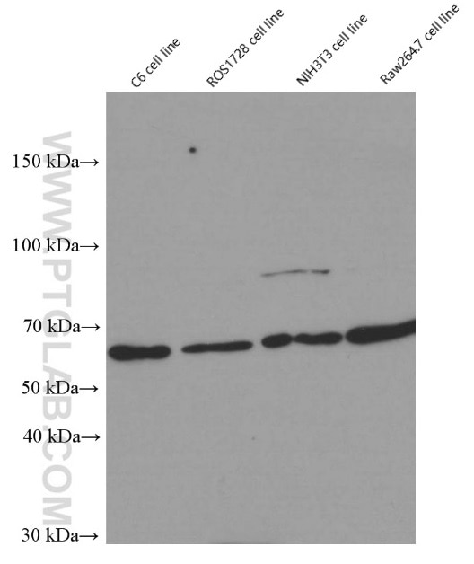
  • Western Blot (WB) analysis of C6 cells using YY1 Monoclonal antibody (66281-1-Ig)

    WB analysis of C6 using 66281-1-Ig

    Western blot of YY1 in rat (C6 and ROS1728) and mouse(NIH3T3 and Raw264.7) cell lines with 66281-1-Ig at dilution of 1:50000 incubated at room temperature for 1.5 hours.

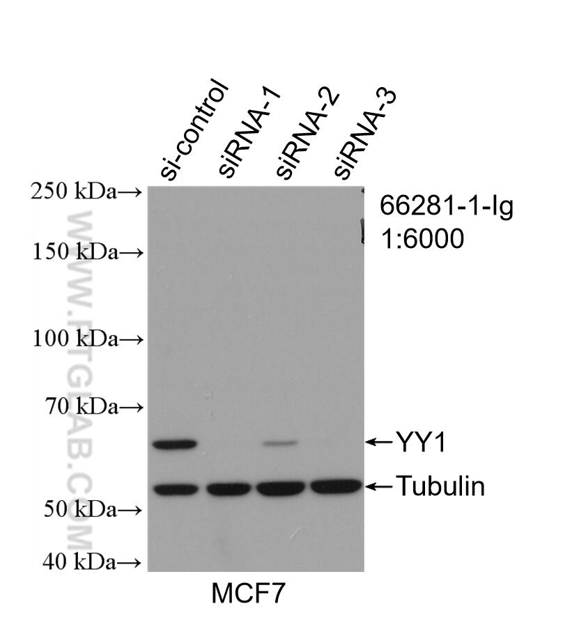
  • Western Blot (WB) analysis of MCF-7 cells using YY1 Monoclonal antibody (66281-1-Ig)

    WB analysis of MCF-7 using 66281-1-Ig

    WB result of YY1 antibody (66281-1-Ig; 1:6000; incubated at room temperature for 1.5 hours) with sh-Control and sh-YY1 transfected MCF-7 cells.

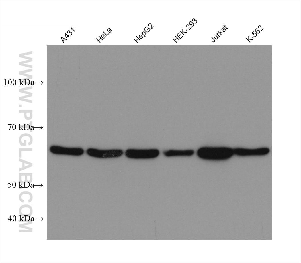
  • Western Blot (WB) analysis of various lysates using YY1 Monoclonal antibody (66281-1-Ig)

    WB analysis using 66281-1-Ig

    Various lysates were subjected to SDS PAGE followed by western blot with 66281-1-Ig (YY1 antibody) at dilution of 1:20000 incubated at room temperature for 1.5 hours.

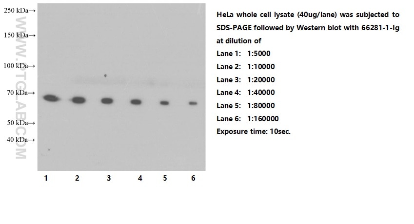
  • Western Blot (WB) analysis of HeLa cells using YY1 Monoclonal antibody (66281-1-Ig)

    WB analysis of HeLa using 66281-1-Ig

    Western blot of Hela cells with mouse anti-YY1 monoclonal antibody (66281-1-Ig) at various dilutions incubated at room temperature for 1.5 hours.

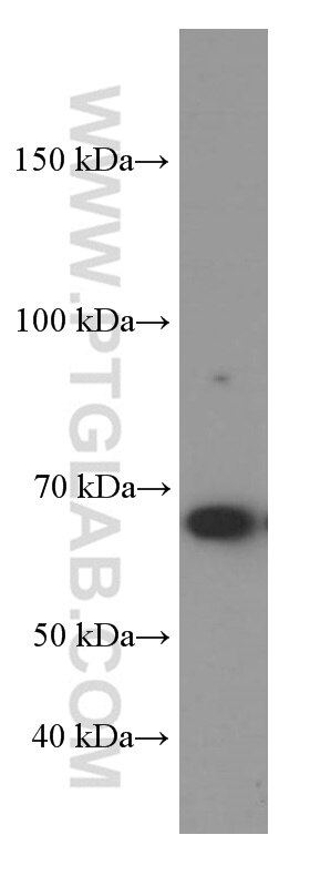
  • Western Blot (WB) analysis of COS-7 cells using YY1 Monoclonal antibody (66281-1-Ig)

    WB analysis of COS-7 using 66281-1-Ig

    COS-7 cells were subjected to SDS PAGE followed by western blot with 66281-1-Ig (YY1 Antibody) at dilution of 1:50000 incubated at room temperature for 1.5 hours.

  • Western Blot (WB) analysis of NIH/3T3 cells using YY1 Monoclonal antibody (66281-1-Ig)

    WB analysis of NIH/3T3 using 66281-1-Ig

    NIH/3T3 cells were subjected to SDS PAGE followed by western blot with 66281-1-Ig (YY1 Antibody) at dilution of 1:50000 incubated at room temperature for 1.5 hours.

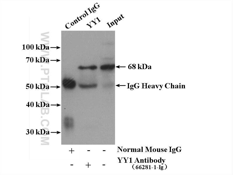
  • Immunoprecipitation (IP) experiment of NIH/3T3 cells using YY1 Monoclonal antibody (66281-1-Ig)

    IP experiment of NIH/3T3 using 66281-1-Ig

    IP result of anti-YY1 (IP:66281-1-Ig, 5ug; Detection:66281-1-Ig 1:1000) with NIH/3T3 cells lysate 4000ug.

  • Immunohistochemistry (IHC) staining of human tonsillitis tissue using YY1 Monoclonal antibody (66281-1-Ig)

    IHC staining of human tonsillitis using 66281-1-Ig

    Immunohistochemical analysis of paraffin-embedded human tonsillitis tissue slide using 66281-1-Ig (YY1 antibody) at dilution of 1:5000 (under 10x lens). Heat mediated antigen retrieval with Tris-EDTA buffer (pH 9.0).

  • Immunohistochemistry (IHC) staining of human tonsillitis tissue using YY1 Monoclonal antibody (66281-1-Ig)

    IHC staining of human tonsillitis using 66281-1-Ig

    Immunohistochemical analysis of paraffin-embedded human tonsillitis tissue slide using 66281-1-Ig (YY1 antibody) at dilution of 1:5000 (under 40x lens). Heat mediated antigen retrieval with Tris-EDTA buffer (pH 9.0).

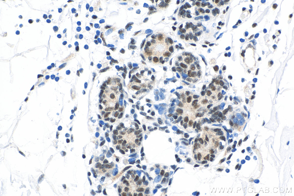
  • Immunohistochemistry (IHC) staining of human breast cancer tissue using YY1 Monoclonal antibody (66281-1-Ig)

    IHC staining of human breast cancer using 66281-1-Ig

    Immunohistochemical analysis of paraffin-embedded human breast cancer tissue slide using 66281-1-Ig (YY1 antibody) at dilution of 1:10000 (under 40x lens). Heat mediated antigen retrieval with Tris-EDTA buffer (pH 9.0).

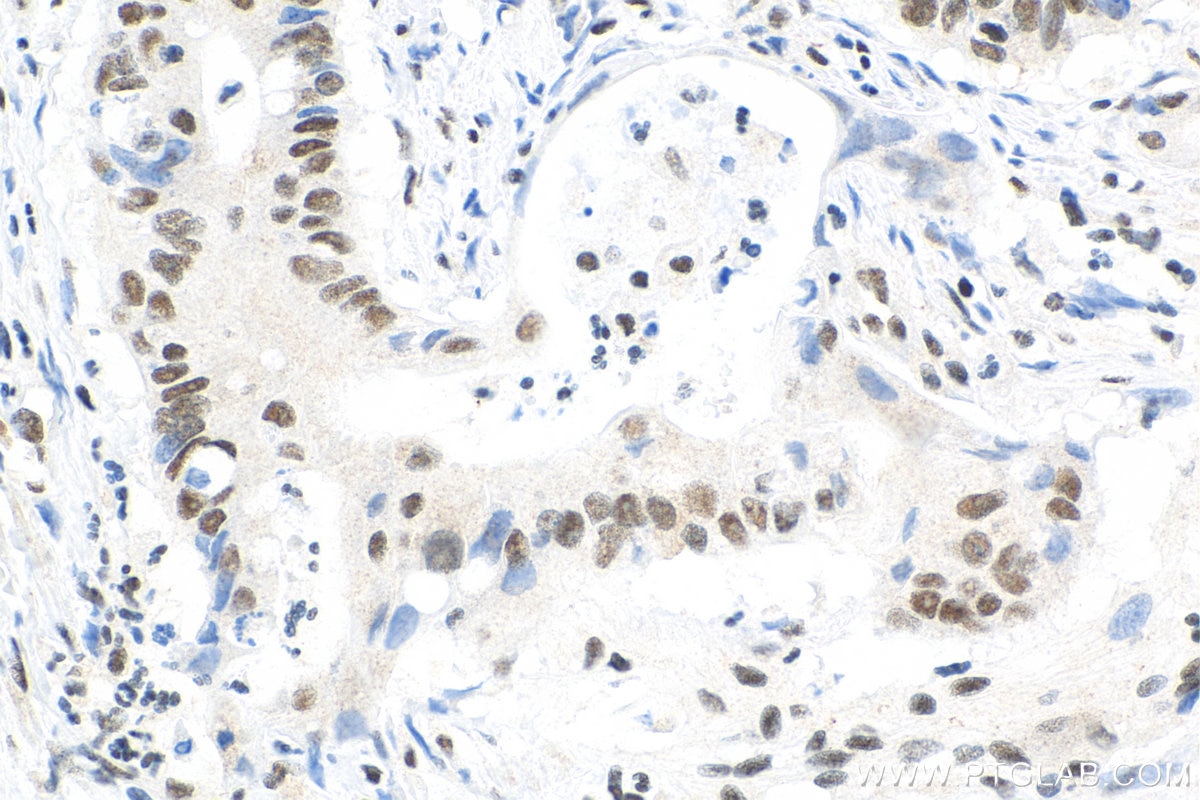
  • Immunohistochemistry (IHC) staining of human colon cancer tissue using YY1 Monoclonal antibody (66281-1-Ig)

    IHC staining of human colon cancer using 66281-1-Ig

    Immunohistochemical analysis of paraffin-embedded human colon cancer tissue slide using 66281-1-Ig (YY1 antibody) at dilution of 1:10000 (under 40x lens). Heat mediated antigen retrieval with Tris-EDTA buffer (pH 9.0).

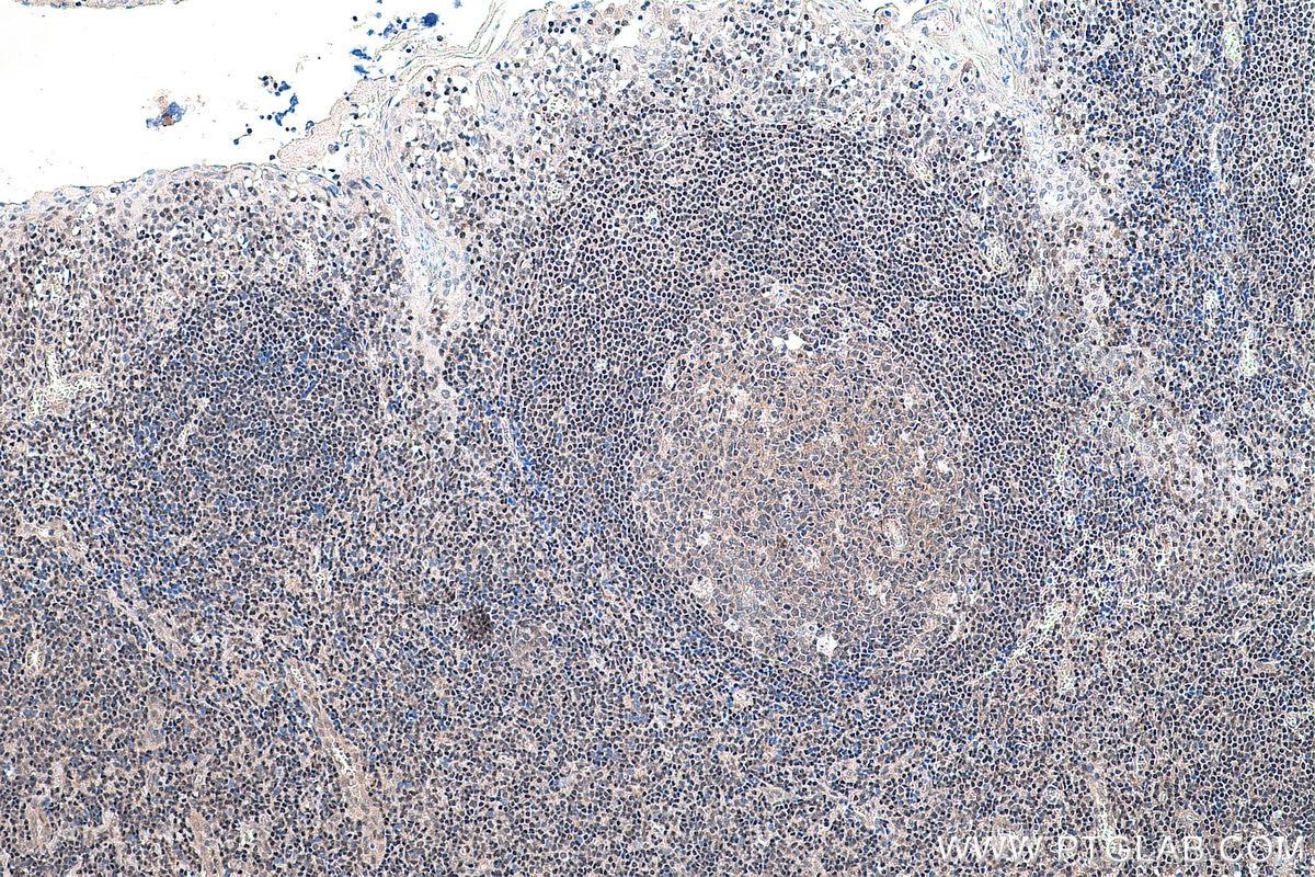
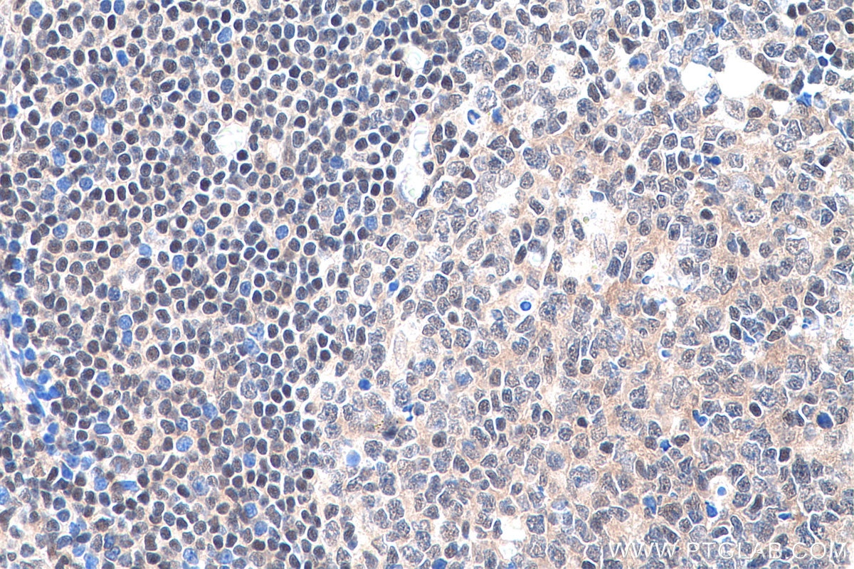
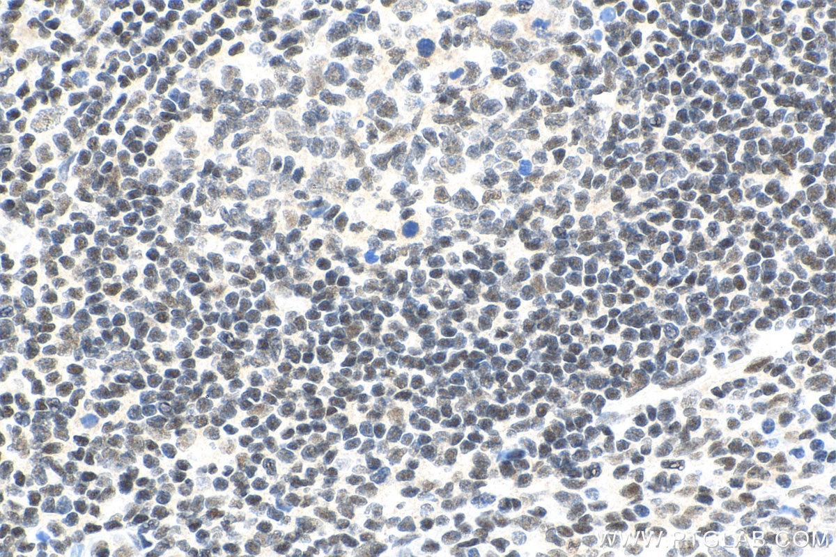
  • Immunohistochemistry (IHC) staining of human tonsillitis tissue using YY1 Monoclonal antibody (66281-1-Ig)

    IHC staining of human tonsillitis using 66281-1-Ig

    Immunohistochemical analysis of paraffin-embedded human tonsillitis tissue slide using 66281-1-Ig (YY1 antibody) at dilution of 1:10000 (under 40x lens). Heat mediated antigen retrieval with Tris-EDTA buffer (pH 9.0).

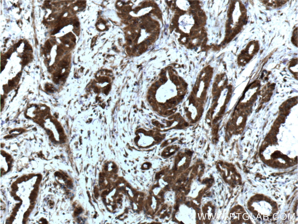
  • Immunohistochemistry (IHC) staining of human breast cancer tissue using YY1 Monoclonal antibody (66281-1-Ig)

    IHC staining of human breast cancer using 66281-1-Ig

    Immunohistochemical analysis of paraffin-embedded human breast cancer tissue slide using 66281-1-Ig (YY1 Antibody) at dilution of 1:200 (under 10x lens). Heat mediated antigen retrieval with Tris-EDTA buffer (pH 9.0).

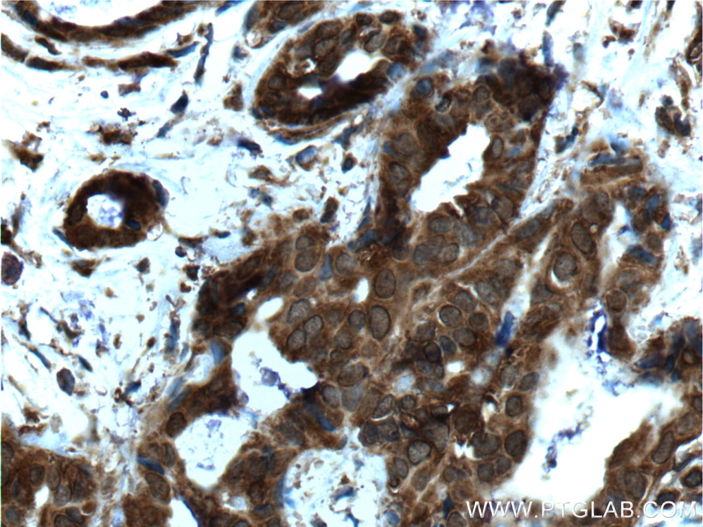
  • Immunohistochemistry (IHC) staining of human breast cancer tissue using YY1 Monoclonal antibody (66281-1-Ig)

    IHC staining of human breast cancer using 66281-1-Ig

    Immunohistochemical analysis of paraffin-embedded human breast cancer tissue slide using 66281-1-Ig (YY1 Antibody) at dilution of 1:200 (under 40x lens). Heat mediated antigen retrieval with Tris-EDTA buffer (pH 9.0).

  • Immunofluorescence (IF) / fluorescent staining of HepG2 cells using YY1 Monoclonal antibody (66281-1-Ig)

    IF Staining of HepG2 using 66281-1-Ig

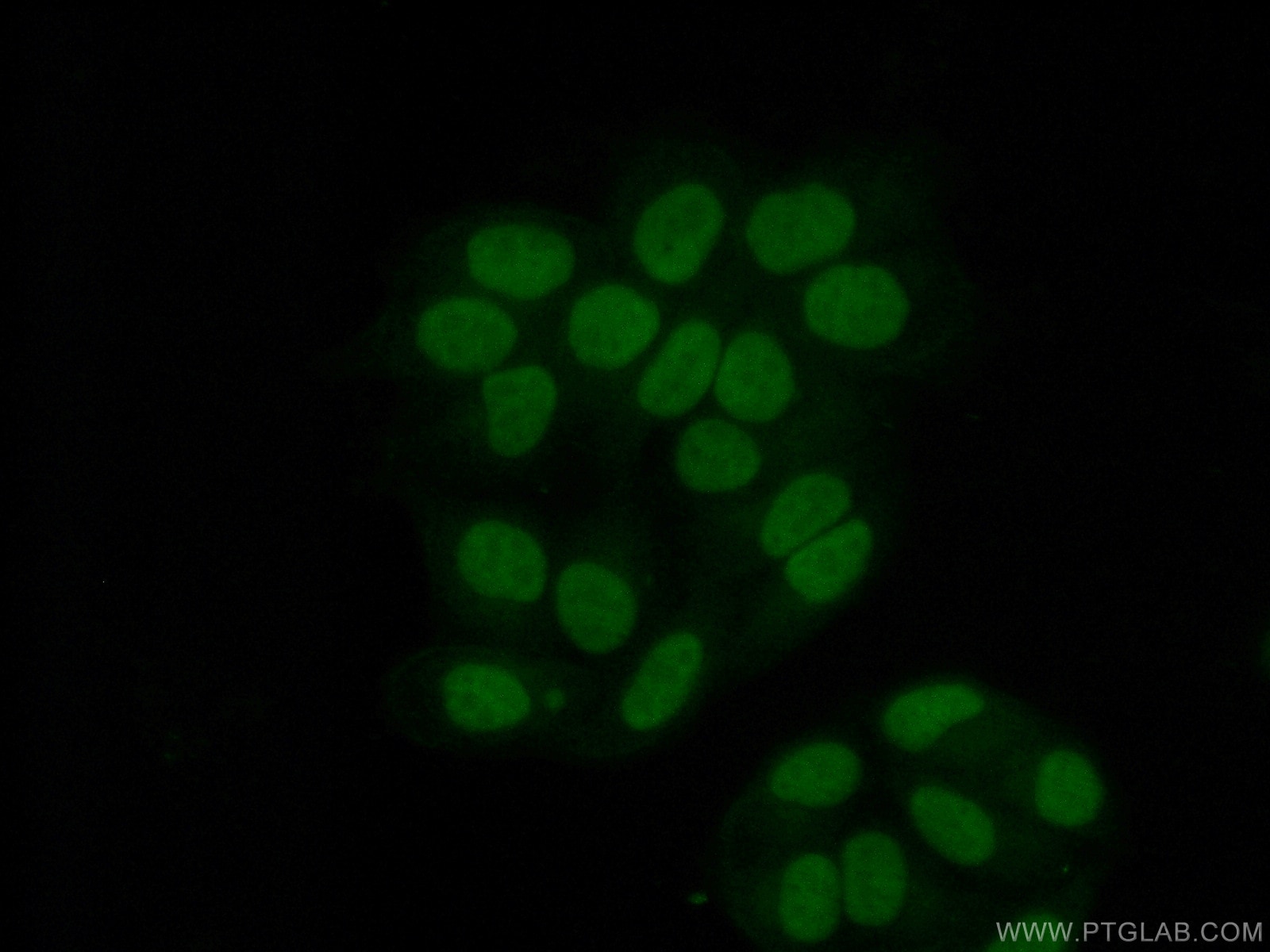
    Immunofluorescent analysis of (4% PFA) fixed HepG2 cells using 66281-1-Ig (YY1 antibody), at dilution of 1:400 and CoraLite®488-Conjugated Goat Anti-Mouse IgG(H+L).

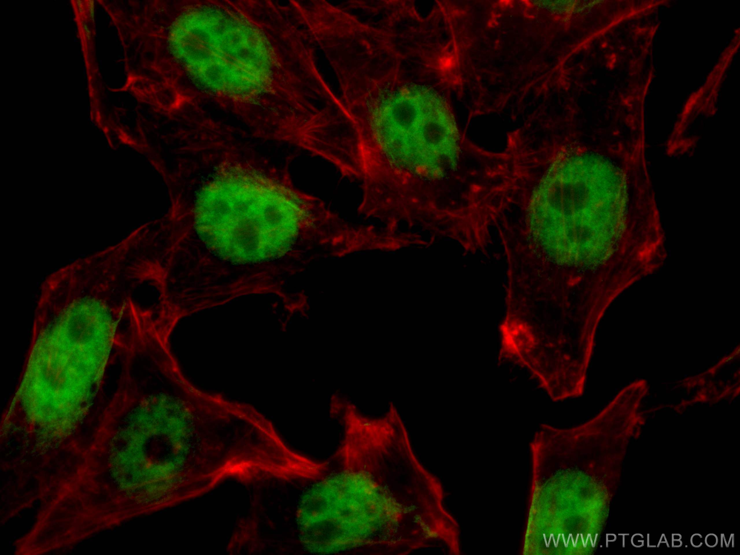
  • Immunofluorescence (IF) / fluorescent staining of HepG2 cells using YY1 Monoclonal antibody (66281-1-Ig)

    IF Staining of HepG2 using 66281-1-Ig

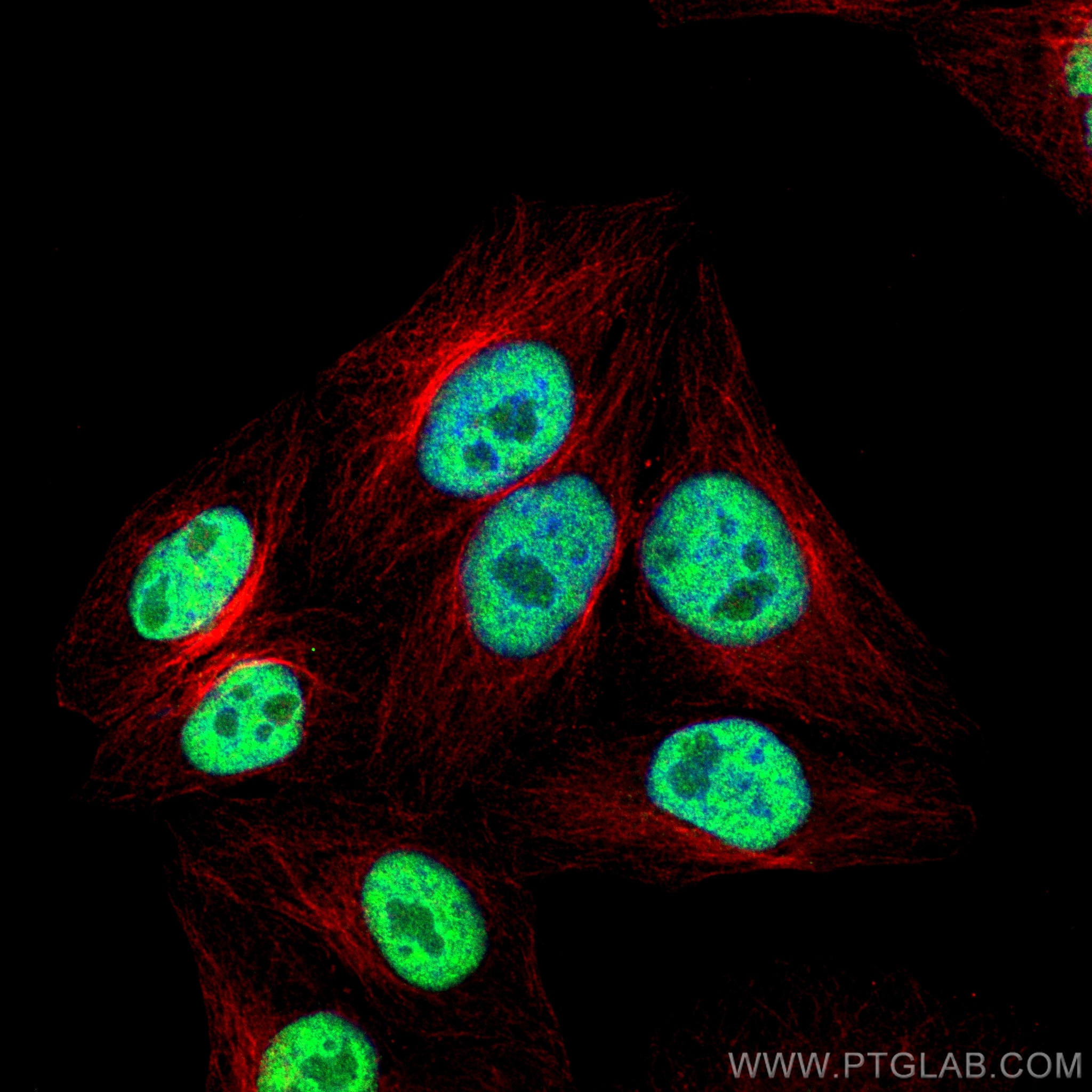
    Immunofluorescent analysis of (4% PFA) fixed HepG2 cells using YY1 antibody (66281-1-Ig, Clone: 2E11C5 ) at dilution of 1:1800 and Multi-rAb CoraLite ® Plus 488-Goat Anti-Mouse Recombinant Secondary Antibody (H+L) (RGAM002), Alpha Tubulin antibody (80762-1-RR, Clone: 1G7, red).

  • Immunofluorescence (IF) / fluorescent staining of HepG2 cells using YY1 Monoclonal antibody (66281-1-Ig)

    IF Staining of HepG2 using 66281-1-Ig

    Immunofluorescent analysis of (10% Formaldehyde ) fixed HepG2 cells using 66281-1-Ig(YY1 antibody) at dilution of 1:100 and Alexa Fluor 488-conjugated Goat Anti-Mouse IgG(H+L).

  • Immunofluorescence (IF) / fluorescent staining of HepG2 cells using YY1 Monoclonal antibody (66281-1-Ig)

    IF Staining of HepG2 using 66281-1-Ig

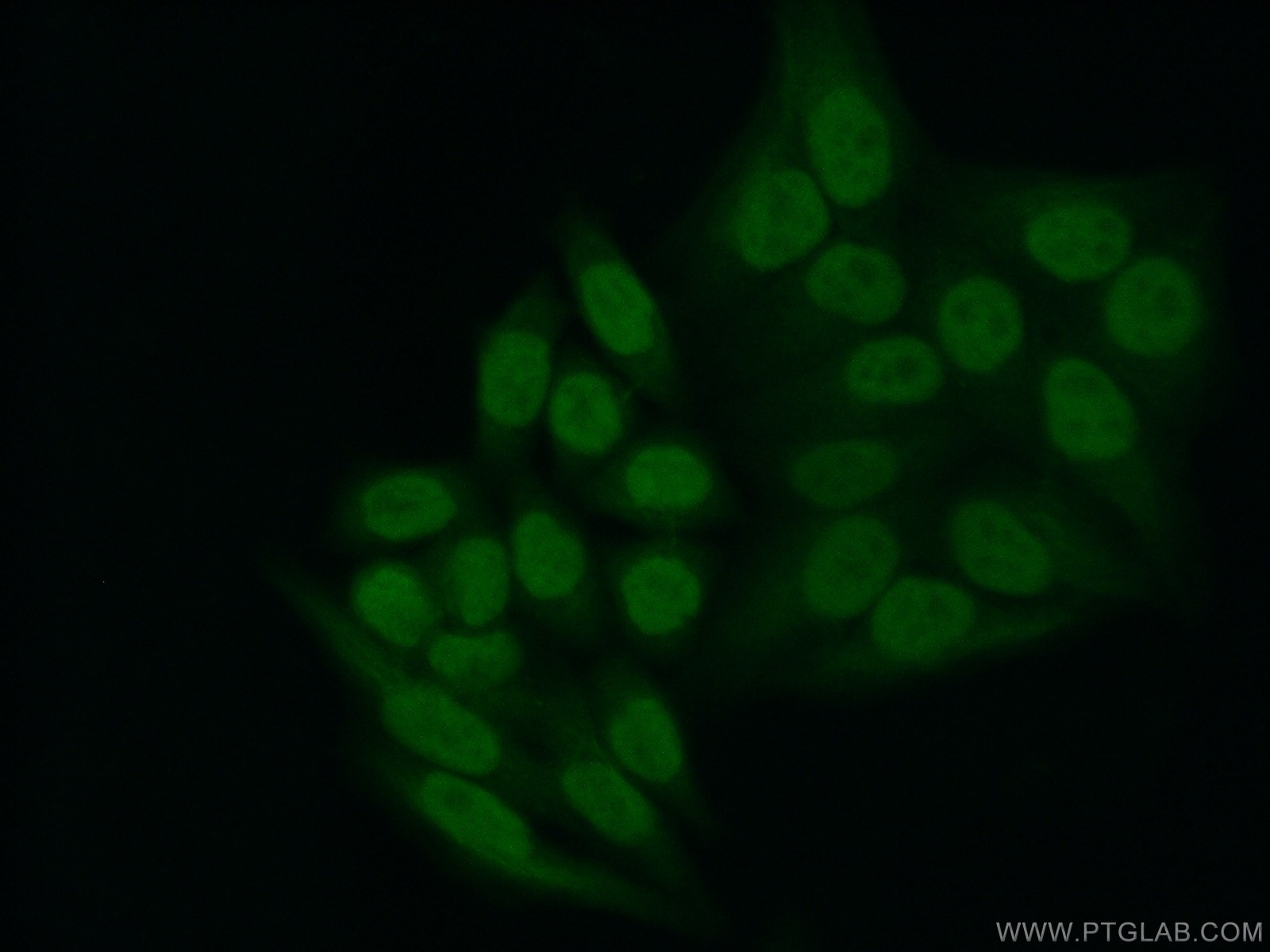
    Immunofluorescent analysis of (10% Formaldehyde ) fixed HepG2 cells using 66281-1-Ig(YY1 antibody) at dilution of 1:800 and Alexa Fluor 488-conjugated Goat Anti-Mouse IgG(H+L).

  • Immunofluorescence (IF) / fluorescent staining of HepG2 cells using YY1 Monoclonal antibody (66281-1-Ig)

    IF Staining of HepG2 using 66281-1-Ig

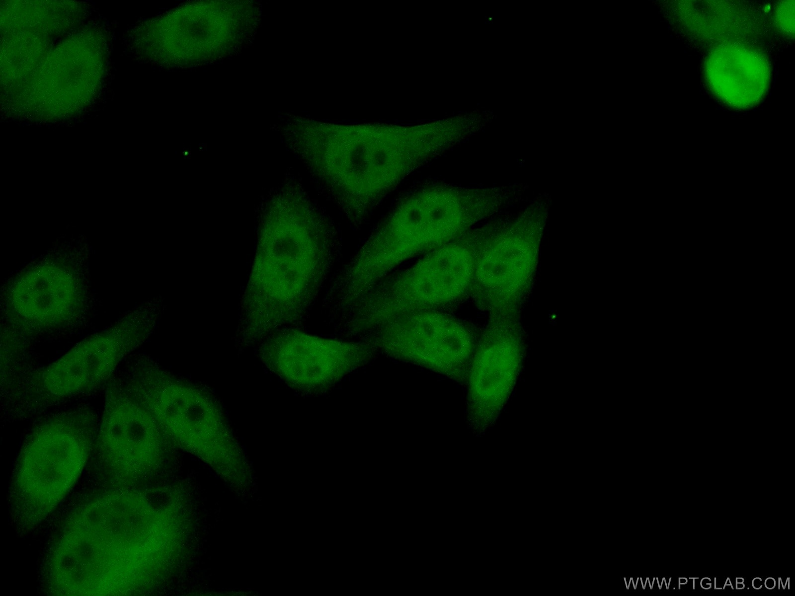
    Immunofluorescent analysis of (10% Formaldehyde ) fixed HepG2 cells using 66281-1-Ig(YY1 antibody) at dilution of 1:400 and Alexa Fluor 488-conjugated Goat Anti-Mouse IgG(H+L).

☆1/31(金)までお得に買える! >> IHC・ELISA・IPキット最大30%OFFキャンペーン


Tested Applications

Positive WB detected inC6 cells, A431 cells, MCF-7 cells, NIH/3T3 cells, HeLa cells, COS-7 cells, HepG2 cells, HEK-293 cells, Jurkat cells, K-562 cells
Positive IP detected inNIH/3T3 cells
Positive IHC detected inhuman breast cancer tissue, human colon cancer tissue, human tonsillitis tissue
Note: suggested antigen retrieval with TE buffer pH 9.0; (*) Alternatively, antigen retrieval may be performed with citrate buffer pH 6.0
Positive IF/ICC detected inHepG2 cells

Recommended dilution

ApplicationDilution
Western Blot (WB)WB : 1:5000-1:50000
Immunoprecipitation (IP)IP : 0.5-4.0 ug for 1.0-3.0 mg of total protein lysate
Immunohistochemistry (IHC)IHC : 1:5000-1:20000
Immunofluorescence (IF)/ICCIF/ICC : 1:200-1:800
It is recommended that this reagent should be titrated in each testing system to obtain optimal results.
Sample-dependent, Check data in validation data gallery.

Product Information

66281-1-Ig targets YY1 in WB, IHC, IF/ICC, IP, CoIP, ChIP, RIP, ELISA applications and shows reactivity with human, mouse, rat, monkey samples.

Tested Reactivity human, mouse, rat, monkey
Cited Reactivityhuman, mouse, rat, pig
Host / Isotype Mouse / IgG2a
Class Monoclonal
Type Antibody
Immunogen YY1 fusion protein Ag17732 相同性解析による交差性が予測される生物種
Full Name YY1 transcription factor
Calculated molecular weight 414 aa, 45 kDa
Observed molecular weight 65-70 kDa
GenBank accession numberBC037308
Gene symbol YY1
Gene ID (NCBI) 7528
RRIDAB_2881664
Conjugate Unconjugated
Form Liquid
Purification MethodProtein A purification
Storage Buffer PBS with 0.02% sodium azide and 50% glycerol pH 7.3.
Storage ConditionsStore at -20°C. Stable for one year after shipment. Aliquoting is unnecessary for -20oC storage.

Background Information

YY1, also named as DELTA, INO80S and NF-E1, contains four C2H2-type zinc fingers and belongs to the YY transcription factor family. YY1 is a multifunctional transcription factor that exhibits positive and negative control on a large number of cellular and viral genes by binding to sites overlapping the transcription start site. YY1 may direct histone deacetylases and histone acetyltransferases to a promoter in order to activate or repress the promoter, thus implicating histone modification in the YY1. The open reading frame of the human YY1 cDNA encodes a protein of 414 amino acids with a predicted molecular weight of 44 kDa. However, YY1 migrates on SDS gels as a 65-68 kDa protein, probably due to the structure of the protein. It is a ubiquitously expressed transcription factor with fundamental roles in embryogenesis, differentiation, replication and proliferation.

Protocols

Product Specific Protocols
WB protocol for YY1 antibody 66281-1-IgDownload protocol
IHC protocol for YY1 antibody 66281-1-IgDownload protocol
IF protocol for YY1 antibody 66281-1-IgDownload protocol
IP protocol for YY1 antibody 66281-1-IgDownload protocol
Standard Protocols
Click here to view our Standard Protocols

Publications

SpeciesApplicationTitle
human

Cell

Pervasive Chromatin-RNA Binding Protein Interactions Enable RNA-Based Regulation of Transcription.

Authors - Rui Xiao
mouseWB

Nat Commun

miR-29b contributes to multiple types of muscle atrophy.

Authors - Jin Li
humanWB,IF,IP

Oncogene

AKIP1 accelerates glioblastoma progression through stabilizing EGFR expression

Authors - Sicheng Wan
mouseWB

Mol Ther Nucleic Acids

Angiotensin II-induced muscle atrophy via PPARγ suppression is mediated by miR-29b.

Authors - Jin Li
humanWB

Cell Death Dis

CircHIPK3 promotes colorectal cancer growth and metastasis by sponging miR-7.

Authors - Kaixuan Zeng
humanWB

Cancers (Basel)

MiR-302b as a Combinatorial Therapeutic Approach to Improve Cisplatin Chemotherapy Efficacy in Human Triple-Negative Breast Cancer.

Authors - Alessandra Cataldo