Validation Data Gallery
Tested Applications
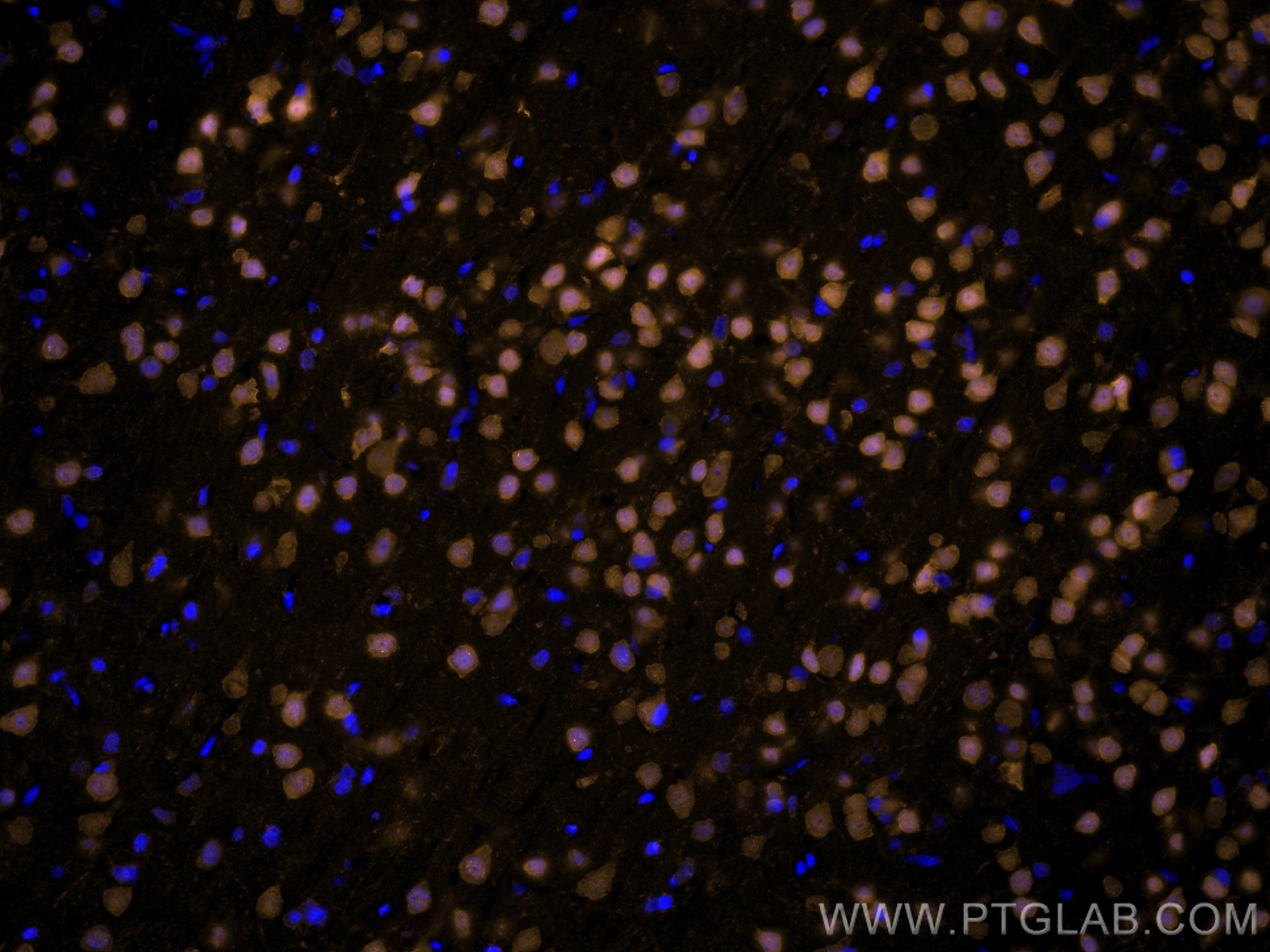
| Positive IF-P detected in | rat brain tissue |
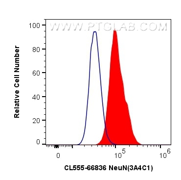
| Positive FC (Intra) detected in | U-87 MG cells |
| Positive FC detected in | U-87 MG cells |
Recommended dilution
| Application | Dilution |
|---|---|
| Immunofluorescence (IF)-P | IF-P : 1:50-1:500 |
| Flow Cytometry (FC) (INTRA) | FC (INTRA) : 0.80 ug per 10^6 cells in a 100 µl suspension |
| Flow Cytometry (FC) | FC : 0.80 ug per 10^6 cells in a 100 µl suspension |
| It is recommended that this reagent should be titrated in each testing system to obtain optimal results. | |
| Sample-dependent, Check data in validation data gallery. | |
Product Information
CL555-66836 targets NeuN in IF-P, FC (Intra) applications and shows reactivity with Human, mouse, rat samples.
| Tested Reactivity | Human, mouse, rat |
| Host / Isotype | Mouse / IgG1 |
| Class | Monoclonal |
| Type | Antibody |
| Immunogen |
CatNo: Ag28016 Product name: Recombinant human NeuN protein Source: e coli.-derived, PET28a Tag: 6*His Domain: 1-100 aa of NM_001082575 Sequence: MAQPYPPAQYPPPPQNGIPAEYAPPPPHPTQDYSGQTPVPTEHGMTLYTPAQTHPEQPGSEASTQPIAGTQTVPQTDEAAQTDSQPLHPSDPTEKQQPKR 相同性解析による交差性が予測される生物種 |
| Full Name | hexaribonucleotide binding protein 3 |
| GenBank accession number | NM_001082575 |
| Gene Symbol | NeuN |
| Gene ID (NCBI) | 146713 |
| RRID | AB_2919697 |
| Conjugate | CoraLite®555 Fluorescent Dye |
| Excitation/Emission maxima wavelengths | 557 nm / 570 nm |
| Form | |
| Form | Liquid |
| Purification Method | Protein G purification |
| UNIPROT ID | A6NFN3 |
| Storage Buffer | PBS with 50% glycerol, 0.05% Proclin300, 0.5% BSA{{ptg:BufferTemp}}7.3 |
| Storage Conditions | Store at -20°C. Avoid exposure to light. Stable for one year after shipment. Aliquoting is unnecessary for -20oC storage. |
Background Information
NeuN, encoded by FOX3, is a neuron-specific nuclear protein. Anti-NeuN stains exclusively neuronal cells in the central and peripheral nervous systems, especially postmitotic and differentiating neurons, as well as terminally differentiated neurons. Anti-NeuN has been used widely as a reliable tool to detect most postmitotic neuronal cell types. The immunohistochemical staining is primarily localized in the nucleus of the neurons with lighter staining in the cytoplasm.
Protocols
| Product Specific Protocols | |
|---|---|
| IF protocol for CL555 NeuN antibody CL555-66836 | Download protocol |
| Standard Protocols | |
|---|---|
| Click here to view our Standard Protocols |


