Use Able AI chat for product recommendations

Validation Data Gallery
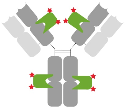
  • Well-defined and characterized immunostaining: Primary anti-rabbit IgG antibody (grey) with 2 copies each of a rabbit Fab- and Fc-specific monoclonal Nanobodies (green) bound. In total, 8 fluorophores (red stars) label the primary rabbit IgG antibody.

    Well-defined and characterized immunostaining: Primary anti-rabbit IgG antibody (grey) with 2 copies each of a rabbit Fab- and Fc-specific monoclonal Nanobodies (green) bound. In total, 8 fluorophores (red stars) label the primary rabbit IgG antibody.

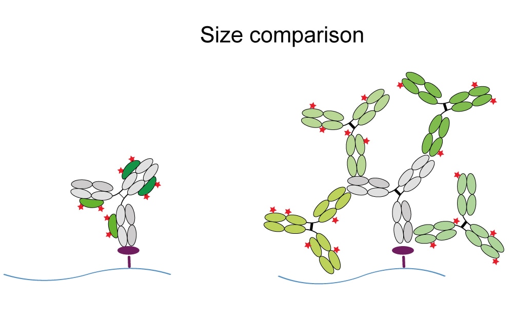
  • Higher resolution with anti-rabbit IgG Nano-Secondaries compared to conventional secondary antibodies: Left: Formation of a small, precise complex of Nanobodies (green) & primary antibody (grey). Right: Formation of a large, arbitrary complex of multiple polyclonal secondaries (green) & primary rabbit antibody.

    Higher resolution with anti-rabbit IgG Nano-Secondaries compared to conventional secondary antibodies: Left: Formation of a small, precise complex of Nanobodies (green) & primary antibody (grey). Right: Formation of a large, arbitrary complex of multiple polyclonal secondaries (green) & primary rabbit antibody.

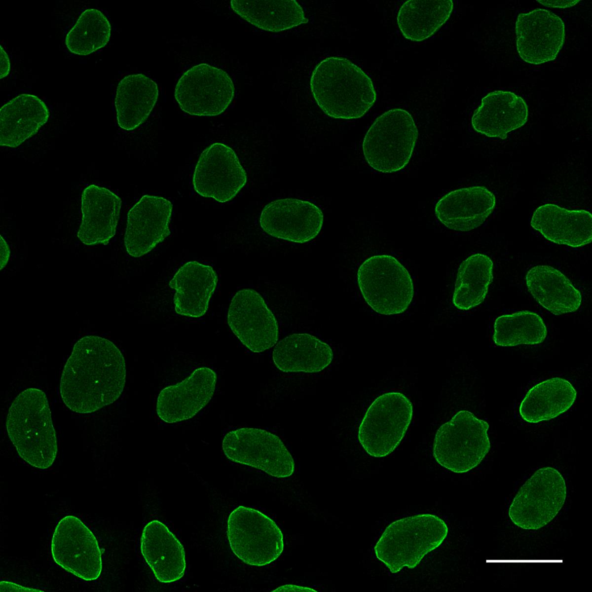
  • Alpaca anti-rabbit IgG VHH Alexa Fluor® 488 was applied in conjunction with rabbit anti-Lamin B1 antibodies for immunostaining of nuclear lamina (green) in HeLa cells. Scale bar, 20 μm. Images were recorded at the Core Facility Bioimaging at the Biomedical Center, LMU Munich.

    Alpaca anti-rabbit IgG VHH Alexa Fluor® 488 was applied in conjunction with rabbit anti-Lamin B1 antibodies for immunostaining of nuclear lamina (green) in HeLa cells. Scale bar, 20 μm. Images were recorded at the Core Facility Bioimaging at the Biomedical Center, LMU Munich.

  • HeLa cells were immunostained with rabbit anti-Lamin B1 antibodies and alpaca anti-rabbit IgG VHH Alexa Fluor® 568 (1:1,000). Confocal and gated STED images were acquired with a Leica TCS SP8 STED 3X microscope, pulsed depletion with a 775 nm laser. Images were recorded at the Core Facility Bioimaging at the Biomedical Center, LMU Munich.

    HeLa cells were immunostained with rabbit anti-Lamin B1 antibodies and alpaca anti-rabbit IgG VHH Alexa Fluor® 568 (1:1,000). Confocal and gated STED images were acquired with a Leica TCS SP8 STED 3X microscope, pulsed depletion with a 775 nm laser. Images were recorded at the Core Facility Bioimaging at the Biomedical Center, LMU Munich.

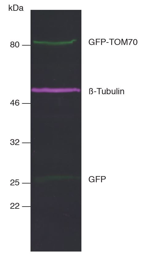
  • Multiple Nano-Secondaries can be applied for multiplex fluorescent Western blotting. This allows multiple targets to be analyzed simultaneously on the same blot at the same time. Multiplex fluorescent Western blot of GFP-TOM70, ®-Tubulin, and GFP in HEK293T cell lysate. Western blot membrane was simultaneously incubated with primary antibodies and Nano-Secondaries. Green: rabbit anti-GFP (ChromoTek PABG1) + alpaca anti-rabbit IgG VHH Alexa Fluor® 488. Magenta: mouse anti-®-Tubulin + alpaca anti-mouse IgG2b VHH Alexa Fluor® 647.

    Multiple Nano-Secondaries can be applied for multiplex fluorescent Western blotting. This allows multiple targets to be analyzed simultaneously on the same blot at the same time. Multiplex fluorescent Western blot of GFP-TOM70, ®-Tubulin, and GFP in HEK293T cell lysate. Western blot membrane was simultaneously incubated with primary antibodies and Nano-Secondaries. Green: rabbit anti-GFP (ChromoTek PABG1) + alpaca anti-rabbit IgG VHH Alexa Fluor® 488. Magenta: mouse anti-®-Tubulin + alpaca anti-mouse IgG2b VHH Alexa Fluor® 647.

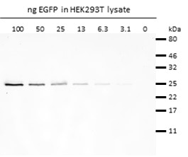
  • Western blot analysis of EGFP (EGFP-250, ChromoTek) added to HEK293T cell lysate. Detection with rabbit anti-GFP antibody PABG1 (ChromoTek) and alpaca anti-rabbit IgG VHH Alexa Fluor® 488.

    Western blot analysis of EGFP (EGFP-250, ChromoTek) added to HEK293T cell lysate. Detection with rabbit anti-GFP antibody PABG1 (ChromoTek) and alpaca anti-rabbit IgG VHH Alexa Fluor® 488.

ChromoTek Nano-Secondary® anti-human IgG/anti-rabbit IgG, recombinant VHH, Alexa Fluor® 488 [CTK0101, CTK0102]

Nano-Secondary® anti-human IgG/anti-rabbit IgG, recombinant VHH is an anti-human IgG and anti-rabbit IgG specific secondary antibody. It consists of of a mixture of 2 Nanobodies that bind to human IgG and rabbit IgG with high affinity & specificity.
Cat No. srbAF488-1

Applications

IF, WB, FC

Reactivity

Rabbit, Human, Macaque

Host

Alpaca

Conjugate

Alexa Fluor® 488

human IgG, rabbit IgG, immunoglobulin Gamma

Size: 

-/ -

Concentration: 0.5 g/ L


ご購入について

国内販売は「コスモ・バイオ株式会社」を通じて行っております。お見積り・ご注文はお近くの販売代理店へご連絡ください。


国内在庫・納期について

約2万点のプロテインテック製品をコスモバイオ社物流センター(国内)に在庫しています。国内在庫の有無はコスモバイオ社ホームページの「品番検索」でカタログ番号を検索して確認できます。


保証・サポートについて

テクニカルサポートまたはご購入後1年間の交換/補填対応を承ります。詳細はこちらをご覧ください。


Product Information

Nano-Secondary® anti-human IgG/anti-rabbit IgG, recombinant VHH is an anti-human IgG and anti-rabbit IgG specific secondary antibody. It consists of of a mixture of 2 Nanobodies that bind to human IgG and rabbit IgG with high affinity & specificity.

DescriptionNano-Secondary® alpaca anti-human IgG/anti-rabbit IgG, recombinant VHH reagent uses a novel class of anti-rabbit IgG and anti-human IgG specific antibodies. This secondary antibody product consists of Nanobodies that bind to rabbit IgG and human IgG with high affinity & specificity. Nano-Secondary reagents enable simultaneous incubation with primary antibodies, thus shortening your experimental. Due to their small size, Nano-Secondary reagents provide enhanced image resolution. No cross-reactivity against serum proteins from other species, details see below.
ApplicationsIF, WB, FC
HostAlpaca
Species ReactivityRabbit, Human, Macaque
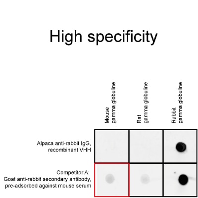
No cross-reactivity to mouse, rat, sheep, goat, and guinea pig IgG
Target/SpecificityFc-fragment of human IgG, Fab- and Fc-fragment of rabbit IgG (co-reactivity)
Physical StateLiquid
ConjugateAlexa Fluor® 488
Suggested DilutionImmunofluorescence 1:1,000
Super-resolution microscopy 1:1,000
Western blot 1:1,000
Purification MethodRecombinant expression, affinity purification IMAC
TypeMixture of 2 monoclonal Nanobodies; Secondary Nanobody
ClassRecombinant
FormatAlpaca single domain antibody, monovalent
Cross-reactivityNo cross-reactivity to goat, guinea pig, mouse, rat, and sheep serum
Cross-reactivity to macaque serum
ImmunogenPurified rabbit IgG
ClonalityBiclonal: mixture of 2 monoclonal Nanobodies
ClonesCTK0101 (VHH0244): Fc-fragment of human IgG, Fc-fragment of rabbit IgG
CTK0102 (VHH0245): Fab-fragment of rabbit IgG
Affinity (KD)CTK0101: KD = 0.2 nM, CTK0102: KD = 1.2 nM
RRIDAB_2827585
Excitation / EmissionExcitation max: 490 nm, Emission max: 525 nm
Storage Buffer10 mM HEPES pH 7.0, 500 mM NaCl, 5 mM EDTA, Preservative: 0.09 % Sodium azide
Storage ConditionStore at -20°C short term or -80°C long term. Aliquot upon delivery. Avoid freeze-thaw cycles.
Size10 μL; 100 μL
NoteThis product is for research use only, not for diagnostic or therapeutic use

Publications

ApplicationTitle

Kidney Int

The small GTPase regulatory protein Rac1 drives podocyte injury independent of cationic channel protein TRPC5

Authors - Onur K Polat
FC

Autophagy

FOXP3+ macrophage represses acute ischemic stroke-induced neural inflammation

Authors - Wei Cai
IF

Nat Commun

The RNA-binding protein landscapes differ between mammalian organs and cultured cells

Authors - Joel I Perez-Perri

Nat Commun

Small molecule APOL1 inhibitors as a precision medicine approach for APOL1-mediated kidney disease

Authors - Brandon Zimmerman
IF

Nat Commun

Engineered macrophages as near-infrared light activated drug vectors for chemo-photodynamic therapy of primary and bone metastatic breast cancer.

Authors - Yanjuan Huang

ACS Nano

M-Sec Overexpressed Stem Cells Growing Abundant Tunneling Nanotubes for Diverse Active Transferring

Authors - Junyi Che
||
New chat

Able™

正在加载,请稍候...